Costello syndrome is a disorder that affects many parts of the body. ... The signs and symptoms of Costello syndrome overlap significantly with those of two other genetic conditions, cardiofaciocutaneous syndrome (CFC syndrome) and Noonan syndrome. ... Mutations that cause Costello syndrome lead to the production of an H-Ras protein that is abnormally turned on (active). ... These individuals may actually have CFC syndrome or Noonan syndrome, which are caused by mutations in related genes. ... Learn more about the gene associated with Costello syndrome HRAS Inheritance Pattern Costello syndrome is considered to be an autosomal dominant condition, which means one copy of the altered gene in each cell is sufficient to cause the disorder.
Costello syndrome shows phenotypic overlap with cardiofaciocutaneous syndrome (CFC; 115150) and Noonan syndrome (see 163950). ... Some similarities to the cardiofaciocutaneous syndrome (CFC; 115150) and Noonan syndrome (163950) were noted. ... Costello (1996) presented a table of manifestations frequently seen in Costello syndrome and also in Noonan syndrome and/or CFC syndrome, as well as a table of manifestations frequently seen in Costello syndrome but infrequent or absent in the other 2 syndromes. ... None of 36 patients with Noonan syndrome or 4 with CFC syndrome had a mutation in the HRAS gene. ... Genotype/Phenotype Correlations Gripp et al. (2007) reported 13 unrelated patients ages 0 to 8 years with a clinical diagnosis of Costello syndrome, Costello-like syndrome, or thought to have either CFC syndrome or Costello syndrome who were negative for mutations in the HRAS gene.
A rare syndrome with intellectual disability, characterized by failure to thrive, short stature, joint laxity, soft skin, and distinctive facial features. Cardiac and neurological involvement is common and there is an increased lifetime risk of certain tumors. Costello syndrome belongs to the RASopathies, a group of conditions resulting from germline derived point mutations affecting the RAS-mitogen activated protein kinase pathway. Epidemiology Costello syndrome (CS) estimated number of patients worldwide is 300. ... Differential diagnosis Costello syndrome shows significant clinical overlap with Noonan syndrome and cardiofaciocutaneous syndrome, which are also RASopathies. Other disorders to consider include Beckwith-Wiedemann, Noonan syndrome with multiple lentigines (formerly known as LEOPARD syndrome) and Simpson-Golabi-Behmel syndromes.
Costello syndrome is a rare condition that affects many different parts of the body. ... Beginning in early childhood, people with Costello syndrome additionally have an increased risk to develop certain cancerous and noncancerous tumors. Costello syndrome is caused by changes (mutations) in the HRAS gene. ... Treatment is based on the signs and symptoms present in each person. Costello syndrome belongs to a group of related conditions called the RASopathies . These conditions have some overlapping features and are all caused by genetic changes that disrupt the body's RAS pathway, affecting growth and development. The features of Costello syndrome overlap significantly with two of the RASopathies, cardiofaciocutaneous (CFC) syndrome and Noonan syndrome .
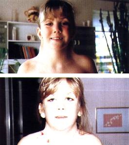
Four girls who attended the 2005 Costello Syndrome Conference in St Louis show several characteristic features, including the friendly, sociable personality associated with Costello syndrome. ... Clinical Characteristics Clinical Description Costello syndrome affects multiple organ systems. ... Differential Diagnosis No other loci have been identified as causative of Costello syndrome [Grant et al 2018]. In earlier series, the 10%-15% of individuals suspected of having Costello syndrome who lacked an HRAS pathogenic variant were subsequently found to have cardiofaciocutaneous (CFC) syndrome [Rauen 2006, Gripp et al 2007] or pathogenic variants in KRAS typical for Noonan syndrome [Lo et al 2009]. Note that while Costello syndrome is difficult to distinguish from CFC syndrome and Noonan syndrome in infants and young children, the distinction between Costello syndrome and Noonan syndrome is clear in older children. Note: The Costello syndrome phenotype would not be mistaken for any known chromosome abnormality syndrome.
For the vascular condition, see Abdominal aortic aneurysm . Triple A syndrome Other names Achalasia–addisonianism–alacrima syndrome or Allgrove syndrome [1] MRI of the brain of 12-year-old boy with triple-A syndrome showing hypoplastic lacrimal glands (yellow arrows.) Specialty Endocrinology Triple-A syndrome or AAA syndrome , is a rare autosomal recessive congenital disorder . ... "Case report of a familial triple: a syndrome and review of the literature" . Medicine . 99 (22): e20474. doi : 10.1097/MD.0000000000020474 . ^ Yadav, Prakarti; Kumar, Deepak; Bohra, Gopal K; Garg, Mahendra K (2020). "Triple A syndrome (Allgrove syndrome) – A journey from clinical symptoms to a syndrome" . ... External links [ edit ] Allgrove (AAA) Syndrome at eMedicine OMIM: 231550 Achalasia Addisonianism Alacrimia syndrome; Triple A syndrome at NIH 's Office of Rare Diseases Classification D ICD - 10 : E27.4 OMIM : 231550 DiseasesDB : 32088 External resources eMedicine : ped/71 Orphanet : 869 v t e Nucleus diseases Telomere Revesz syndrome Nucleolus Treacher Collins syndrome Spinocerebellar ataxia 7 Cajal body : Spinal muscular atrophy Centromere CENPJ Seckel syndrome 4 Other AAAS Triple-A syndrome Laminopathy SMC1A / SMC3 Cornelia de Lange Syndrome SETBP1 Schinzel–Giedion syndrome see also nucleus
A number sign (#) is used with this entry because alacrima, achalasia, and mental retardation syndrome (AAMR) is caused by homozygous mutation in the GMPPA gene (615495) on chromosome 2q35. Description Alacrima, achalasia, and mental retardation syndrome (AAMR) is an autosomal recessive disorder characterized by onset of these 3 main features at birth or in early infancy. ... The disorder shows similarity to the triple A syndrome (231550), but patients with AAMR do not have adrenal insufficiency (summary by Koehler et al., 2013). ... Koehler et al. (2013) noted the considerable clinical overlap with triple A syndrome, but none of the patients had mutations in the AAAS gene (605378).
The association of adrenal and neurologic disease in the triple-A syndrome is similar to that in X-linked adrenoleukodystrophy (300100). ... Lanes et al. (1980) suggested that a degenerative process of progressive nature may be responsible for the 3 features of this syndrome of alacrima-achalasia-addisonianism, and suggested that the 'triple-A syndrome' may be a useful designation. ... They pointed out that the triple-A syndrome has some features of a contiguous gene syndrome. ... Gazarian et al. (1995) proposed that autonomic neuropathy be considered an integral feature of this disorder, making it the 4A syndrome rather than the triple-A syndrome. Chu et al. (1996) reported orthostatic hypotension with compensatory tachycardia in 2 unrelated Hispanic teenagers with Allgrove syndrome. They suggested that Allgrove syndrome could be explained by a 'progressive loss of cholinergic function.'
Triple A syndrome is a very rare multisystem disease characterized by adrenal insufficiency with isolated glucocorticoid deficiency, achalasia, alacrima, autonomic dysfunction and neurodegeneration. ... Clinical description The onset of Triple A syndrome varies between infancy and adulthood. ... Neurological manifestations are diverse: dysautonomia results in dyshidrosis and digestive, sexual, circulatory and urinary dysfunction; pyramidal syndrome and peripheral neuropathy lead to walking difficulties and sometimes to sensory deficit; and bulbar and facial deficiencies are responsible for velar insufficiency, tongue amyotrophy or paresis, orbicularis oris dysfunction and oropharyngeal dysphagia. ... Management and treatment Treatment for Triple A syndrome includes hydrocortisone substitutive therapy, esophageal dilatation or myotomy of the lower esophageal sphincter and artificial tear drops. ... Prognosis If untreated, triple A syndrome may have a high morbidity and prognosis can be severe.
The third major feature of triple A syndrome is a reduced or absent ability to secrete tears (alacrima). Most people with triple A syndrome have all three of these features, although some have only two. Many of the features of triple A syndrome are caused by dysfunction of the autonomic nervous system. ... Many of the neurological symptoms of triple A syndrome worsen over time. People with triple A syndrome frequently develop a thickening of the outer layer of skin (hyperkeratosis) on the palms of their hands and the soles of their feet. ... Frequency Triple A syndrome is a rare condition, although its exact prevalence is unknown.
Triple A syndrome is an inherited condition characterized by three specific features: achalasia , Addison disease , and alacrima (a reduced or absent ability to secrete tears). Most people with triple A syndrome have all three of these features, although some have only two. Several authors published descriptions of a more global autonomic disturbance associated with the original three characteristics, leading one author to suggest the name 4A syndrome (adrenal insufficiency, achalasia, alacrima, autonomic abnormalities). Specific autonomic disturbances described in this syndrome include abnormal pupillary reflexes, poor heart rate variability, and orthostatic hypotension . ... Many of the neurological symptoms of triple A syndrome worsen over time. Triple A syndrome is caused by mutations in the AAAS gene and is inherited in an autosomal recessive pattern.
Campomelic dysplasia is a rare genetic disorder that affects the development of the skeleton, reproductive system, and face. Symptoms of campomelic dysplasia may include bowing of the legs, dislocated hips, small lungs and chest, and external genitalia that do not look clearly male or clearly female (ambiguous genitalia). In addition, infants with campomelic dysplasia have distinctive facial features including a small chin with cleft palate, prominent eyes, flat face, and a large head. Many infants die at an early age due to breathing problems. Campomelic dysplasia usually results from a new genetic change ( DNA variant) in or the near the SOX9 gene. Diagnosis is based on physical findings and x-ray (radiograph) findings and may be confirmed by genetic testing.
The facies in CD resembles the type 2 collagen disorders, such as Stickler syndrome, with marked micrognathia. In the newborn period, the midface is hypoplastic and the eyes are prominent. ... Histologic pancreatic abnormalities have been described in three newborns who died at term from CD; however, pancreatic dysfunction has not been seen in survivors with CD [Piper et al 2002]. Ischiopubic-patella syndrome (IPP). The phenotypic description of IPP is limited to findings in the pelvis and legs including hypoplastic patellae, hypoplastic lesser trochanters, and defective ischiopubic ossification. ... The milder type 2 collagenopathy, Stickler syndrome, may also be considered in the differential as the facial features are very similar.
Clinical Features In a 5-generation family with multiple musculoskeletal anomalies, Stalker and Zori (1997) described an apparently new malformation syndrome with autosomal dominant inheritance. ... Stalker and Zori (1997) considered the syndrome in this family to be distinct from other reported syndromes, including the disorder reported by Chitayat et al. (1991) in 2 half brothers (see 311895).
Campomelic dysplasia is a severe disorder that affects development of the skeleton, reproductive system, and other parts of the body. This condition is often life-threatening in the newborn period. The term "campomelic" comes from the Greek words for "bent limb." Affected individuals are typically born with bowing of the long bones in the legs, and occasionally, bowing in the arms. Bowing can cause characteristic skin dimples to form over the curved bone, especially on the lower legs. People with campomelic dysplasia usually have short legs, dislocated hips, underdeveloped shoulder blades, 11 pairs of ribs instead of 12, bone abnormalities in the neck, and inward- and upward-turning feet (clubfeet ).
Clinical Features Caffey (1947) reported a congenital syndrome with prenatal bowing and thickening of tubular bones with multiple cutaneous dimples in the arms and legs. ... Severe anomalies of the lower cervical spine may lead to an appearance of pterygium colli. Pterygium syndrome was a referral diagnosis in at least 1 case. ... Houston et al. (1983) reported 17 cases of the campomelic syndrome and a follow-up of one of the original patients (by then 17 years old) of Maroteaux et al. (1971). ... In angiographic studies in 4 patients with the campomelic syndrome, Rodriguez (1993) found striking abnormalities, particularly absence or marked deficiency of the anterior tibial artery. One of the 4, a phenotypic female with a 46,XY karyotype, lacked lower limb bowing and the talipes equinovarus typical of campomelic syndrome. This infant was thought to constitute a further example of campomelic syndrome without campomelia.
It also includes dislocated hips, short bowed femura and tibiae (lower limbs more frequently than upper limbs), pretibial skin dimples (bowing of the lower leg is often associated with a skin dimple over the apex of curve) and clubfeet. A few cases of a variant syndrome, referred to as ``acampomelic campomelic dysplasia'' have been described. ... Differential diagnosis In the prenatal period, differential diagnoses include osteogenesis imperfecta type 2 and 3, hypophosphatasia and thanatophoric dysplasia and Stuve-Wiedemann syndrome; after birth, spondyloepiphyseal dysplasia congenita (SEDC), diastrophic dysplasia and Larsen syndrome may be considered.
Khajavi et al. (1976) recognized three varieties of campomelic syndrome: (1) the long-limb form, in which the bent bones are of normal width and only slightly shortened and the arms are rarely involved; (2) the short-limbed form, in which the bent bones are short and wide; and (3) a short-limbed form with associated cloverleaf skull deformity ('Kleeblattschaedel'). ... Mellows et al. (1980) described XX female sibs with camptomelic syndrome of the long-limbed variety. Both infants died soon after birth.
A number sign (#) is used with this entry because autosomal recessive dyskeratosis congenita-2 (DKCB2) is caused by homozygous or compound heterozygous mutation in the NOLA2 gene (606470), also known as NHP2, on chromosome 5q35. Description Dyskeratosis congenita is a multisystem disorder caused by defective telomere maintenance. Clinical manifestations include mucocutaneous abnormalities, bone marrow failure, and an increased predisposition to cancer, among other variable features (summary by Vulliamy et al., 2008). For a discussion of genetic heterogeneity of dyskeratosis congenita, see DKCA1 (127550). Clinical Features Vulliamy et al. (2008) reported a Turkish man with nail dystrophy, thrombocytopenia, testicular atrophy, opportunistic infections, growth and mental retardation, liver cirrhosis, and intracranial calcification.
A number sign (#) is used with this entry because autosomal recessive dyskeratosis congenita-3 (DKCB3) is caused by compound heterozygous mutation in the TCAB1 gene (WRAP53; 612661) on chromosome 17p13. Description Dyskeratosis congenita is a genetic disorder of defective tissue maintenance, impaired stem cell function, and cancer predisposition caused by short telomeres resulting from a defect in telomerase. Clinical manifestations may be seen in the skin as leukoplakia, nail dystrophy, and reticular pigmentation, in the bone marrow as pancytopenia, and in the lung as pulmonary fibrosis, as well as in other tissues (summary by Zhong et al., 2011). For a discussion of genetic heterogeneity of dyskeratosis congenita, see DKCA1 (127550). Clinical Features Zhong et al. (2011) reported 2 unrelated patients with the classic triad of DKC, including oral leukoplakia, abnormal skin pigmentation, and nail dystrophy.
People with dyskeratosis congenita also have an increased risk of developing several life-threatening conditions, including pulmonary fibrosis, bone marrow failure, aplastic anemia, myelodysplastic syndrome, leukemia, and other cancers. The severity of dyskeratosis congenita varies widely among affected individuals.
Description Dyskeratosis congenita is a bone marrow failure syndrome classically characterized by the triad of abnormal skin pigmentation, nail dystrophy, and leukoplakia of the oral mucosa. ... Autosomal dominant cases had a milder variant of the syndrome, the median age at diagnosis being 28 years and the median survival longer than 50 years.
., 2007 and Bessler et al., 2010). Hoyeraal-Hreidarsson syndrome (HHS) refers to a clinically severe variant of DKC that is characterized by multisystem involvement and early onset in utero. ... Genetic Heterogeneity of Dyskeratosis Congenita and Hoyeraal-Hreidarsson Syndrome Dyskeratosis congenita is a genetically heterogeneous disorder, showing autosomal recessive, autosomal dominant, and X-linked inheritance. ... X-linked recessive DKCX (305000) is caused by mutation in the dyskerin gene (DKC1; 300126) on Xq28. Hoyeraal-Hreidarsson syndrome, the severe clinical variant of DKC, can be caused by mutation in several different DKC-associated genes; see, e.g., DKC1 (300136), TINF2 (604319), TERT (187270), and RTEL1 (608833). ... Patients with dyskeratosis congenita have a premature aging syndrome that can be caused by mutations in the RNA or catalytic component of telomerase (see TERC, 602322 and TERT, 187270).
A rare ectodermal dysplasia syndrome that often presents with the classic triad of nail dysplasia, skin pigmentary changes, and oral leukoplakia associated with a high risk of bone marrow failure (BMF) and cancer. ... Patients are at high risk of progressive BMF and may develop myelodysplastic syndrome or acute myelogenous leukemia at any age (the risk increasing with age). ... Differential diagnosis Differential diagnosis includes palmoplantar keratoderma-spastic paralysis syndrome, nail-patella syndrome, autosomal dominant nail dysplasia, poikiloderma with netropenia, Fanconi anemia, Diamond-Blackfan anemia, Shwachman-Diamond Anemia, idiopathic aplastic anemia, idiopathic pulmonary fibrosis, Coats plus syndrome.
Two forms of DC with more severe manifestations have been identified: Hoyeraal Hreidarsson syndrome and Revesz syndrome (see Severe Forms of DC). ... Intrauterine growth retardation has been noted in children with the more severe Hoyeraal Hreidarsson syndrome or Revesz syndrome variants. Developmental delay may be present in some. It can be more pronounced in persons with the Hoyeraal Hreidarsson syndrome or Revesz syndrome variants. Ophthalmic. ... Although most persons with DC have normal psychomotor development and normal neurologic function, significant developmental delay is present in the Hoyeraal Hreidarsson syndrome and Revesz syndrome variants. Cerebellar hypoplasia is present in the Hoyeraal Hreidarsson syndrome variant (Figure 2) and intracranial calcifications have been reported in the Revesz syndrome variant. ... Persons with DKC1 or TINF2 pathogenic variants may have Hoyeraal Hreidarsson syndrome. Persons with Revesz syndrome may have TINF2 pathogenic variants.
Lowry (1983) discussed the overlap of the Smith-Lemli-Opitz syndrome (270400) and the Meckel syndrome (249000). The 2 syndromes had apparently not been seen together in the same sibship until the report by Casamassima et al. (1987). These workers described a new syndrome in 3 members of 1 sibship: in the first 2 children the disorder resembled the Smith-Lemli-Opitz syndrome and in the third it resembled the Meckel syndrome. In addition, all 3 children had cerebellar defects similar to those described in the Joubert syndrome (213300). The affected children were 2 sisters and a brother with nonconsanguineous parents of mixed European ancestry. ... The third patient, whose condition resembled Meckel syndrome, had a large occipital meningoencephalocele, postaxial hexadactyly, immature glomeruli and dilated collecting ducts in the kidney, and periportal fibrosis and bile duct accentuation in the liver.
Hepatic fibrosis-renal cysts-intellectual disability syndrome is a rare, syndromic intellectual disability characterized by early developmental delay with failure to thrive, intellectual disability, congenital hepatic fibrosis, renal cystic dysplasia, and dysmorphic facial features (bilateral ptosis, anteverted nostrils, high arched palate, and micrognathia).
In addition to agenesis, other degrees of callosal defects exist, including hypoplasia (underdevelopment or thinness), hypogenesis (partial agenesis) or dysgenesis (malformation). [1] ACC is found in many syndromes and can often present alongside hypoplasia of the cerebellar vermis . ... Other characteristics sometimes associated with callosal disorders include seizures , spasticity , early feeding difficulties and/or gastric reflux, hearing impairments, abnormal head and facial features, and intellectual disability. [1] Associated brain anomalies [ edit ] Brain anomalies that can sometimes occur in syndromes that cause callosal disorders include: [1] [5] Aplasia of the cerebellar vermis Chiari malformation Colpocephaly Dandy–Walker syndrome Holoprosencephaly Hydrocephalus Neuronal migration disorders such as grey matter heterotopia Schizencephaly Associated syndromes and conditions [ edit ] Some syndromes that frequently include ACC are: Acrocallosal syndrome Aicardi syndrome Andermann syndrome Donnai–Barrow syndrome Dwarfism FG syndrome L1CAM syndrome Microcephalic osteodysplastic primordial dwarfism type II Mowat–Wilson syndrome Oculocerebrocutaneous syndrome Saal Bulas syndrome Septo-optic dysplasia ( optic nerve hypoplasia ) Shapiro syndrome Vici syndrome Some conditions that can sometimes be associated with ACC or other callosal disorders include: 1p36 deletion syndrome 13q deletion syndrome CDK13-related disorder Craniofacial abnormalities and other oral and maxillofacial pathologies Fetal alcohol syndrome Fetal warfarin syndrome Genitopatellar syndrome Gomez-Lopez-Hernandez syndrome Joubert syndrome Lujan–Fryns syndrome Marden–Walker syndrome Maternal nutritional deficiencies or infections Metabolic disorders Okamoto syndrome Opitz G/BBB syndrome Pascual-Castroviejo syndrome Pitt–Hopkins syndrome Sensenbrenner syndrome Strømme syndrome Triploid syndrome Trisomy 9 Xia-Gibbs syndrome Causes [ edit ] Agenesis of the corpus callosum is caused by disruption to development of the fetal brain between the 3rd and 12th weeks of pregnancy. [6] In most cases, it is not possible to know what caused an individual to have ACC or another callosal disorder. ... The cilia defects adversely affect "numerous critical developmental signaling pathways" essential to cellular development and thus offer a plausible hypothesis for the often multi-symptom nature of a large set of syndromes and diseases. Known ciliopathies include primary ciliary dyskinesia , Bardet–Biedl syndrome , polycystic kidney and liver disease , nephronophthisis , Alström syndrome , Meckel–Gruber syndrome and some forms of retinal degeneration . [7] Cocaine and other street drugs [ edit ] In utero exposure to cocaine , heroin , amphetamines and phenylpropanolamine can lead to agenesis of corpus callosum. [8] Diagnosis [ edit ] Callosal disorders can be diagnosed through brain imaging studies or during autopsy. [6] They may be diagnosed through an MRI , CT scan , Sonography , prenatal ultrasound , or prenatal MRI. [1] Treatment [ edit ] There are currently no specific medical treatments for callosal disorders, but individuals with ACC and other callosal disorders may benefit from a range of developmental therapies, educational support, and services. ... "Clinical, genetic and imaging findings identify new causes for corpus callosum development syndromes" . Brain . 137 (6): 1579–1613. doi : 10.1093/brain/awt358 . ... External links [ edit ] Classification D OMIM : 217990 MeSH : D061085 DiseasesDB : 29900 External resources eMedicine : radio/193 Patient UK : Agenesis of the corpus callosum v t e Congenital malformations and deformations of nervous system Brain Neural tube defect Anencephaly Acephaly Acrania Acalvaria Iniencephaly Encephalocele Chiari malformation Other Microcephaly Congenital hydrocephalus Dandy–Walker syndrome other reduction deformities Holoprosencephaly Lissencephaly Microlissencephaly Pachygyria Hydranencephaly Septo-optic dysplasia Megalencephaly Hemimegalencephaly CNS cyst Porencephaly Schizencephaly Polymicrogyria Bilateral frontoparietal polymicrogyria Spinal cord Neural tube defect Spina bifida Rachischisis Other Currarino syndrome Diastomatomyelia Syringomyelia
The former, probably the most common type of ACC, occurs in all ACC syndromes in which Probst bundles are seen. ... A second type of confusion with ACC occurs when there is degeneration or atrophy of the corpus callosum resulting in striking thinning, as in the Lyon syndrome (225740) and other syndromes in which the corpus callosum is thin but not foreshortened. Molecular Genetics Dobyns (1996) reviewed the genetics of agenesis of the corpus callosum and noted that ACC has been reported in more than 20 autosomal malformation syndromes. These include Miller-Dieker syndrome (MDLS; 247200), Rubinstein-Taybi syndrome (RSTS; 180849), acrocallosal syndrome (ACLD; 200990), and Joubert syndrome (JBTS; 213300). Dobyns (1996) also identified 17 X-linked malformation syndromes with ACC, including pyruvate dehydrogenase deficiency (312170). ... One of the best documented of these is del(4p16), or Wolf-Hirschhorn syndrome (194190). ACC malformation syndromes have also been associated with cytogenetic deletions involving Xp22.3 and Xq13-q21 (Dobyns, 1996).
This birth defect can occur as an isolated condition or combined with other cerebral abnormalities, including Arnold-Chiari malformation , Dandy-Walker syndrome , schizencephaly (clefts or deep divisions in brain tissue), and holoprosencephaly (failure of the forebrain to divide into lobes.) It may also occur as part of other diseases such as Aicardi syndrome , (which only affect girls, includes corpus callosum agenesis, and other problems) or Andermann syndrome or it can also be associated with malformations in other parts of the body, such as midline facial defects.
GRACILE syndrome Other names Growth delay-aminoaciduria-cholestasis-iron overload-lactic acidosis-early death (GRACILE) syndrome, Finnish lethal neonatal metabolic syndrome (FLNMS), lactic acidosis, Finnish, with hepatic hemosiderosis, Fellman syndrome This condition is inherited in an autosomal recessive manner. GRACILE syndrome is a very rare lethal autosomal recessive genetic disorder, one of the Finnish heritage diseases . ... "GRACILE syndrome, a lethal metabolic disorder with iron overload, is caused by a point mutation in BCS1L" . ... CS1 maint: uses authors parameter ( link ) ^ a b "GRACILE syndrome - Conditions - GTR - NCBI" . www.ncbi.nlm.nih.gov . ... Retrieved 2020-04-03 . ^ a b c Reference, Genetics Home. "GRACILE syndrome" . Genetics Home Reference . Retrieved 2020-04-03 .
GRACILE syndrome is a severe disorder that begins before birth. ... Within the first day of life, infants with GRACILE syndrome have a buildup of a chemical called lactic acid in the body (lactic acidosis). ... Frequency GRACILE syndrome is found almost exclusively in Finland, where it is estimated to affect 1 in 47,000 infants. At least 32 affected infants have been described in the medical literature. Causes GRACILE syndrome is caused by a mutation in the BCS1L gene. ... As a result, complex III activity and oxidative phosphorylation are decreased in these organs in people with GRACILE syndrome. Without energy, these organs become damaged, leading to many of the features of GRACILE syndrome.
Epidemiology The typical GRACILE syndrome is prevalent in Finland, where it has an incidence of about 1/50,000 births. ... The hearing response assessed with BAEP has been abnormal. Etiology GRACILE syndrome is caused by a mutation in BCS1L , located on chromosome 2q35, encoding a protein essential in the assembly of complex III in the mitochondrial respiratory chain. ... Differential diagnosis Other mitochondrial hepatopathies such as Pearson syndrome should be excluded. Disorders of mitochondrial fatty acid oxidation and Krebs cycle disorders may also mimic GRACILE syndrome. Antenatal diagnosis In families with a previous case of GRACILE syndrome or renal tubulopathy, encephalopathy, liver failure, the causative BCS1L mutation can be investigated antenatally. Genetic counseling GRACILE syndrome is inherited autosomal recessively so genetic counseling should be offered to affected families.
GRACILE syndrome is an inherited metabolic disease. ... Those that do survive this period generally do not live past 4 months despite receiving treatment. GRACILE syndrome is caused by a mutation in the BCS1L gene, and it is inherited in an autosomal recessive pattern.
A number sign (#) is used with this entry because GRACILE syndrome is caused by homozygous mutation in the BCS1L gene (603647) on chromosome 2q35. ... Molecular Genetics In Finnish patients with GRACILE syndrome, Visapaa et al. (2002) identified a homozygous mutation in the BCS1L gene that resulted in a ser78-to-gly (S78G; 603647.0005) substitution. They also identified 5 different mutations in the BCS1L gene in 3 British infants, originally reported by Morris et al. (1995), with symptoms resembling those of GRACILE syndrome but with the additional features of complex III deficiency and neurologic symptoms. ... Nomenclature Fellman (2001) suggested the acronymic designation GRACILE syndrome for this disorder based on the cardinal clinical findings: growth retardation, amino aciduria, cholestasis, iron overload, lactic acidosis, and early death. History Because the ABCB6 gene (605452) maps to the same region of 2q33-q37 as the GRACILE syndrome and is involved in iron homeostasis, it was considered a highly probable candidate gene for this syndrome (Mitsuhashi et al., 2000; Lill and Kispal, 2001).
Turner syndrome is a chromosomal condition that affects development in females. The most common feature of Turner syndrome is short stature, which becomes evident by about age 5. ... A small percentage of females with Turner syndrome retain normal ovarian function through young adulthood. ... Women with Turner syndrome caused by X chromosome mosaicism are said to have mosaic Turner syndrome. ... The loss of one copy of this gene likely causes short stature and skeletal abnormalities in women with Turner syndrome. Learn more about the gene and chromosome associated with Turner syndrome SHOX x chromosome Inheritance Pattern Most cases of Turner syndrome are not inherited.
Turner syndrome may be diagnosed before birth (prenatally), during infancy or in early childhood. ... Girls and women with Turner syndrome need ongoing medical care from a variety of specialists. ... The genetic changes of Turner syndrome may be one of the following: Monosomy. ... Complications Turner syndrome can affect the proper development of several body systems, but this varies greatly among individuals with the syndrome. ... Peer groups for girls with Turner syndrome can help reinforce self-esteem and provide a social network of people who understand how to live with Turner syndrome.
Turner syndrome is a chromosomal disorder associated with the complete or partial absence of an X chromosome. ... Etiology X-chromosome monosomy is responsible for less than half of the cases of Turner syndrome and a large majority of cases are caused by the presence of a mosaicism (45,X) and/or an abnormal X or Y chromosome (deletion, isochromosome X, dicentric chromosome).
They presented 7 patients with 5p deletion syndrome, with ages ranging from 16 to 47 years. ... Clinical Variability Ladekarl (1968) reported a patient with features of cri-du-chat syndrome and Goldenhar syndrome (164210) associated with a 5q deletion. ... The association of Goldenhar syndrome and cri-du-chat syndrome in this patient suggested that the chromosome 5p14 locus may harbor a gene implicated with Goldenhar syndrome. Population Genetics The cri-du-chat syndrome appears to be one of the most common human deletion syndromes, with an incidence varying between 1 in 20,000 to 1 in 50,000 births (Niebuhr, 1978). ... A thrombospondin-like gene and 3 other cDNAs were considered candidate genes for the cri-du-chat contiguous gene deletion syndrome. Cerruti Mainardi et al. (2001) studied 80 patients with cri-du-chat syndrome.
Differential diagnosis Differential diagnosis includes Mowat-Wilson and Wolf-Hirschhorn syndromes, 1p36 monosomy, 17q21 microdeletion, and other chromosomal anomalies. ... Noninvasive prenatal testing (NIPT) is currently commercially available in some countries and can be used as a screening method for some microdeletion syndromes, including Cri-du-chat syndrome. ... Genetic counseling Genetic counseling, including communication of risk of recurrence, family orientation, and anticipatory supervision for common complications, is possible for cases with a diagnosis of Cri-du-chat syndrome. Parents should be tested in order to rule out a balanced translocation.
Cri du chat syndrome is present from birth and affects growth and development. ... People with this condition typically have intellectual disability, developmental and speech delay, and behavioral issues. Cri du chat syndrome is due to a missing piece (deletion) of a specific part of chromosome 5 known as the 'p' arm. ... This deletion occurs very early in the development of an embryo and cri du chat syndrome is usually not inherited in families.
Cri-du-chat (cat's cry) syndrome, also known as 5p- (5p minus) syndrome, is a chromosomal condition that results when a piece of chromosome 5 is missing . ... Affected individuals also have distinctive facial features, including widely set eyes (hypertelorism), low-set ears, a small jaw, and a rounded face. Some children with cri-du-chat syndrome are born with a heart defect. Frequency Cri-du-chat syndrome occurs in an estimated 1 in 20,000 to 50,000 newborns. ... The signs and symptoms of cri-du-chat syndrome are probably related to the loss of multiple genes on the short arm of chromosome 5. ... They are working to determine how the loss of other genes in this region contributes to the characteristic features of cri-du-chat syndrome. Learn more about the gene and chromosome associated with Cri-du-chat syndrome CTNND2 chromosome 5 Inheritance Pattern Most cases of cri-du-chat syndrome are not inherited. ... About 10 percent of people with cri-du-chat syndrome inherit the chromosome abnormality from an unaffected parent.
Goal 2 Review the genetic causes of FGFR craniosynostosis syndromes. Goal 3 Provide an evaluation strategy to identify the genetic cause of an FGFR craniosynostosis syndrome in a proband. ... In primary craniosynostosis, abnormal biology of the suture causes premature suture closure, as in FGFR craniosynostosis syndromes. Primary craniosynostosis can be isolated or part of a syndrome. ... Among individuals with apparently nonsyndromic unicoronal craniosynostosis the prevalence of any syndrome was 17%; Muenke syndrome was identified in 10%. ... Among individuals with apparently isolated bicoronal craniosynostosis, Muenke syndrome was diagnosed in 38%; no other syndromes were identified. ... Additional syndromes that should be considered are included in Table 2.
Sertoli cell-only syndrome (SCO syndrome) is a cause of male infertility . In SCO syndrome, only Sertoli cells (cells that nurture the immature sperm) line the seminiferous tubules (tubes inside the testicles where sperm develop). ... Other signs and symptoms are rare, but in some cases there could be an underlying cause of SCO syndrome that causes other symptoms, such as Klinefelter syndrome . ... Other causes include exposure to chemicals or toxins, history of radiation therapy, and history of severe trauma. Diagnosis of SCO syndrome is confirmed with testicular biopsy . Although there is currently no effective treatment, assisted reproductive technology may assist some men with SCO syndrome in being able to have children.
Description In the evaluation of male infertility, the Sertoli cell-only (SCO) syndrome is diagnosed on testicular biopsy when either no germ cells are visible in any seminiferous tubules (SCO type I) or germ cells are present in a minority of tubules (SCO type II). ... There is evidence that Sertoli cell-only syndrome can be caused by interstitial deletions in the 'azoospermia factor' (AZF) region on the long arm of the Y chromosome (SPGFY1; 400042). ... The authors suggested that in this boy they had an opportunity to observe the Del Castillo syndrome at an earlier stage than had previously been possible. They suggested that, as in other similar conditions such as the testicular feminization syndrome (300068) and the Reifenstein syndrome (312300), the inheritance is either X-linked or male-limited autosomal dominant. ... In 8 of 111 patients (7.2%) with Sertoli cell-only syndrome, Stouffs et al. (2005) found the same 3 changes in the nucleotide sequence of the USP26 gene, all on the same allele: an insertion, 370-371insACA, and 2 transitions, 494T-C and 1423C-T.
Hermansky–Pudlak syndrome Other names Albinism with hemorrhagic diathesis and pigmented reticuloendothelial cells, Delta storage pool disease Hermansky–Pudlak syndrome is inherited via autosomal recessive manner Specialty Endocrinology Heřmanský–Pudlák syndrome (often written Hermansky–Pudlak syndrome or abbreviated HPS ) is an extremely rare autosomal recessive [1] disorder which results in oculocutaneous albinism (decreased pigmentation ), bleeding problems due to a platelet abnormality ( platelet storage pool defect ), and storage of an abnormal fat-protein compound ( lysosomal accumulation of ceroid lipofuscin). ... "Genetic testing for oculocutaneous albinism type 1 and 2 and Hermansky–Pudlak syndrome type 1 and 3 mutations in Puerto Rico" . ... PMID 12923531 . ^ "CIGNA - Hermansky–Pudlak Syndrome" . Retrieved 24 November 2008 . ^ "Hermansky-Pudlak Syndrome: Background, Pathophysiology, Epidemiology" . 13 June 2017. Cite journal requires |journal= ( help ) ^ Hermansky–Pudlak Syndrome . University of Washington, Seattle. 1993 . ... "Hermansky–Pudlak syndrome in a Swiss population" (Free full text) .
A number sign (#) is used with this entry because Hermansky-Pudlak syndrome-3 (HPS3) is caused by homozygous or compound heterozygous mutation in the HPS3 gene (606118) on chromosome 3q24. For a phenotypic description and a discussion of genetic heterogeneity of Hermansky-Pudlak syndrome, see HPS1 (203300). Clinical Features Hazelwood et al. (1997) ascertained 2 individuals with Hermansky-Pudlak syndrome from central Puerto Rico who lacked the 16-bp duplication in the HPS1 gene (604982.0001), exhibited significant amounts of normal-sized HPS1 gene mRNA by Northern blot analysis, and had haplotypes in the HPS1 region of chromosome 10 that were different from the haplotype of every 16-bp duplication patient.
Hermansky-Pudlak syndrome (HPS) affects multiple body systems and includes bleeding and visual problems, and abnormally light coloring of the skin, hair, and eyes ( oculocutaneous albinism ).
Genetic Heterogeneity of Hermansky-Pudlak Syndrome HPS2 (608233) is caused by mutation in the AP3B1 gene (603401) on chromosome 5q14. ... Two families, each with 2 sibs affected with this syndrome, had come to the attention of Hermansky (1963). This syndrome is clearly different from the Chediak-Higashi syndrome (214500) because no qualitative changes of leukocytes are found in Hermansky syndrome and no pigmented macrophages are found in Chediak-Higashi syndrome. ... Kinnear and Tuddenham (1985) reported 4 cases of Hermansky-Pudlak syndrome. Cutaneous malignant melanoma developed in 1. ... Hermansky-Pudlak syndrome due to mutation in a gene on 3q24 (606118) is known as HPS3 (Anikster et al., 2001).
Hermansky-Pudlak syndrome (HSP) is a multi-system disorder characterized by oculocutaneous albinism, bleeding diathesis and, in some cases, neutropenia, pulmonary fibrosis, or granulomatous colitis. ... Differential diagnosis Differential diagnoses include other forms/causes of oculocutaneous albinism, i.e. , X-linked ocular albinism, Chediak-Higashi syndrome, Griscelli syndrome, Cross syndrome, pulmonary fibrosis and hemophagocytic lymphohistiocytosis.
A number sign (#) is used with this entry because Hermansky-Pudlak syndrome-2 (HPS2) is caused by homozygous or compound heterozygous mutation in the gene encoding the beta-3A subunit of the AP3 complex (AP3B1; 603401) on chromosome 5q14. For a general phenotypic description and a discussion of genetic heterogeneity of Hermansky-Pudlak syndrome, see HPS1 (203300). Description Hermansky-Pudlak syndrome (HPS) is a rare autosomal recessive disease characterized by platelet defects and oculocutaneous albinism. ... Inheritance The transmission pattern of Hermansky-Pudlak syndrome in the family reported by Kotzot et al. (1994) was consistent with autosomal recessive inheritance. ... Lack of the beta-3A subunit of this complex causes Hermansky-Pudlak syndrome type 2 (Fontana et al., 2006). ... Taken together, these observations suggested that type 2 Hermansky-Pudlak syndrome is characterized by defects of innate immunity.
A number sign (#) is used with this entry because of evidence that Hermansky-Pudlak syndrome-9 (HPS9) is caused by homozygous mutation in the PLDN gene (BLOC1S6; 604310), which encodes pallidin, on chromosome 15q21. ... For a general phenotypic description and discussion of genetic heterogeneity of Hermansky-Pudlak syndrome, see HPS1 (203300). Clinical Features Badolato et al. (2012) studied a 17-year-old northern Italian girl with oculocutaneous albinism, nystagmus, and normal neurologic development who presented with recurrent cutaneous infections but without hemorrhagic episodes. ... Molecular Genetics In a 17-year-old Italian girl with an HPS-like primary immunodeficiency syndrome, Badolato et al. (2012) performed whole-exome sequencing and identified homozygosity for the same Q78X mutation in the PLDN gene found by Cullinane et al. (2011) in a patient with HPS. ... History Cullinane et al. (2011) reported a child with an HPS-like syndrome and a homozygous Q78X mutation in the PLDN gene; however, this article was retracted based on the finding of the United States Office of Research Integrity that Andrew R.
A number sign (#) is used with this entry because Hermansky-Pudlak syndrome (HPS4) is caused by homozygous or compound heterozygous mutation in the HPS4 gene (606682) on chromosome 22q12. Description Hermansky-Pudlak syndrome-4 (HPS4) is characterized by oculocutaneous albinism in association with easy bruising or a bleeding tendency and absence of platelet dense bodies. ... For a general phenotypic description and a discussion of genetic heterogeneity of Hermansky-Pudlak syndrome, see HPS1 (203300). Clinical Features Anderson et al. (2003) reported 7 patients with HPS and mutations in the HPS4 gene. ... Mapping In the mouse, more than 15 loci manifest mutant phenotypes similar to human Hermansky-Pudlak syndrome (HPS), including 'pale ear' (ep), the mouse homolog of Hermansky-Pudlak syndrome caused by mutation in the HPS1 gene (604982).
A number sign (#) is used with this entry because of evidence that Hermansky-Pudlak syndrome-5 (HPS5) is caused by homozygous mutation in the HPS5 gene (607521) on chromosome 11p14. Description Hermansky-Pudlak syndrome-5 (HPS5) is characterized by oculocutaneous albinism, a bleeding diathesis, and lack of platelet dense bodies. HPS5 appears to be a milder form of the syndrome because the complications present in other forms of HPS, such as pulmonary fibrosis, granulomatous colitis, and neutropenia, have not been reported in HPS5 patients (Ringeisen et al., 2013). For a general phenotypic description and a discussion of genetic heterogeneity of Hermansky-Pudlak syndrome, see HPS1 (203300). Clinical Features Zhang et al. (2003) described a patient with Hermansky-Pudlak syndrome who had a mutation in the HPS5 gene (see MOLECULAR GENETICS).
A number sign (#) is used with this entry because of evidence that Hermansky-Pudlak syndrome-7 (HPS7) is caused by homozygous mutation in the DTNBP1 gene (607145) on chromosome 6p22. For a phenotypic description and a discussion of genetic heterogeneity of Hermansky-Pudlak syndrome, see HPS1 (203300). Clinical Features Li et al. (2003) identified a 48-year-old Portuguese woman with Hermansky-Pudlak syndrome. ... In a 48-year-old Portuguese woman with Hermansky-Pudlak syndrome, Li et al. (2003) identified homozygosity for a nonsense mutation in the DTNBP1 gene (Q103X; 607145.0001). ... Li et al. (2003) showed that dysbindin is a component of the biogenesis of lysosome-related organelles complex-1 (BLOC1), which regulates trafficking to lysosome-related organelles and includes the proteins pallidin (604310), muted (607289), and cappuccino (605695), all of which are associated with Hermansky-Pudlak syndrome in mice. INHERITANCE - Autosomal recessive HEAD & NECK Eyes - Decreased visual acuity - Ocular albinism - Nystagmus Nose - Epistaxis RESPIRATORY Lung - Decreased lung compliance (1 patient) ABDOMEN Gastrointestinal - Granulomatous colitis (1 patient) SKIN, NAILS, & HAIR Skin - Pale skin - Cutaneous albinism Hair - Pale hair HEMATOLOGY - Bleeding tendency due to platelet defect - Defective platelet aggregation - Lack of dense granules in platelets MISCELLANEOUS - Onset in childhood - Two unrelated patients have been reported (last curated January 2015) MOLECULAR BASIS - Caused by mutation in the dystrobrevin-binding protein 1 gene (DTNBP1, 607145.0001 ) ▲ Close
Summary Clinical characteristics. Hermansky-Pudlak syndrome (HPS) is characterized by oculocutaneous albinism, a bleeding diathesis, and, in some individuals, pulmonary fibrosis, granulomatous colitis, or immunodeficiency. ... Diagnosis Suggestive Findings Hermansky-Pudlak syndrome (HPS) should be suspected in a proband with the following clinical and laboratory features. ... Reviewed in Gunay-Aygun et al [2004], these disorders include the following: Chediak-Higashi syndrome (CHS) , caused by biallelic pathogenic variants in LYST . ... Without bone marrow transplantation, individuals with classic Chediak-Higashi syndrome die in childhood. Griscelli syndrome including GS1 (OMIM 214450), GS2 (OMIM 607624), and GS3 (OMIM 609227). ... GS1, GS2, and GS3 are inherited in an autosomal recessive manner. Note: Elejalde syndrome (OMIM 256710) is considered a type of Griscelli syndrome in which neurologic involvement (rather than immunodeficiency and lymphohistiocytosis) occurs.
A number sign (#) is used with this entry because Hermansky-Pudlak syndrome-6 (HPS6) is caused by homozygous or compound heterozygous mutation in the HPS6 gene (607522) on chromosome 10q24. For a phenotypic description and a discussion of genetic heterogeneity of Hermansky-Pudlak syndrome, see HPS1 (203300). Clinical Features Zhang et al. (2003) described a 39-year-old Belgian woman with Hermansky-Pudlak syndrome. ... Schreyer-Shafir et al. (2006) studied a large consanguineous Israeli Bedouin family in which the Hermansky-Pudlak syndrome phenotype was characterized mainly by oculocutaneous albinism. ... In affected members of a large consanguineous Israeli Bedouin family with Hermansky-Pudlak syndrome, who exhibited primarily oculocutaneous albinism, Schreyer-Shafir et al. (2006) identified homozygosity for a 1-bp insertion in the HPS6 gene (607522.0002). Huizing et al. (2009) identified homozygous or compound heterozygous mutations (607522.0003-607522.0007) in the HPS6 gene in 4 unrelated patients with Hermansky-Pudlak syndrome. All mutations except 1 resulted in a truncated protein.
A number sign (#) is used with this entry because of evidence that Hermansky-Pudlak syndrome-8 (HPS8) is caused by homozygous mutation in the BLOC1S3 gene (609762) on chromosome 19q13. For a phenotypic description and a discussion of genetic heterogeneity of Hermansky-Pudlak syndrome, see HPS1 (203300). Clinical Features Morgan et al. (2006) described a large consanguineous Pakistani family in which affected individuals displayed features of incomplete oculocutaneous albinism and platelet dysfunction.
Malpuech facial clefting syndrome Specialty Medical genetics Malpuech facial clefting syndrome , also called Malpuech syndrome or Gypsy type facial clefting syndrome , [1] is a rare congenital syndrome . ... Termed "3MC syndrome", this proposed spectrum includes Malpuech, Michels and Mingarelli-Carnevale (OSA) syndromes. [7] [8] Mutations in the COLLEC11 and MASP1 genes are believed to be a cause of these syndromes. [9] The incidence of Malpuech syndrome is unknown. ... The bilateral type of cleft lip has been reported in Malpuech syndrome. Malpuech syndrome is congenital, being apparent at birth. ... Malpuech syndrome, as with the other disorders within the 3MC syndrome consideration, is caused by mutations in the COLLEC11 and MASP1 genes. ... PMID 11310992 . ^ a b Titomanlio, L.; Bennaceur, S.; Bremond-Gignac, D.; Baumann, C.; Dupuy, O.; Verloes, A. (Sep 2005). "Michels syndrome, Carnevale syndrome, OSA syndrome, and Malpuech syndrome: Variable expression of a single disorder (3MC syndrome)?"
Description The term '3MC syndrome' encompasses 4 rare autosomal recessive disorders that were previously designated the Carnevale, Mingarelli, Malpuech, and Michels syndromes, respectively. ... Reardon et al. (2001) reported a 9-year-old boy whose clinical presentation resembled Malpuech syndrome, but who had radiologic features similar to those seen in patients with Juberg-Hayward syndrome (216100). ... Reardon et al. (2001) suggested that the Malpuech and Juberg-Hayward syndromes may be allelic. Galan-Gomez et al. (2004) described a brother and sister with a constellation of anomalies, some of which suggested Kabuki syndrome (147920) and others Malpuech syndrome. ... Titomanlio et al. (2005) reviewed the phenotypic similarities between Michels syndrome (257920), Malpuech syndrome, and Carnevale syndrome (265050), and suggested that they may represent a single recessive spectrum rather than separate disorders. Titomanlio et al. (2005) proposed that the combined entity could be referred to as the '3MC syndrome' (Malpuech-Michels-Mingarelli-Carnevale syndrome); Mingarelli et al. (1996) had described 2 sisters with what the authors called ocular-skeletal-abdominal (OSA) syndrome (see 265050).
3MC syndrome describes a rare developmental disorder, that unifies the overlapping autosomal recessive disorders previously known as Carnevale, Mingarelli, Malpuech and Michels syndromes, characterized by a spectrum of developmental anomalies that include distinctive facial dysmorphism (i.e. hypertelorism, blepharophimosis, blepharoptosis, highly arched eyebrows), cleft lip and/or palate, craniosynostosis, learning disability, radioulnar synostosis and genital and vesicorenal anomalies.
Mismatch repair cancer syndrome Other names Brain tumor-polyposis syndrome, Glioma-polyposis syndrome This condition is inherited in an autosomal recessive manner Specialty Oncology Mismatch repair cancer syndrome (MMRCS) is a cancer syndrome associated with biallelic DNA mismatch repair mutations. [1] It is also known as Turcot syndrome (after Jacques Turcot, who described the condition in 1959) and by several other names. [1] In MMRCS, neoplasia typically occurs in both the gut and the central nervous system (CNS). [1] In the large intestine , multiple colonic polyps develop; in the CNS, brain tumors . ... You can help by adding to it . ( July 2017 ) History [ edit ] OMIM currently includes "Turcot syndrome" under Mismatch repair cancer syndrome. Turcot syndrome is the association between familial polyposis of the colon and brain tumors [8] like medulloblastoma , malignant glioma . ... "Constitutional mismatch repair-deficiency syndrome: have we so far seen only the tip of an iceberg?". ... PMID 18273873 . ^ "Constitutional Mismatch Repair Deficiency Syndrome" . www.stjude.org . Retrieved 2020-03-10 . ^ " Turcot syndrome " at Dorland's Medical Dictionary ^ Turcot J, Despres JP, St Pierre F (1959).
Turcot syndrome is a condition characterized by multiple adenomatous colon polyps, an increased risk of colorectal cancer, and an increased risk of brain cancer. It may be associated with familial adenomatous polyposis (FAP) or Lynch syndrome (also known as hereditary non-polyposis colorectal cancer or HNPCC). The molecular basis of most Turcot syndrome is either a mutation in APC associated with FAP or a mutation in one of the mismatch repair genes associated with Lynch syndrome ( MLH1 and PMS2 ) . The brain tumors in individuals with APC mutations are typically medulloblastoma , whereas those with mismatch repair mutations are usually glioblastoma multiforme . Turcot syndrome typically follows an autosomal dominant inheritance pattern.
Primary immune deficiency disorder Hyper IgM syndrome type 2 Immunoglobulin M Types Hyper-IgM syndrome type 1,2,3,4 and 5 [1] [2] [3] [4] [5] Diagnostic method MRI, Chest radiography and genetic testing [6] Treatment Allogeneic hematopoietic cell transplantation [7] Hyper IgM Syndrome Type 2 is a rare disease . Unlike other hyper-IgM syndromes , Type 2 patients identified thus far did not present with a history of opportunistic infections. One would expect opportunistic infections in any immunodeficiency syndrome. The responsible genetic lesion is in the AICDA gene found at 12p13. [8] Contents 1 Hyper IgM syndromes 2 Signs and symptoms 3 Cause 4 Pathophysiology 5 Diagnosis 6 Treatment 7 References Hyper IgM syndromes [ edit ] Hyper IgM syndromes is a group of primary immune deficiency disorders characterized by defective CD40 signaling; via B cells affecting class switch recombination (CSR) and somatic hypermutation . ... As a consequence, people with HIGM have an increased susceptibility to infections. [9] [7] [10] Signs and symptoms [ edit ] Hyper IgM syndrome can have the following syndromes: [6] [11] Infection / Pneumocystis pneumonia (PCP), which is common in infants with hyper IgM syndrome, is a serious illness. [9] PCP is one of the most frequent and severe opportunistic infections in people with weakened immune systems. ... Classification D ICD - 10 : D80.5 OMIM : 608106 External resources Orphanet : 101092 v t e Lymphoid and complement disorders causing immunodeficiency Primary Antibody / humoral ( B ) Hypogammaglobulinemia X-linked agammaglobulinemia Transient hypogammaglobulinemia of infancy Dysgammaglobulinemia IgA deficiency IgG deficiency IgM deficiency Hyper IgM syndrome ( 1 2 3 4 5 ) Wiskott–Aldrich syndrome Hyper-IgE syndrome Other Common variable immunodeficiency ICF syndrome T cell deficiency ( T ) thymic hypoplasia : hypoparathyroid ( Di George's syndrome ) euparathyroid ( Nezelof syndrome Ataxia–telangiectasia ) peripheral: Purine nucleoside phosphorylase deficiency Hyper IgM syndrome ( 1 ) Severe combined (B+T) x-linked: X-SCID autosomal: Adenosine deaminase deficiency Omenn syndrome ZAP70 deficiency Bare lymphocyte syndrome Acquired HIV/AIDS Leukopenia : Lymphocytopenia Idiopathic CD4+ lymphocytopenia Complement deficiency C1-inhibitor ( Angioedema / Hereditary angioedema ) Complement 2 deficiency / Complement 4 deficiency MBL deficiency Properdin deficiency Complement 3 deficiency Terminal complement pathway deficiency Paroxysmal nocturnal hemoglobinuria Complement receptor deficiency
Primary immune deficiency disorders Hyper IgM syndrome Immunoglobulin M Specialty Immunology Symptoms Chronic diarrhea [1] Types Hyper-IgM syndrome type 1,2,3,4 and 5 [2] [3] [4] [5] [6] Diagnostic method MRI, Chest radiography [1] Treatment Allogeneic hematopoietic cell transplantation [7] Hyper IgM syndrome describes a group of primary immune deficiency disorders characterized by defective CD40 signaling; via B cells affecting class switch recombination (CSR) and somatic hypermutation . ... Certain insults, usually from encapsulated bacteria and toxin, then have a greater opportunity to damage the body. [1] Diagnosis [ edit ] The diagnosis of hyper IgM syndrome can be done via the following methods and tests: [1] MRI Chest radiography Pulmonary function test Lymph node test Laboratory test (to measure CD40) Types [ edit ] Five types of hyper IgM syndrome have been characterized: Hyper-IgM syndrome type 1 (X-linked), characterized by mutations of the CD40LG gene. ... In this type, B cells cannot receive the signal from T cells to switch classes. [4] Hyper-IgM syndrome type 4 which is a defect in class switch recombination downstream of the AICDA gene that does not impair Somatic Hypermutation. [13] Hyper-IgM syndrome type 5 characterized by mutations of the UNG gene. [5] Treatment [ edit ] In terms of treatment for hyper IgM syndrome, there is the use of allogeneic hematopoietic cell transplantation. ... "The Hyper IgM Syndrome—An Evolving Story" . Pediatric Research . 56 (4): 519–525. doi : 10.1203/01.PDR.0000139318.65842.4A . ... "Update on the hyper immunoglobulin M syndromes" . British Journal of Haematology . 149 (2): 167–180. doi : 10.1111/j.1365-2141.2010.08077.x .
A number sign (#) is used with this entry because autosomal recessive immunodeficiency with hyper-IgM type 2 (HIGM2) is caused by homozygous or compound heterozygous mutation in the gene encoding activation-induced cytidine deaminase (AICDA; 605257) on chromosome 12p13. Description Hyper-IgM syndrome type 2 (HIGM2) is a rare immunodeficiency characterized by normal or elevated serum IgM levels with absence of IgG, IgA, and IgE, resulting in a profound susceptibility to bacterial infections. ... Clinical Features Revy et al. (2000) described 18 patients from 12 unrelated families who were diagnosed with hyper-IgM syndrome defined by markedly diminished serum levels of IgG and IgA with normal or increased serum level of IgM.
Hyper-IgM syndrome without susceptibility to opportunistic infections is a rare, genetic, primary immunodeficiency due to a defect in adaptive immunity disorder characterized by normal or elevated IgM serum levels with low or absent IgG, IgA and IgE serum concentrations, which manifests with recurrent bacterial sinopulmonary and gastrointestinal infections, with frequent lymphoid hyperplasia (peripheral lymphadenopathy, tonsillar hypertrophy), with no increased susceptibility to opportunistic infections.
Reginato et al. (1982) reported 3 sibs with primary hypertrophic osteoarthropathy and widely open cranial sutures and fontanelles. The syndrome was apparent after birth and included digital clubbing, subperiosteal bone formation, and painful soft tissue swelling over bones.
The specific sacral anomaly is distinct to this syndrome. Of 10 affected members in the family reported by O'Riordain et al. (1991), only 4 were symptomatic, 3 of these had the full Currarino syndrome (sacral agenesis, presacral mass, and anorectal malformation), and the fourth had an anterior meningocele with no anorectal problems. ... Lynch et al. (2000) provided a review of the clinical features and molecular basis of Currarino syndrome. Kim et al. (2007) reported 2 Korean families with Currarino syndrome. ... Diagnosis Prenatal Diagnosis Cretolle et al. (2007) reported prenatal diagnosis of Currarino syndrome by ultrasound in a second pregnancy at 22 weeks' gestation. ... In the families they studied, some individuals had the typical scimitar, or sickle-shaped, hemisacrum but were asymptomatic, whereas others had the full Currarino syndrome. Hagan et al. (2000) identified mutations in the HLXB9 gene in 20 of 21 patients with familial Currarino syndrome and in 2 of 7 patients with sporadic Currarino syndrome. ... In affected members of a 3-generation family segregating Currarino syndrome, Urioste et al. (2004) identified a frameshift mutation in the HLXB9 gene (142994.0009).
Currarino syndrome (CS) is a rare congenital disease characterized by the triad of anorectal malformations (ARMs) (usually anal stenosis), presacral mass (commonly anterior sacral meningocele (ASM) or teratoma) and sacral anomalies (i.e. total or partial agenesis of the sacrum and coccyx or deformity of the sacral vertebrae).
Currarino triad or syndrome is an autosomal dominant hereditary condition which is characterized by the triad of sacral agenesis abnormalities (abnormally developed lower spine), anorectal malformation (most commonly in the form of anorectal stenosis ) and presacral mass consisting of a teratoma , anterior sacral meningocele or both.
Cranio-osteoarthropathy (COA) is a form of primary hypertrophic osteoarthropathy (see this term) characterized by delayed closure of the cranial sutures and fontanels, digital clubbing, arthropathy, and periostosis. Epidemiology Prevalence is unknown. To date, about 30 cases have been reported. Clinical description COA presents in childhood with features of primary hypertrophic osteoarthropathy including digital clubbing and clinodactyly of the fingers, eczematous skin eruption, arthropathy and periosteal new bone formation as well as poor neurocranium ossification with delayed closure of the cranial sutures and fontanels, resulting in wide fontanels, and an increased number of wormian bones. COA may also be associated with congenital heart disease. Etiology It is caused by mutations in the HPGD gene (4q33-q34) and is inherited as an autosomal recessive trait; however, heterozygous carriers can have a mild phenotype.
Aplasia cutis congenita is a condition in which there is congenital (present from birth) absence of skin, with or without the absence of underlying structures such as bone. It most commonly affects the scalp, but any location of the body can be affected. While most people with aplasia cutis congenita have no other abnormalities, some people have congenital malformations involving the cardiovascular (heart), gastrointestinal, genitourinary, and central nervous systems. The cause of this condition is unclear and appears to be multifactorial (many different factors appear to play a role); contributing factors may include teratogens, genes, trauma, and compromised blood flow to the skin.
Cutaneous aplasia of the scalp vertex also occurs in Johanson-Blizzard syndrome (243800) and Adams-Oliver syndrome (AOS; 100300). A defect in the scalp is sometimes found in cases of trisomy 13 and in about 15% of cases of deletion of the short arm of chromosome 4, the Wolf-Hirschhorn syndrome (WHS; 194190) (Hirschhorn et al., 1965; Fryns et al., 1973). ... The authors suggested that the family had ACC or a mild form of Adams-Oliver syndrome (see 100300) with preaxial polydactyly as a feature.
Clinical Features Freire-Maia et al. (1980) described congenital absence of skin in the upper or lower limbs or both in 6 members (5 males and 1 female) of 3 inbred sibships of the same kindred. The lesions usually healed spontaneously leaving a clinical appearance like that of the hypotrichotic scar of an old burn. In 6 instances, an affected father married to a relative had an affected child (pseudodominance). There was no trunk or scalp involvement. Portnoy and Metzker (1981) described a disorder affecting a brother and sister with Yemenite parents who were presumed to be unrelated. Clinically, the disorder resembled the Bart type of epidermolysis bullosa dystrophica (132000), which is inherited as an autosomal dominant, but may, in fact, represent prenatal manifestation of epidermolysis bullosa of either the Koebner type (131900), due to a keratin defect, or epidermolysis bullosa dystrophica, due to a defect in COL7A1 (120120).
However, the characteristic skin lesions can occur as one of many symptoms in other conditions, including Johanson-Blizzard syndrome and Adams-Oliver syndrome. These instances are described as syndromic aplasia cutis congenita.
Diagnosis Suggestive Findings Nijmegen breakage syndrome (NBS) should be suspected in individuals with the following clinical and supportive laboratory findings. ... Two reports of gonadal dysfunction in affected females with chromosome instability disorder / syndrome appeared in 1986 [Maraschio et al 1986]. ... Nomenclature The Nijmegen breakage syndrome was described by Weemaes et al [1981]. Three Czech families with Seemanová syndrome [Seemanová et al 1985] were later identified as having NBS. ... The report of Jaspers et al [1988] noted a strong similarity between NBS cells and ataxia-telangiectasia (A-T) cells; however, they also described the NBS cells as genetically distinct from A-T, grouping individuals with either Nijmegen breakage syndrome or Czech breakage syndrome into A-T variant group V1 and Germans with "Berlin breakage syndrome" [Wegner et al 1999] into A-T variant group V2 [Jaspers et al 1988].
Nijmegen breakage syndrome is a rare genetic disease presenting at birth with microcephaly, dysmorphic facial features, becoming more noticeable with age, growth delay, and later-onset complications such as malignancies and infections. ... Differential diagnosis Differential diagnosis includes Fanconi anemia, Bloom syndrome, NBS-like disorder, ataxia-telangectasia-like disorder, LIG4 syndrome, NHEJ1 syndrome and Seckel syndrome (see these terms).
About half of individuals with Nijmegen breakage syndrome develop non-Hodgkin lymphoma, usually before age 15. ... People with Nijmegen breakage syndrome are 50 times more likely to develop cancer than people without this condition. ... Most women with Nijmegen breakage syndrome are unable to have biological children (infertile). ... Causes Mutations in the NBN gene cause Nijmegen breakage syndrome. The NBN gene provides instructions for making a protein called nibrin. ... A buildup of these breaks lead to errors in DNA that can trigger cells to grow and divide abnormally, increasing the risk of cancer in people with Nijmegen breakage syndrome. Additionally, medical radiation or chemotherapy, which is often used to treat cancers, can cause further DNA damage in people with Nijmegen breakage syndrome.
Description The Nijmegen breakage syndrome and the phenotypically indistinguishable Berlin breakage syndrome are autosomal recessive chromosomal instability syndromes characterized by microcephaly, growth retardation, immunodeficiency, and predisposition to cancer. Ataxia-telangiectasia variant-1 is the designation applied to the Nijmegen breakage syndrome and AT variant-2 is the designation for the Berlin breakage syndrome, which differ only in complementation studies. ... Conley et al. (1986) described a 21-year-old woman with growth failure, immunodeficiency, and chromosomal breakage syndrome involving chromosomes 7 and 14. ... Der Kaloustian et al. (1996) suggested that the boy described by Woods et al. (1995) as a patient with Seckel syndrome might have the same variant of Nijmegen breakage syndrome. ... Population Genetics Some of the patients studied by Saar et al. (1997) were Germans in whom the Berlin breakage syndrome had been described and others were Slavic patients in whom the Seemanova syndrome (a synonym for NBS) had been described.