It is distinguished from measles or forms of rubella , though it was considered as a form of viral rash . [2] Although Dukes identified it as a separate entity, it is thought not to be different from scarlet fever caused by exotoxin-producing Streptococcus pyogenes after Keith Powell proposed equating it with the condition currently known as staphylococcal scalded skin syndrome in 1979. [2] [4] It was never associated with a specific pathogen, [5] and the terminology is no longer in use. [2] However, a mysterious rash of unknown cause in school children often gives rise to the question of whether it could be Dukes' disease. [6] Contents 1 Signs and symptoms 2 Diagnosis 3 References 4 External links Signs and symptoms [ edit ] Signs and symptoms may include fever , nausea , vomiting , and diarrhea , along with typical viral symptoms of sensitivity to light , enlarged lymph nodes , sore throat , and possibly brain inflammation . ... External links [ edit ] Classification D ICD - 10 : B09 ICD - 9-CM : 057.8 v t e Skin infections , symptoms and signs related to viruses DNA virus Herpesviridae Alpha HSV Herpes simplex Herpetic whitlow Herpes gladiatorum Herpes simplex keratitis Herpetic sycosis Neonatal herpes simplex Herpes genitalis Herpes labialis Eczema herpeticum Herpetiform esophagitis Herpes B virus B virus infection VZV Chickenpox Herpes zoster Herpes zoster oticus Ophthalmic zoster Disseminated herpes zoster Zoster-associated pain Modified varicella-like syndrome Beta Human herpesvirus 6 / Roseolovirus Exanthema subitum Roseola vaccinia Cytomegalic inclusion disease Gamma KSHV Kaposi's sarcoma Poxviridae Ortho Variola Smallpox Alastrim MoxV Monkeypox CPXV Cowpox VV Vaccinia Generalized vaccinia Eczema vaccinatum Progressive vaccinia Buffalopox Para Farmyard pox : Milker's nodule Bovine papular stomatitis Pseudocowpox Orf Sealpox Other Yatapoxvirus : Tanapox Yaba monkey tumor virus MCV Molluscum contagiosum Papillomaviridae HPV Wart / plantar wart Heck's disease Genital wart giant Laryngeal papillomatosis Butcher's wart Bowenoid papulosis Epidermodysplasia verruciformis Verruca plana Pigmented wart Verrucae palmares et plantares BPV Equine sarcoid Parvoviridae Parvovirus B19 Erythema infectiosum Reticulocytopenia Papular purpuric gloves and socks syndrome Polyomaviridae Merkel cell polyomavirus Merkel cell carcinoma RNA virus Paramyxoviridae MeV Measles Togaviridae Rubella virus Rubella Congenital rubella syndrome ("German measles" ) Alphavirus infection Chikungunya fever Picornaviridae CAV Hand, foot, and mouth disease Herpangina FMDV Foot-and-mouth disease Boston exanthem disease Ungrouped Asymmetric periflexural exanthem of childhood Post-vaccination follicular eruption Lipschütz ulcer Eruptive pseudoangiomatosis Viral-associated trichodysplasia Gianotti–Crosti syndrome v t e Numbered Diseases of Childhood Diseases First Disease (Measles) Second Disease (Scarlet Fever) Third Disease (Rubella) Fourth Disease (Dukes' Disease) Fifth Disease (Erythema Infectiosum) Sixth Disease (Roseola)
Rhabdoid tumor predisposition syndrome (RTPS) is characterized by a high risk of developing cancerous (malignant) growths called rhabdoid tumors. ... Learn more about the genes associated with Rhabdoid tumor predisposition syndrome SMARCA4 SMARCB1 Inheritance Pattern This condition is inherited in an autosomal dominant pattern, which means one copy of the altered gene in each cell is sufficient to predispose the affected individual to rhabdoid tumors.
Genetic counseling ATRT can occur sporadically or as part of a RT predisposition syndrome (familial RT; see this term). Management and treatment No standard of care exists for ATRT.
A number sign (#) is used with this entry because of evidence that urocanase deficiency is caused by mutation in the UROC1 gene (613012) on chromosome 3q21. One such family has been reported. Clinical Features Yoshida et al. (1971) reported a case of urocanase deficiency. Kalafatic et al. (1980) reported 2 sisters with this deficiency. Both had severe mental retardation, short stature, blond hair, and blue eyes. They showed periods of aggression and periods of exaggerated affection-seeking. The family lived near Zagreb, Yugoslavia. The paternal grandmother and great-grandmother were regarded as 'strange' and died at 35 and 42 years, respectively.
Encephalopathy due to urocanase deficiency is an extremely rare histidine metabolism disorder characterized by urocanic aciduria and other variable manifestations including intellectual deficit and intermittent ataxia in the 4 cases reported to date.
A rare inborn error of metabolism characterized by abnormal urinary excretion of D-glyceric acid due to D-glycerate kinase deficiency. Reported manifestations are highly variable and include a severe encephalopathic picture, chronic metabolic acidosis, developmental delay, intellectual disability, microcephaly, seizures, behavioral abnormalities, as well as only mild speech delay and apparently normal development.
EEG showed hypsarrhythmia, consistent with West syndrome. Laboratory studies at age 11 months showed D-glyceric aciduria. ... A female infant was born prematurely of a Mexican mother and had multiple problems due to prematurity, including neonatal respiratory distress syndrome. She also had optic nerve hypoplasia, severe failure to thrive, microcephaly, and seizures.
A number sign (#) is used with this entry because aminoacylase-1 deficiency (ACY1D) is caused by homozygous or compound heterozygous mutation in the ACY1 gene (104620) on chromosome 3p21. Description Aminoacylase-1 deficiency (ACY1D) is a rare autosomal recessive inborn error of metabolism characterized by increased urinary excretion of specific N-actyl amino acids. Most patients show neurologic abnormalities such as intellectual disability, seizures, hypotonia, and motor delay (summary by Ferri et al., 2014). Clinical Features Van Coster et al. (2005) reported an infant with aminoacylase-1 deficiency. He presented neonatally with an acute encephalopathy with onset on the third day of life and duration of about 2 weeks.
An inborn error of metabolism marked by a characteristic pattern of urinary N-acetyl amino acid excretion and neurologic symptoms. Epidemiology Prevalence is unknown but less than 20 cases have been reported in the literature so far. Clinical description Most individuals with ACY1D identified so far are children who underwent selective screening tests for inborn errors of metabolism prompted mainly by delayed psychomotor development or by the occurrence of seizures. However, there is a considerable phenotypic variability between ACY1D individuals. Etiology ACY1D is caused by biallelic mutations in the ACY1 gene (3p21.2).
Aminoacylase 1 deficiency is an inherited disorder that can cause neurological problems; the pattern and severity of signs and symptoms vary widely among affected individuals. Individuals with this condition typically have delayed development of mental and motor skills (psychomotor delay). They can have movement problems, reduced muscle tone (hypotonia), mild intellectual disability, and seizures. However, some people with aminoacylase 1 deficiency have no health problems related to the condition. A key feature common to all people with aminoacylase 1 deficiency is high levels of modified protein building blocks (amino acids), called N -acetylated amino acids, in the urine.
A number sign (#) is used with this entry because congenital erythropoietic porphyria (CEP) is caused by homozygous or compound heterozygous mutation in the uroporphyrinogen III synthase gene (UROS; 606938) on chromosome 10q26. Description The porphyrias are diseases caused by defects in heme synthesis, resulting in the accumulation and increased excretion of porphyrins or porphyrin precursors. They are classified as erythropoietic or hepatic, depending on whether the enzyme deficiency occurs in red blood cells or in the liver (Gross et al., 2000). Desnick and Astrin (2002) provided a comprehensive review of congenital erythropoietic porphyria pathogenesis and treatment. One patient with a phenotype suggestive of congenital erythropoietic anemia was found to have a mutation in the GATA1 gene (305371.0010) that affected UROS expression (see XLTT, 314050).
Congenital erythropoietic porphyria (CEP) is the rarest type of porphyria and is commonly seen in infancy. It is characterized by severe skin photosensitivity that may lead to scarring, blistering, and increased hair growth at the face and back of the hands. Photosensitivity and infection may cause the loss of fingers and facial features. Symptoms of CEP range from mild to severe and may include excessive hair growth throughout the body ( hypertrichosis ), reddish discoloration of the teeth, anemia, and reddish-colored urine. In CEP, there is a defect in the synthesis of heme within the red blood cells of bone marrow.
Disorders to Consider in the Differential Diagnosis of Congenital Erythropoietic Porphyria View in own window Disease Name Gene(s) MOI Clinical Features Overlapping Distinguishing Porphyria cutanea tarda (PCT) type I (OMIM 176090) See footnote 1 Cutaneous photosensitivity w/blistering & friability of skin in sun-exposed areas Facial hypertrichosis Discolored urine Usually manifests in adulthood Distinct biochemical porphyrin profile Porphyria cutanea tarda (PCT) type II UROD AD Hepato-erythropoietic porphyria UROD AR Phenotype similar to PCT Manifests in early childhood Discolored urine Photosensitivity Distinct biochemical porphyrin profile Developmental delay (in some) Hereditary coproporphyria CPOX AD 20% of affected individuals experience photosensitivity w/skin blistering in sun-exposed areas Acute (hepatic) porphyria Acute attacks of abdominal or generalized pain; can be associated w/neurologic symptoms Incompletely penetrant in absence of environmental inducers Usually manifests after puberty Variegate porphyria PPOX AD Myeloid malignancy Elderly adults w/myelodysplastic syndrome may exhibit features of CEP 2, 3 Epidermolysis bullosa simplex (EBS) KRT5 KRT14 AD 4 Fragility of skin resulting in nonscarring blisters caused by little/no trauma Major & minor subtypes share common feature of blistering above dermal-epidermal junction at the ultrastructural level Junctional epidermolysis bullosa (JEB) LAMA3 LAMB3 LAMC2 COL17A1 AR Fragility of skin & mucous membranes, manifest by blistering w/little or no trauma Herlitz JEB (classic severe form): blisters present at birth or become apparent in neonatal period Non-Herlitz JEB: may be mild w/blistering localized to hands, feet, knees, elbows Dystrophic epidermolysis bullosa COL7A1 AR Blisters affecting whole body may be present in neonatal period Oral involvement Corneal erosions Esophageal erosions Severe nutritional deficiency & secondary problems "Mitten" hands & feet >90% lifetime risk of aggressive squamous cell carcinoma AD Blistering, often mild & limited to hands, feet, knees, elbows; heals w/scarring Dystrophic nails possibly the only manifestation AD = autosomal dominant; AR = autosomal recessive; MOI = mode of inheritance 1. 80% of cases are sporadic or acquired (type I PCT).
Congenital erythropoietic porphyria, or Günther disease, is a form of erythropoietic porphyria characterized by very severe and mutilating photodermatosis. Epidemiology Since its description at the end of the 19th century, about 200 cases have been reported in the literature. Clinical description The disease most often manifests at birth with extreme cutaneous photosensitivity that is severe and mutilating. The principle signs include cutaneous lesions that are bullous and rapidly erosive on the surface of skin exposed to the sun and light (hands, face, feet). Urine is often very red/brown and colors the diaper of affected infants.
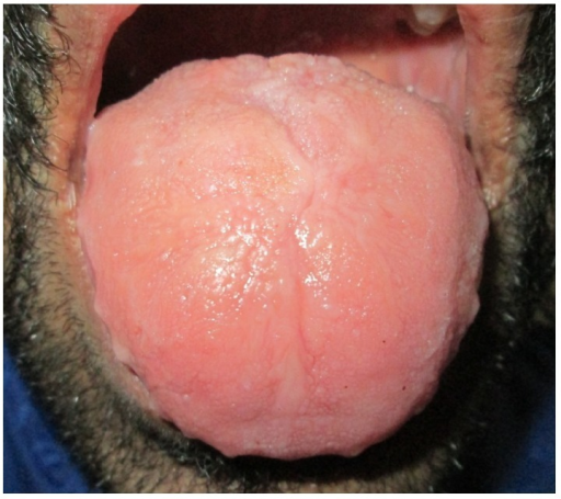
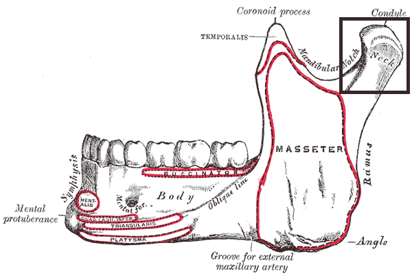
Description The criteria for the hemifacial type of congenital hypertrophy are (1) unilateral enlargement of the viscerocranium bounded superiorly by the frontal bone (not including the eye), inferiorly by the inferior border of the mandible, medially by the midline of the face, and laterally by the ear, the pinna being included within the hypertropic area, and (2) enlargement of all tissues--teeth, bone, and soft tissue--within this area (Rowe, 1962). Clinical Features Rowe (1962) reported 4 cases of hemifacial hypertrophy and reviewed 30 previously reported cases. The left and right sides were almost equally affected. Histologic examination in one case demonstrated that the enlargement resulted from an increased number of cells rather than an increase in cell size. Burchfield and Escobar (1980) described a family in which several members showed mandibular asymmetry and maxillary hypoplasia. Several instances of male-to-male transmission were observed. This was classified by the authors as facial hemihypertrophy.
It may be isolated or related to some syndromes (e.g. Beckwith-Wiedemann, Proteus, Klippel-Trenaunay-Weber, McCune-Albright syndrome, Neurofibromatosis type 1).
SRTD encompasses Ellis-van Creveld syndrome (EVC) and the disorders previously designated as Jeune syndrome or asphyxiating thoracic dystrophy (ATD), short rib-polydactyly syndrome (SRPS), and Mainzer-Saldino syndrome (MZSDS). ... There is phenotypic overlap with the cranioectodermal dysplasias (Sensenbrenner syndrome; see CED1, 218330). For a discussion of genetic heterogeneity of short-rib thoracic dysplasia with or without polydactyly, see SRTD1 (208500). Clinical Features Alby et al. (2015) described 4 unrelated families with a lethal ciliopathy syndrome. In a consanguineous Lebanese family, the mother had 4 spontaneous abortions before 10 weeks' gestation, followed by a female infant born at term who died a few hours after birth. ... Molecular Genetics In a consanguineous Lebanese family in which 2 fetuses exhibited features similar to those of hydrolethalus syndrome (see 236680), including severe hydrocephaly, polydactyly, and skeletal abnormalities, Alby et al. (2015) sequenced the HYLS1 (610693) and KIF7 (611254) genes but found no mutations.
A number sign (#) is used with this entry because of evidence that hypotonia, ataxia, and delayed development syndrome (HADDS) is caused by heterozygous mutation in the EBF3 gene (607407) on chromosome 10q26. Description Hypotonia, ataxia, and delayed development syndrome (HADDS) is a neurodevelopmental syndrome characterized by congenital hypotonia, delayed psychomotor development, variable intellectual disability with speech delay, variable dysmorphic facial features, and ataxia, often associated with cerebellar hypoplasia. ... Molecular Genetics In 2 sibs with hypotonia, ataxia, and delayed development syndrome, Harms et al. (2017) identified a heterozygous mutation in the EBF3 gene (607407.0001).
A number sign (#) is used with this entry because autosomal dominant aplasia of lacrimal and salivary glands (ALSG) is caused by heterozygous mutation in the FGF10 gene (602115) on chromosome 5p12. Lacrimoauriculodentodigital syndrome (LADD; 149730) is an allelic disorder with a more severe phenotype. ... In affected individuals, the misdiagnosis is often made of the more prevalent disorder Sjogren syndrome (270150), an autoimmune condition characterized by keratoconjunctivitis sicca and xerostomia. ... Wiedemann (1991) suggested that the patient of Milunsky et al. (1990) had LADD syndrome (149730). Ferreira et al. (2000) described a Brazilian family in which members of 3 generations had congenital absence of lacrimal puncta and salivary glands in an autosomal dominant pedigree pattern. ... In a mother with ALSG and her daughter with LADD syndrome, Milunsky et al. (2006) identified a heterozygous mutation in the FGF10 gene (602115.0005). The findings in this family indicated that ALSG and LADD syndrome are allelic disorders. The authors suggested that differences in modifier genes may explain the less severe ALSG phenotype in the mother versus the LADD syndrome phenotype in her daughter.
A rare autosomal dominant disorder characterized by aplasia, atresia or hypoplasia of the lacrimal and salivary glands leading to varying features since infancy such as recurrent eye infections, irritable eyes, epiphora, xerostomia, dental caries, dental erosion and oral inflammation.
Contents 1 Cancer risk 2 Serrated polyposis syndrome 3 Histopathology 3.1 Mucin-rich type 3.2 Goblet cell-rich type 3.3 Epithelial misplacement 3.4 Cellular structure 3.5 Immunohistochemistry 3.6 Differential diagnoses 4 References Cancer risk [ edit ] Most hyperplastic polyps are found in the distal colon and rectum . [2] They have no malignant potential, [2] which means that they are no more likely than normal tissue to eventually become a cancer. [ citation needed ] Hyperplastic polyps on the right side of the colon do exhibit a malignant potential. ... This leads to microsatellite instability which can eventually lead to malignant transformation in polyps on the right side of the colon. [ citation needed ] Serrated polyposis syndrome [ edit ] Main article: Serrated polyposis syndrome Serrated polyposis syndrome is a rare condition that has been defined by the World Health Organization as either: [3] ≥5 serrated lesions/polyps proximal to the rectum, all ≥ 5 mm in size, with two lesions ≥10 mm >20 serrated lesions/polyps of any size distributed throughout the large bowel with 5 proximal to the rectum. ... ISBN 978-1-4160-3121-5 . ^ Dekker, E; Bleijenberg, A; Balaguer, F; Dutch-Spanish-British Serrated Polyposis Syndrome, collaboration. (May 2020). "Update on the World Health Organization Criteria for Diagnosis of Serrated Polyposis Syndrome" . ... Last updated 6/2/2015 v t e Digestive system neoplasia GI tract Upper Esophagus Squamous cell carcinoma Adenocarcinoma Stomach Gastric carcinoma Signet ring cell carcinoma Gastric lymphoma MALT lymphoma Linitis plastica Lower Small intestine Duodenal cancer Adenocarcinoma Appendix Carcinoid Pseudomyxoma peritonei Colon/rectum Colorectal polyp : adenoma , hyperplastic , juvenile , sessile serrated adenoma , traditional serrated adenoma , Peutz–Jeghers Cronkhite–Canada Polyposis syndromes: Juvenile MUTYH-associated Familial adenomatous / Gardner's Polymerase proofreading-associated Serrated polyposis Neoplasm: Adenocarcinoma Familial adenomatous polyposis Hereditary nonpolyposis colorectal cancer Anus Squamous cell carcinoma Upper and/or lower Gastrointestinal stromal tumor Krukenberg tumor (metastatic) Accessory Liver malignant : Hepatocellular carcinoma Fibrolamellar Hepatoblastoma benign : Hepatocellular adenoma Cavernous hemangioma hyperplasia : Focal nodular hyperplasia Nodular regenerative hyperplasia Biliary tract bile duct : Cholangiocarcinoma Klatskin tumor gallbladder : Gallbladder cancer Pancreas exocrine pancreas : Adenocarcinoma Pancreatic ductal carcinoma cystic neoplasms : Serous microcystic adenoma Intraductal papillary mucinous neoplasm Mucinous cystic neoplasm Solid pseudopapillary neoplasm Pancreatoblastoma Peritoneum Primary peritoneal carcinoma Peritoneal mesothelioma Desmoplastic small round cell tumor
Ternaux et al. (1979) found serum 5-hydroxytryptamine (5-HT) to be markedly decreased in Down syndrome whereas cerebrospinal fluid levels of 5-hydroxytryptamine and of 5-hydroxyindoleacetic acid were increased. It had generally been agreed that 5-HT is decreased in platelets in Down syndrome. Their measurements reflected this low platelet 5-HT.
Four of these also had congenital cataract and 3 had increased corneal diameter. Possibly the Rieger syndrome (see 180500) should be considered. Eyes - Persistent pupillary membrane remnants - Congenital cataract - Increased corneal diameter Inheritance - Autosomal dominant - ? same as Rieger syndrome ▲ Close
A rare myelodysplastic/myeloproliferative neoplasm characterized by clinical, laboratory, and morphological features of both myelodysplastic syndrome and myeloproliferative neoplasm at onset, in the absence of recent cytotoxic or growth factor therapy, and without Philadelphia chromosome, BCR-ABL1 or PCM1-JAK2 fusion, or rearrangement of PDGFRA, PDGFRB, or FGFR1. Cases of a previously well-defined myeloproliferative neoplasm developing dysplastic features are excluded, and the criteria for any other myelodysplastic/myeloproliferative neoplasm, myelodysplastic syndrome, or myeloproliferative neoplasm are not met.