Though most cases are found impacted within the jaw there are instances where odontomas have erupted into the oral cavity. [8] Contents 1 Types 2 Histopathology 3 Presentation 4 Aetiology 4.1 Gardner's syndrome 5 Treatment 6 Epidemiology 7 References 8 External links Types [ edit ] There are two main types: compound and complex. [9] A compound odontoma consists of the four separate dental tissues ( enamel , dentine , cementum and pulp ) embedded in fibrous connective tissue and surrounded by a fibrous capsule. ... However, odontomas have been related to local trauma, inflammatory and/or infectious processes, hereditary anomalies such as Gardener's syndrome and Hermanns syndrome, odontoblastic hyperactivity, mature odontoblasts and dental lamina remnants (Cell Rests of Serres). [12] Gardner's syndrome [ edit ] Gardner's syndrome is a subtype of Familial adenomatous polyposis . The clinical presentation of this syndrome includes multiple odontomas. This condition has a high risk of malignancy through adenocarcinoma of the bowel. [10] Treatment [ edit ] Most common treatment is surgical enucleation due to well-encapsulated nature of odontomas allowing separation from surrounding bone. [10] [6] If left untreated can result in a dentigerous cyst . [6] [12] Epidemiology [ edit ] Odontomas are thought to be the second most frequent type of odontogenic tumor worldwide (after ameloblastoma ), accounting for about 20% of all cases within this relatively uncommon tumor category which shows large geographic variations in incidence.
Contents 1 Classification 2 Signs and symptoms 3 Causes 4 Diagnosis 5 Prevention 5.1 Controversy 6 Treatment 7 References 8 External links Classification [ edit ] VKDB is classified as early, classical or late depending on when it first starts with each having somewhat different types of bleeding and underlying cause: Classification of vitamin K deficiency in the newborn (VKDB) [2] Syndrome Time of onset Common sites of bleeding Potential causes Early First 24 hours Scalp, skin, brain, chest, abdomen Maternal medications Classical 1-7 days Gut, umbilicus, skin, nose, circumcision Idiopathic , breast feeding Late After day 8 Brain, skin, gut Idiopathic, breast feeding, cholestasis Signs and symptoms [ edit ] VKDB presents typically in the first month of life with bleeding which can be from various locations. ... External links [ edit ] Classification D ICD - 11 : KA8F.0 ICD - 10 : P53 ICD - 9-CM : 776.0 MeSH : D006475 DiseasesDB : 29544 SNOMED CT : 12546009 External resources MedlinePlus : 007320 eMedicine : article/974489 Patient UK : Vitamin K deficiency bleeding v t e Conditions originating in the perinatal period / fetal disease Maternal factors complicating pregnancy, labour or delivery placenta Placenta praevia Placental insufficiency Twin-to-twin transfusion syndrome chorion / amnion Chorioamnionitis umbilical cord Umbilical cord prolapse Nuchal cord Single umbilical artery presentation Breech birth Asynclitism Shoulder presentation Growth Small for gestational age / Large for gestational age Preterm birth / Postterm pregnancy Intrauterine growth restriction Birth trauma scalp Cephalohematoma Chignon Caput succedaneum Subgaleal hemorrhage Brachial plexus injury Erb's palsy Klumpke paralysis Affected systems Respiratory Intrauterine hypoxia Infant respiratory distress syndrome Transient tachypnea of the newborn Meconium aspiration syndrome Pleural disease Pneumothorax Pneumomediastinum Wilson–Mikity syndrome Bronchopulmonary dysplasia Cardiovascular Pneumopericardium Persistent fetal circulation Bleeding and hematologic disease Vitamin K deficiency bleeding HDN ABO Anti-Kell Rh c Rh D Rh E Hydrops fetalis Hyperbilirubinemia Kernicterus Neonatal jaundice Velamentous cord insertion Intraventricular hemorrhage Germinal matrix hemorrhage Anemia of prematurity Gastrointestinal Ileus Necrotizing enterocolitis Meconium peritonitis Integument and thermoregulation Erythema toxicum Sclerema neonatorum Nervous system Perinatal asphyxia Periventricular leukomalacia Musculoskeletal Gray baby syndrome muscle tone Congenital hypertonia Congenital hypotonia Infections Vertically transmitted infection Neonatal infection rubella herpes simplex mycoplasma hominis ureaplasma urealyticum Omphalitis Neonatal sepsis Group B streptococcal infection Neonatal conjunctivitis Other Miscarriage Perinatal mortality Stillbirth Infant mortality Neonatal withdrawal
They include the classical Huntington's disease 'mimic' or phenocopy syndromes, called Huntington's disease-like syndrome types 1, 2 and 3; inherited prion disease , the spinocerebellar ataxias type 1, 3 and 17, neuroacanthocytosis , dentatorubral-pallidoluysian atrophy (DRPLA), brain iron accumulation disorders , Wilson's disease , benign hereditary chorea , Friedreich's ataxia , mitochondrial disease and Rett syndrome . [2] Acquired causes [ edit ] The most common acquired causes of chorea are cerebrovascular disease and, in the developing world, HIV infection—usually through its association with cryptococcal disease . [2] Sydenham's chorea occurs as a complication of streptococcal infection. ... The symptoms then progressively disappear in the next few days following the delivery. [2] Chorea may also be caused by drugs (commonly levodopa , anti-convulsants and anti-psychotics ). [2] Other acquired causes include CSF leak , [3] systemic lupus erythematosus , antiphospholipid syndrome , thyrotoxicosis , polycythaemia rubra vera , [2] transmissible spongiform encephalopathies and coeliac disease . [4] Treatment [ edit ] There is no standard course of treatment for chorea. ... External links [ edit ] Chorea Gravidarum~clinical at eMedicine Classification D ICD - 10 : G25.5 ICD - 9-CM : 333.5 MeSH : D002819 DiseasesDB : 16662 External resources eMedicine : neuro/62 Patient UK : Chorea v t e Diseases of the nervous system , primarily CNS Inflammation Brain Encephalitis Viral encephalitis Herpesviral encephalitis Limbic encephalitis Encephalitis lethargica Cavernous sinus thrombosis Brain abscess Amoebic Brain and spinal cord Encephalomyelitis Acute disseminated Meningitis Meningoencephalitis Brain / encephalopathy Degenerative Extrapyramidal and movement disorders Basal ganglia disease Parkinsonism PD Postencephalitic NMS PKAN Tauopathy PSP Striatonigral degeneration Hemiballismus HD OA Dyskinesia Dystonia Status dystonicus Spasmodic torticollis Meige's Blepharospasm Athetosis Chorea Choreoathetosis Myoclonus Myoclonic epilepsy Akathisia Tremor Essential tremor Intention tremor Restless legs Stiff-person Dementia Tauopathy Alzheimer's Early-onset Primary progressive aphasia Frontotemporal dementia / Frontotemporal lobar degeneration Pick's Dementia with Lewy bodies Posterior cortical atrophy Vascular dementia Mitochondrial disease Leigh syndrome Demyelinating Autoimmune Inflammatory Multiple sclerosis For more detailed coverage, see Template:Demyelinating diseases of CNS Episodic/ paroxysmal Seizures and epilepsy Focal Generalised Status epilepticus For more detailed coverage, see Template:Epilepsy Headache Migraine Cluster Tension For more detailed coverage, see Template:Headache Cerebrovascular TIA Stroke For more detailed coverage, see Template:Cerebrovascular diseases Other Sleep disorders For more detailed coverage, see Template:Sleep CSF Intracranial hypertension Hydrocephalus Normal pressure hydrocephalus Choroid plexus papilloma Idiopathic intracranial hypertension Cerebral edema Intracranial hypotension Other Brain herniation Reye syndrome Hepatic encephalopathy Toxic encephalopathy Hashimoto's encephalopathy Both/either Degenerative SA Friedreich's ataxia Ataxia–telangiectasia MND UMN only: Primary lateral sclerosis Pseudobulbar palsy Hereditary spastic paraplegia LMN only: Distal hereditary motor neuronopathies Spinal muscular atrophies SMA SMAX1 SMAX2 DSMA1 Congenital DSMA Spinal muscular atrophy with lower extremity predominance (SMALED) SMALED1 SMALED2A SMALED2B SMA-PCH SMA-PME Progressive muscular atrophy Progressive bulbar palsy Fazio–Londe Infantile progressive bulbar palsy both: Amyotrophic lateral sclerosis
Both a dominant (see 118700) and a recessive form may exist. Nutting et al. (1969) described 3 affected sibs out of 5 with phenotypically normal, nonconsanguineous parents. Chun et al. (1973) described 4 affected sibs out of 7, again with normal, unrelated parents. Reduced penetrance in 1 parent is possible. Damasio et al. (1977) described the disorder in a brother and sister with normal parents. Neuro - Chorea Inheritance - Autosomal recessive ▲ Close
A number sign (#) is used with this entry because some patients with benign hereditary chorea (BHC or BCH) have mutations in the NKX2-1 gene (600635) encoding thyroid transcription factor-1 (TITF1). See also choreoathetosis, congenital hypothyroidism, and neonatal respiratory distress (610978), an allelic disorder with a more severe phenotype. Clinical Features Pincus and Chutorian (1967) and Haerer et al. (1967) described an early-onset, nonprogressive form of chorea not associated with intellectual deterioration. Bird et al. (1976) pointed out that hereditary benign chorea is a socially embarrassing condition and, perhaps for that reason, may be associated with behavioral problems and learning difficulties. For purposes of genetic counseling and prognostication, it is important to distinguish BCH from Huntington disease (HD; 143100).
A rare, genetic, movement disorder characterized by early-onset, very slowly progressive choreiform movements that may involve variable parts of the body, typically aggravated by stress or anxiety, in various members of a family. Additional variable manifestations include hypotonia, often resulting in psychomotor delay (including gait disturbances) and dysarthria, as well as myoclonus, dystonia, behavioral symptoms (ADHD, obsessive-compulsive disorder), learning difficulties (particularly in writing) and spasticity with hyperreflexia and/or flexor/extensor plantar reflexes.
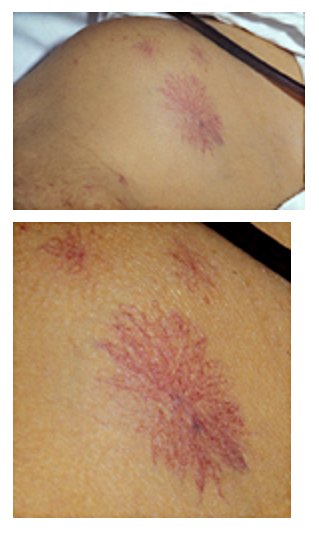
Weakness in elderly person Frailty syndrome A woman supporting herself with a walking frame . Specialty Geriatrics Frailty is a common geriatric syndrome that embodies an elevated risk of catastrophic declines in health and function among older adults . ... It is also a marker of a more widespread syndrome of frailty, with associated weakness, slowing, decreased energy, lower activity, and, when severe, unintended weight loss. ... "The association between obesity and the frailty syndrome in older women: the Women's Health and Aging Studies" (PDF) . ... "Association of low vitamin D levels with the frailty syndrome in men and women" . The Journals of Gerontology.
Both pharmacological agents have vasoconstrictive effects on coronary arteries. [19] However, in the clinical setting, provocative testing is not routinely performed. [4] The reason for this is due to the adverse effects of these pharmacological agents. [4] Complications [ edit ] Depending on how long the occlusion lasts, a spectrum of different myocardial ischemic syndromes can occur. Shorter episodes of occlusion can lead to what is referred to as silent myocardial ischemia due to its asymptomatic nature. [1] These episodes can also be accompanied by arrhythmias. [1] Longer episodes of occlusion can lead to stable or unstable angina, myocardial infarction, and sudden cardiac death. [1] References [ edit ] ^ a b c d e f g h Hung, Ming-Jui; Hu, Patrick; Hung, Ming-Yow (2014). ... External links [ edit ] Classification D ICD - 9-CM : 413.1 MeSH : D003329 External resources MedlinePlus : 000159 eMedicine : med/447 v t e Cardiovascular disease (heart) Ischaemic Coronary disease Coronary artery disease (CAD) Coronary artery aneurysm Spontaneous coronary artery dissection (SCAD) Coronary thrombosis Coronary vasospasm Myocardial bridge Active ischemia Angina pectoris Prinzmetal's angina Stable angina Acute coronary syndrome Myocardial infarction Unstable angina Sequelae hours Hibernating myocardium Myocardial stunning days Myocardial rupture weeks Aneurysm of heart / Ventricular aneurysm Dressler syndrome Layers Pericardium Pericarditis Acute Chronic / Constrictive Pericardial effusion Cardiac tamponade Hemopericardium Myocardium Myocarditis Chagas disease Cardiomyopathy Dilated Alcoholic Hypertrophic Tachycardia-induced Restrictive Loeffler endocarditis Cardiac amyloidosis Endocardial fibroelastosis Arrhythmogenic right ventricular dysplasia Endocardium / valves Endocarditis infective endocarditis Subacute bacterial endocarditis non-infective endocarditis Libman–Sacks endocarditis Nonbacterial thrombotic endocarditis Valves mitral regurgitation prolapse stenosis aortic stenosis insufficiency tricuspid stenosis insufficiency pulmonary stenosis insufficiency Conduction / arrhythmia Bradycardia Sinus bradycardia Sick sinus syndrome Heart block : Sinoatrial AV 1° 2° 3° Intraventricular Bundle branch block Right Left Left anterior fascicle Left posterior fascicle Bifascicular Trifascicular Adams–Stokes syndrome Tachycardia ( paroxysmal and sinus ) Supraventricular Atrial Multifocal Junctional AV nodal reentrant Junctional ectopic Ventricular Accelerated idioventricular rhythm Catecholaminergic polymorphic Torsades de pointes Premature contraction Atrial Junctional Ventricular Pre-excitation syndrome Lown–Ganong–Levine Wolff–Parkinson–White Flutter / fibrillation Atrial flutter Ventricular flutter Atrial fibrillation Familial Ventricular fibrillation Pacemaker Ectopic pacemaker / Ectopic beat Multifocal atrial tachycardia Pacemaker syndrome Parasystole Wandering atrial pacemaker Long QT syndrome Andersen–Tawil Jervell and Lange-Nielsen Romano–Ward Cardiac arrest Sudden cardiac death Asystole Pulseless electrical activity Sinoatrial arrest Other / ungrouped hexaxial reference system Right axis deviation Left axis deviation QT Short QT syndrome T T wave alternans ST Osborn wave ST elevation ST depression Strain pattern Cardiomegaly Ventricular hypertrophy Left Right / Cor pulmonale Atrial enlargement Left Right Athletic heart syndrome Other Cardiac fibrosis Heart failure Diastolic heart failure Cardiac asthma Rheumatic fever
Gatti et al. (1982) concluded that MPS IIID cannot be distinguished clinically from the other forms of Sanfilippo syndrome. Autosomal recessive inheritance was considered confirmed. ... Jansen et al. (2007) reported a 15-year-old Turkish English girl with Sanfilippo syndrome type D with a homozygous mutation in the GNS gene (607664.0004). ... Biochemical Features Kresse et al. (1980) found that cultured skin fibroblasts from 2 patients with clinical features of Sanfilippo syndrome accumulated excessive amounts of heparan sulfate and were unable to release sulfate from N-acetylglucosamine-6-sulfate linkages in heparan sulfate-derived oligosaccharides. ... The activity directed against heparan sulfate is deficient in this form of Sanfilippo syndrome, designated type D by Kresse et al. (1980). ... Animal Model Thompson et al. (1992) described type D Sanfilippo syndrome in a Nubian goat. INHERITANCE - Autosomal recessive GROWTH Height - Normal stature HEAD & NECK Face - Coarse facial features - Prominent forehead Ears - Hearing loss - Low-set ears Eyes - Clear corneas - Synophrys - Decreased vision Nose - Flat nasal bridge - Anteverted nares Mouth - Thick lips - Wide mouth - Drooling Neck - Short neck CARDIOVASCULAR Heart - Asymmetric septal hypertrophy RESPIRATORY Nasopharynx - Frequent upper respiratory tract infections CHEST Ribs Sternum Clavicles & Scapulae - Thick ribs ABDOMEN Liver - Mild hepatomegaly Spleen - Mild splenomegaly Gastrointestinal - Diarrhea - Dysphagia - Drooling SKELETAL - Mild dysotosis multiplex - Joint contractures Spine - Ovoid thoracolumbar vertebrae Limbs - Mild joint stiffness SKIN, NAILS, & HAIR Hair - Hirsutism - Coarse hair - Thick eyebrows - Synophrys NEUROLOGIC Central Nervous System - Mental retardation - Hyperactivity - Seizures - Lack of speech - Dysarthria - Slowing mental development by 1.5 to 3 years of age - Sleep disturbances common Behavioral Psychiatric Manifestations - Severe behavioral problems beginning at 3 to 4 years of age LABORATORY ABNORMALITIES - N-acetylglucosamine-6-sulfate sulfatase deficiency - Heparan sulfate excretion in urine - Metachromasia of white blood cells and fibroblasts MISCELLANEOUS - Four clinically indistinguishable biochemically distinct forms (see 252900 , 252920 , 252930 ) - Progressive disorder MOLECULAR BASIS - Caused by mutation in the N-acetylglucosamine-6-sulfatase gene (GNS, 607664.0001 ) ▲ Close
Mucopolysaccharidosis type IIID (MPS IIID) is an genetic disorder that makes the body unable to break down large sugar molecules called glycosaminoglycans (GAGs, formerly called mucopolysaccharides). Specifically, people with this condition are unable to break down a GAG called heparan sulfate. Affected individuals can have severe neurological symptoms, including progressive dementia, aggressive behavior, hyperactivity, seizures, deafness, loss of vision, and an inability to sleep for more than a few hours at a time. MPS IIID is caused by the missing or deficient enzyme N -acetylglucosamine 6-sulfatase. MPS IIID is inherited in an autosomal recessive manner. There is no specific treatment for this condition.
A number sign (#) is used with this entry because of evidence that a syndrome of microcephaly, seizures, spasticity, and brain calcifications (MISSBC) is caused by homozygous mutation in the PCDH12 gene (605622) on chromosome 5q31. ... The severity of the neurologic features and their perinatal onset differentiated the syndrome from the more common autosomal recessive microcephaly. ... There were 2 unaffected sibs, 1 male and 1 female, in this sibship. Sibs with the syndrome of microcephaly and severe myoclonic seizures, profound mental retardation, hypertonia, and spasticity were also described by Schinzel and Litschgi (1984). Straussberg et al. (1998) described an infant with the same syndrome born to a consanguineous couple of Palestinian origin. ... On reevaluation of the family, Bundey et al. (1991) concluded that the disorder may represent the Allan-Herndon syndrome (300523). INHERITANCE - Autosomal recessive GROWTH Other - Intrauterine growth retardation (IUGR) HEAD & NECK Head - Microcephaly, progressive (up to -7.5 SD) Eyes - Visual impairment, severe NEUROLOGIC Central Nervous System - Delayed psychomotor development, profound - Intellectual disability, profound - Early-onset seizures - Axial hypotonia - Spastic quadriplegia - Dystonia - Hyperreflexia - Early-onset seizures - Midbrain-hypothalamus dysplasia - Perithalamic and periventricular calcifications MISCELLANEOUS - Onset in infancy MOLECULAR BASIS - Caused by mutation in the protocadherin 12 gene (PCDH12, 605622.0001 ) ▲ Close
A number sign (#) is used with this entry because it represents a contiguous gene deletion syndrome on chromosome 6q. Description The cardinal features of chromosome 6q11-q14 interstitial deletions include hypotonia, short stature, skeletal/limb anomalies, umbilical hernia, and urinary tract anomalies, as well as characteristic facial features including upslanting palpebral fissures, low-set and/or dysplastic ears, and high-arched palate (summary by Wang et al., 2009). ... INHERITANCE - Isolated cases GROWTH Height - Short stature HEAD & NECK Ears - Low-set ears - Dysplastic ears Eyes - Upslanting palpebral fissures - Epicanthic folds - Hypertelorism - Hypotelorism (in some patients) Nose - Short nose - Broad nasal tip - Long philtrum Mouth - High-arched palate - Thin upper lip Neck - Short neck ABDOMEN External Features - Umbilical hernia - Inguinal hernia (in some patients) SKELETAL Hands - Single palmar crease NEUROLOGIC Central Nervous System - Hypotonia - Psychomotor delay MISCELLANEOUS - Contiguous gene deletion syndrome MOLECULAR BASIS - Caused by deletion of 3.7Mb on chromosome 6q13-q14 ▲ Close
There are more than 4,000 types of birth defects. [2] Contents 1 Causes 1.1 Difficult labor (dystocia) 1.2 External causes 1.3 Genetic mutations 1.4 Infection 1.5 Intrauterine hypoxia 1.6 Maternal health issues 1.7 Pregnancy complications 2 Common types of birth injury 2.1 Brachial plexus injury 2.2 Brain damage 2.3 Bruising 2.4 Bone fractures 2.5 Meconium aspiration syndrome 3 Legal issues 4 References Causes [ edit ] Difficult labor (dystocia) [ edit ] Difficult labor, also known as dystocia or obstructed labor, occurs when the child cannot easily pass through the birth canal. ... Fracture of the clavicle is the most common birth injury. [17] Meconium aspiration syndrome [ edit ] Meconium is a sticky substance that usually makes up the child’s first bowel movement. ... If it gets into the child's airways or lungs, it can cause meconium aspiration syndrome . Serious cases may result in pneumonia or a collapsed lung . [18] Legal issues [ edit ] Birth injuries may be unavoidable or they may be attributable to medical malpractice .
Citrullinemia type 1 is an inherited disorder that causes ammonia and other toxic substances to accumulate in the blood. There are four types. The classic, most severe form, occurs in newborns, while a milder, later-onset form occurs in children or adults. There's also a form that occurs during or after pregnancy, and a form with no symptoms. In the classic form, symptoms occur right after birth and include excessive sleepiness, poor appetite, vomiting, and irritability. As ammonia builds up, muscle weakness, seizures, coma, and death can occur.
Citrullinemia is an autosomal recessively inherited disorder of urea cycle metabolism and ammonia detoxification (see this term) characterized by elevated concentrations of serum citrulline and ammonia. The disease presents with a large range of manifestations including neonatal hyperammonemic encephalopathy with lethargy, seizures and coma; hepatic dysfunction in all age groups; episodes of hyperammonemia and neuropsychiatric symptoms in children or adults, or, can be asymptomatic in some cases (detected in newborn screening programs). Citrullinemia is divided into two main groups that are encoded by different genes: citrullinemia type I (comprised of acute neonatal citrullinemia type I and adult-onset citrullinemia type I) and citrin deficiency (comprised of adult-onset citrullinemia type II and neonatal intrahepatic cholestasis due to citrin deficiency) (see these terms).
In other words, repeated exposure to the flight environment decreases an individual's susceptibility to subsequent airsickness. [1] Recently, several devices have been introduced that are intended to reduce motion sickness through stimulation of various body parts (usually the wrist). [7] Alternative medicine [ edit ] Ginger . [8] See also [ edit ] Acclimatization Airsickness bag Motion sickness Space adaptation syndrome References [ edit ] ^ a b c d e f g Benson AJ (2002). "35". ... External links [ edit ] Classification D ICD - 10 : T75.3 ICD - 9-CM : 994.6 v t e Consequences of external causes Temperature Elevated Hyperthermia Heat syncope Reduced Hypothermia Immersion foot syndromes Trench foot Tropical immersion foot Warm water immersion foot Chilblains Frostbite Aerosol burn Cold intolerance Acrocyanosis Erythrocyanosis crurum Radiation Radiation poisoning Radiation burn Chronic radiation keratosis Eosinophilic, polymorphic, and pruritic eruption associated with radiotherapy Radiation acne Radiation-induced cancer Radiation recall reaction Radiation-induced erythema multiforme Radiation-induced hypertrophic scar Radiation-induced keloid Radiation-induced morphea Air Hypoxia / Asphyxia Barotrauma Aerosinusitis Decompression sickness High altitude Altitude sickness Chronic mountain sickness Death zone HAPE HACE Food Starvation Maltreatment Physical abuse Sexual abuse Psychological abuse Travel Motion sickness Seasickness Airsickness Space adaptation syndrome Adverse effect Hypersensitivity Anaphylaxis Angioedema Allergy Arthus reaction Adverse drug reaction Other Electrical injury Drowning Lightning injuries Ungrouped skin conditions resulting from physical factors Dermatosis neglecta Pinch mark Pseudoverrucous papules and nodules Sclerosing lymphangitis Tropical anhidrotic asthenia UV-sensitive syndrome environmental skin conditions Electrical burn frictional/traumatic/sports Black heel and palm Equestrian perniosis Jogger's nipple Pulling boat hands Runner's rump Surfer's knots Tennis toe Vibration white finger Weathering nodule of ear Wrestler's ear Coral cut Painful fat herniation Uranium dermatosis iv use Skin pop scar Skin track Slap mark Pseudoacanthosis nigricans Narcotic dermopathy v t e Motion sickness Types Airsickness Seasickness Simulator sickness Ski sickness Space adaptation syndrome Virtual reality sickness Medicine treatment Bonine Cinnarizine Dramamine Marezine Promethazine Transdermscop Related Bárány chair Sickness bag
Nasopharyngeal linguatuliasis appears to be quite prevalent throughout the Middle East where it is often known as the Halzoun syndrome after religious festivals in which infected raw meat is consumed . [7] In Sudan nasopharyngeal linguatuliasis is known as Marrara syndrome; Marrara is a popular local dish prepared from raw offal . It has been suggested that up to 20% of the population in some parts of Sudan may be affected by this syndrome at some stage of their lives . [8] Transmission [ edit ] Eating raw or semi-cooked infected liver or lymph nodes infected with nymphal L. serrata causes severe symptoms in the human nasopharynx . ... These symptoms are well recognized as a disease called “ halzoun syndrome ” in Lebanon and nearby countries. ... M. El Hassan (1996). "The Marrara syndrome: a hypersensitivity reaction of the upper respiratory tract and buccopharyngeal mucosa to nymphs of Linguatula serrata ".
About 40% of patients with sexual headaches in one study also experienced headaches from non-sexual exertion. [1] A pressor response to exercise has been suggested as a mechanism. [4] For other patients, the pain appears to be specifically activated by sexual excitement and contraction of facial and neck muscles . [1] Sporadic case studies have linked sexual headaches to the use of certain drugs, including amiodarone , pseudoephedrine , birth control pills , and cannabis . [1] It may be secondary to another condition, such as reversible cerebral vasoconstriction syndrome . [1] It is associated with migraines . [1] Treatment [ edit ] A physician may recommend engaging in sexual activity less strenuously. [1] Case series have found indomethacin and beta blockers to be successful in treating these headaches. [1] [5] Propranolol , Bellergal, and triptans have also been used with success. [1] Anecdotal and indirect evidence suggests a trial of magnesium supplementation may improve symptoms (in subjects with known or suspected low Mg levels). [6] Epidemiology [ edit ] These headaches are estimated to appear in roughly 1% of the population. [1] They can occur with sexual activity at any age. [2] It is more common in men than women, with studies putting the gender ratio between 1.2:1 and 3:1. [2] See also [ edit ] Postorgasmic illness syndrome (POIS) Intra-axial hematoma Thunderclap headache References [ edit ] ^ a b c d e f g h i j k l m n o Cutrer, F. ... PMID 22684099 . v t e Post-orgasmic diseases Illnesses Dhat syndrome Post-coital tristesse (PCT) Postorgasmic illness syndrome (POIS) Sexual headache Specialties Andrology Allergy Endocrinology Psychiatry General Orgasm Ejaculation La petite mort v t e Headache Primary ICHD 1 Migraine Familial hemiplegic Retinal migraine ICHD 2 Tension Mixed tension migraine ICHD 3 Cluster Chronic paroxysmal hemicrania SUNCT ICHD 4 Hemicrania continua Thunderclap headache Sexual headache New daily persistent headache Hypnic headache Secondary ICHD 5 Migralepsy ICHD 7 Ictal headache Post-dural-puncture headache ICHD 8 Hangover Medication overuse headache ICHD 13 Trigeminal neuralgia Occipital neuralgia External compression headache Cold-stimulus headache Optic neuritis Postherpetic neuralgia Tolosa–Hunt syndrome Other Vascular
Description The International Headache Society (1988) classifies headache associated with sexual activity (HSA) as an idiopathic headache under 'miscellaneous headaches unassociated with structural lesions.' Based on initial descriptions, 3 subtypes are differentiated: type 1 is a dull ache in the head and neck that slowly intensifies as sexual excitement increases, and is believed to be caused by muscle contraction similar to tension-type headache; type 2, also called 'vascular-type,' is a sudden severe, explosive headache occurring at orgasm, which may be due to increased intracranial pressure; type 3, the most uncommon type, is a postural headache resembling that caused by decreased CSF pressure, perhaps due to a meningeal tear during coitus (summary by Frese et al., 2003). Clinical Features Johns (1986) reported 4 sisters with the vascular type of benign sexual headache (type 2 HSA). The most severely affected patient was successfully treated with beta-blocker prophylaxis. In a review of the literature, the author found that 28% of those affected had either personal history or family history of migraine (see 157300), suggesting that type 2 HSA is a migraine variant.