Specialty Nephrology Symptoms Hematuria [2] Types Type I, II and III [3] Diagnostic method Serum analysis [2] Treatment Corticosteroids Rapidly progressive glomerulonephritis ( RPGN ) is a syndrome of the kidney that is characterized by a rapid loss of kidney function, [4] [5] (usually a 50% decline in the glomerular filtration rate (GFR) within 3 months) [5] with glomerular crescent formation seen in at least 50% [5] or 75% [4] of glomeruli seen on kidney biopsies. ... In 50% of cases, RPGN is associated with an underlying disease such as Goodpasture syndrome , systemic lupus erythematosus or granulomatosis with polyangiitis ; the remaining cases are idiopathic . Regardless of the underlying cause, RPGN involves severe injury to the kidneys' glomeruli , with many of the glomeruli containing characteristic glomerular crescents ( crescent -shaped scars ). [ medical citation needed ] Contents 1 Signs and symptoms 2 Pathophysiology 3 Diagnosis 3.1 Classification 3.1.1 Type I 3.1.2 Type II 3.1.3 Type III 4 Treatment 5 Epidemiology 6 References 7 Further reading 8 External links Signs and symptoms [ edit ] Most types of RPGN are characterized by severe and rapid loss of kidney function with marked hematuria ; red blood cell casts in the urine; and proteinuria sometimes exceeding three grams in twenty-four hours, a range associated with nephrotic syndrome . Some patients also experience hypertension and edema . ... Some cases are associated with antibodies directed against the basement membrane of lung alveoli , producing Goodpasture syndrome . The majority of type I disease, however, features anti-GBM antibodies alone; these cases are considered idiopathic. [2] Type II [ edit ] Characterized by deposition of immune complexes in glomerular tissues, type II RPGN accounts for 25% of cases. ... External links [ edit ] Classification D ICD - 10 : N01 DiseasesDB : 3165 External resources eMedicine : med/881 med/890 Orphanet : 280569 Scholia has a topic profile for Rapidly progressive glomerulonephritis . v t e Disease of the kidney glomerules Primarily nephrotic Non-proliferative Minimal change Focal segmental Membranous Proliferative Mesangial proliferative Endocapillary proliferative Membranoproliferative/mesangiocapillary By condition Diabetic Amyloidosis Primarily nephritic , RPG Type I RPG / Type II hypersensitivity Goodpasture syndrome Type II RPG / Type III hypersensitivity Post-streptococcal Lupus diffuse proliferative IgA Type III RPG / Pauci-immune Granulomatosis with polyangiitis Microscopic polyangiitis Eosinophilic granulomatosis with polyangiitis General glomerulonephritis glomerulonephrosis
Familial pityriasis rubra pilaris is a rare genetic condition that affects the skin. The name of the condition reflects its major features: The term "pityriasis" refers to scaling; "rubra" means redness; and "pilaris" suggests the involvement of hair follicles in this disorder. Affected individuals have a salmon-colored skin rash covered in fine scales. This rash occurs in patches all over the body, with distinct areas of unaffected skin between the patches. Affected individuals also develop bumps called follicular keratoses that occur around hair follicles.
Pityriasis rubra pilaris is a rare chronic papulosquamous disorder of unknown etiology characterized by small follicular papules, scaly red-orange patches, and palmoplantar hyperkeratosis, which may progress to plaques or erythroderma. Although most of the cases are sporadic and acquired, a familial form of the disease exists.
A number sign (#) is used with this entry because of evidence that pityriasis rubra pilaris (PRP) is caused by heterozygous mutation in the CARD14 gene (607211) on chromosome 17q25. Description Pityriasis rubra pilaris is an uncommon skin disorder characterized by the appearance of keratotic follicular papules, well-demarcated salmon-colored erythematous plaques covered with fine powdery scales interspersed with distinct islands of uninvolved skin, and palmoplantar keratoderma. Most cases are sporadic, although up to 6.5% of PRP-affected individuals report a positive family history. The rare familial cases show autosomal dominant inheritance with incomplete penetrance and variable expression: the disorder is usually present at birth or appears during the first years of life and is characterized by prominent follicular hyperkeratosis, diffuse palmoplantar keratoderma, and erythema, with only a modest response to treatment (summary by Fuchs-Telem et al., 2012). Clinical Features Pityriasis rubra pilaris is 'characterized by scaly and horny productions situated chiefly in the sebaceous follicles and by a more or less generalized hyperemia' to use the words of DeVergie who first described it (Zeisler, 1923) in a man and his son and 2 daughters.
Pityriasis rubra pilaris (PRP) refers to a group of skin conditions that cause constant inflammation and scaling of the skin. People with PRP have reddish, scaly patches that may occur everywhere on the body, or only on certain areas. Some people with PRP also develop thickened skin on the underside of the hands and feet ( palmoplantar keratoderma ), various nail abnormalities, and/or thinning of the hair. There are several types of PRP classified by age when symptoms begin, body areas involved, and whether other conditions are present. This condition occurs in adults (adult onset PRP) as well as children (juvenile onset PRP).
Palindromic rheumatism Specialty Rheumatology Palindromic rheumatism ( PR ) is a syndrome characterised by recurrent, self-resolving inflammatory attacks in and around the joints, consists of arthritis or periarticular soft tissue inflammation. [1] The course is often acute onset, with sudden and rapidly developing attacks or flares. ... Contents 1 Presentation 2 Causes 3 Diagnosis 4 Management 5 Etymology 6 References 7 External links Presentation [ edit ] The exact prevalence of palindromic rheumatism in general population is unknown, and this condition is often considered a rare disease by nonrheumatologists. [2] However, recent Canadian study showed that the incidence of PR in a cohort of incident arthritis was one case of PR for every 1.8 cases of rheumatoid arthritis (RA). [3] The incidence of PR is less than that of RA but is not as rare as that was thought to be. Palindromic rheumatism is a syndrome presented with inflammatory para-arthritis (soft tissue rheumatism) and inflammatory arthritis both of which cause sudden inflammation in one or several joints or soft tissue around joints. ... One study showed an average age of onset of 49. [3] A population cohort study in Taiwan suggested that patients with PR had an increased risk of developing rheumatoid arthritis, systemic lupus erythematosus, Sjogren's syndrome, systemic sclerosis, and polymyositis. [5] Causes [ edit ] Palindromic rheumatism is a disease of unknown cause. ... It is important to note that a person may experience more than one autoimmune disorder at the same time, as overlap syndrome . Laboratory findings are usually normal. ... PMID 1341421 . ^ Arthritis Foundation [1] External links [ edit ] Classification D ICD - 10 : M12.3 ICD - 9-CM : 719.3 MeSH : C538103 C538103, C538103 DiseasesDB : 9508 v t e Diseases of joints General Arthritis Monoarthritis Oligoarthritis Polyarthritis Symptoms Joint pain Joint stiffness Inflammatory Infectious Septic arthritis Tuberculosis arthritis Crystal Chondrocalcinosis CPPD (Psudogout) Gout Seronegative Reactive arthritis Psoriatic arthritis Ankylosing spondylitis Other Juvenile idiopathic arthritis Rheumatoid arthritis Felty's syndrome Palindromic rheumatism Adult-onset Still's disease Noninflammatory Hemarthrosis Osteoarthritis Heberden's node Bouchard's nodes Osteophyte
Palindromic rheumatism (PR) is a type of recurrent arthritis characterized by episodes or "attacks" of joint inflammation, sequentially affecting one to several joint areas for hours to days. A PR attack often occurs suddenly without any obvious triggers or warning symptoms. Any joint(s) may be affected, but finger joints, wrists, and knees are most commonly affected. Symptoms during episodes may include pain, swelling, stiffness, and redness in and around the joints. Some people may have a fever and other systemic symptoms. Between episodes, people with PR have no symptoms.
A number sign (#) is used with this entry because of evidence that familial cylindromatosis is caused by heterozygous mutation in the CYLD gene (605018) on chromosome 16q12. See also Brooke-Spiegler syndrome (BRSS; 605041) and multiple familial trichoepithelioma-1 (MFT1; 601606), which are allelic disorders with overlapping phenotypes. Description The disorders classically referred to as familial cylindromatosis, Brooke-Spiegler syndrome, and multiple familial trichoepithelioma were originally described as distinct clinical entities. ... Clinical Features Ancell (1842) and Spiegler (1899) described a familial syndrome characterized by tumors of skin appendages, now known as cylindromas (Lee et al., 2005). ... Young et al. (2006) identified a heterozygous mutation in the CYLD gene (605018.0008) in a 73-year-old man with cylindromatosis and turban tumor syndrome and in his 2 children with multiple familial trichoepitheliomas without cylindromas. ... INHERITANCE - Autosomal dominant SKIN, NAILS, & HAIR Skin - Cylindromas, multiple (face, trunk and extremities) - Cylindromas usually occur on the scalp may coalesce into large 'turban tumors' Skin Histology - Mosaic-like masses of epithelial cells surrounded by thin layers of PAS-positive stroma - Cells appear to be of glandular origin NEOPLASIA - Cylindromas may show malignant transformation MISCELLANEOUS - Onset in early adulthood - Allelic disorder to multiple familial trichoepithelioma 1 (MFT1, 601606 ) and Brooke-Spiegler syndrome (BSS, 605041 ) MOLECULAR BASIS - Caused by mutation in the CYLD gene ( 605018.0001 ) ▲ Close
Hypocalcemia, Autosomal Dominant 1, With Bartter Syndrome In a 19-year-old man and an unrelated 26-year-old woman, both of whom presented soon after birth with tetany and hypocalcemia and demonstrated features of Bartter syndrome (see 607364), including hypomagnesemia, hypokalemia, metabolic alkalosis, hyperreninemia, and hyperaldosteronemia, Watanabe et al. (2002) identified heterozygous missense mutations in the CASR gene (601199.0034 and 601199.0035, respectively). The authors noted that in rats it had been shown that activation of this calcium-sensing receptor by higher concentrations of extracellular calcium ions inhibits the activity of the renal outer-medullary potassium channel (KCNJ1; 600359) (see Brown and MacLeod, 2001); the KCNJ1 gene is mutated in type 2 Bartter syndrome (241200). Watanabe et al. (2002) suggested that other molecules that inhibit activity of Bartter syndrome-associated genes might be additional causes of Bartter and related syndromes. Vargas-Poussou et al. (2002) reported a boy who had severe autosomal dominant hypocalcemia associated with Bartter syndrome-like features, characterized by a decrease in the distal tubular fractional chloride reabsorption rate and negative NaCl balance with secondary hyperaldosteronism and hypokalemia. ... In a follow-up study, Vezzoli et al. (2006) reported that the twins developed Bartter syndrome-like features at 22 years of age, with mild hypokalemia, mild hyperreninemia and hyperaldosteronism, but no alkalosis; the authors designated the disorder 'type 5 Bartter syndrome.' Vezzoli et al. (2006) noted that all 4 CASR mutations causing hypocalcemia associated with Bartter syndrome-like features were highly activating, with EC50 values less than 1.5 mmol/L, whereas the EC50 values for other CASR mutations causing autosomal dominant hypocalcemia but not Bartter syndrome ranged between 1.5 and 3 mmol/L.
A number sign (#) is used with this entry because of evidence that autosomal dominant hypocalcemia-2 (HYPOC2) is caused by heterozygous mutation in the GNA11 gene (139313) on chromosome 19p13. For a discussion of genetic heterogeneity of autosomal dominant hypocalcemia, see HYPOC1 (601198). Clinical Features Hunter et al. (1981) reported a large 4-generation family of English Canadian origin segregating what they designated 'autosomal dominant hypoparathyroidism.' There were 8 affected members of the family, 2 of whom were asymptomatic. The proband was evaluated at 6 years old for 'febrile convulsions' and found to have positive Trousseau and Chvostek signs, possible laryngospasm, and low serum calcium (6.3 mg/dl) and phosphorus (7.8 mg/dl) levels, with a parathyroid hormone (PTH) level in the low-normal range (177 pg Eq/ml).
A number sign (#) is used with this entry because of evidence that X-linked hypoparathyroidism (HYPX) is caused by an interstitial deletion/insertion on chromosome Xq27.1, which may have a position effect on expression of SOX3 (313430). Clinical Features Peden (1960) reported a family in which multiple males had neonatal idiopathic hypoparathyroidism in an X-linked pattern of inheritance. No affected males reproduced. Peden (1960) suggested that most familial cases with early onset are of the X-linked type, the autosomal type (146200) having a later onset. Whyte and Weldon (1981) performed extensive studies of a second kindred from Missouri (where Peden's family also lived) with neonatal or infantile onset of X-linked isolated hypoparathyroidism. No ancestor common to the 2 kindreds could be identified. Whyte et al. (1986) reported the autopsy findings in a member of the family of Peden (1960) who had died as a teenager after an automobile crash.
Congenital absence of the parathyroid and thymus glands (III and IV pharyngeal pouch syndrome, or DiGeorge syndrome, 188400) is usually a sporadic condition (Taitz et al., 1966). ... Yumita et al. (1986) suggested that progressive sensorineural deafness, which was present in both families, was an integral part of the hypoparathyroidism syndrome. However, in the second family, it appears to have been segregating, probably as an autosomal dominant, independently of the hypoparathyroidism. ... The patients had no cardiac abnormality or T-cell deficiency and had no cleft palate, features that occur in the DiGeorge syndrome (188400) and the velocardiofacial syndrome (VCFS; 192430).
Familial isolated hypoparathyroidism (FIH) is a rare heterogeneous group of metabolic disorders characterized by abnormal calcium metabolism due to deficient secretion of parathormone (PTH), without other endocrine disorders or developmental defects. Clinical description It can occur at any age (from the newborn period to adulthood) but generally starts within the first decade of life. Diagnosis is made when hypocalcemia, hyperphosphoremia, and low or undetectable PTH levels are observed. The clinical signs are mainly those of hypocalcemia: myopathy, muscular weakness, cramps, tetany, lenticular cataracts, teeth anomalies and short stature. Etiology FIH may be due to an activating mutation of the calcium-sensing receptor ( CASR ) gene.
Overview An aortic aneurysm is a bulge that occurs in the wall of the body's main artery, called the aorta. The aorta carries blood from the heart to the body. Aortic aneurysms can occur anywhere in the aorta. They may be tube shaped or round. Aortic aneurysms include: Abdominal aortic aneurysm. An abdominal aortic aneurysm occurs along the part of the aorta that passes through the belly area. Thoracic aortic aneurysm. A thoracic aortic aneurysm occurs along the part of the aorta that passes through the chest cavity.
These include: Prior heart attack or other heart condition that caused scarring of heart tissue (structural heart disease) Poor blood flow to the heart muscle due to coronary artery disease Congenital heart diseases, including long QT syndrome Imbalance of substances in the blood called electrolytes — such as potassium, sodium, calcium and magnesium Medication side effects Use of stimulants such as cocaine or methamphetamine Sometimes, the exact cause of ventricular tachycardia can't be determined (idiopathic ventricular tachycardia).
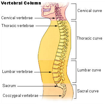
Other causes include obesity, hyperkyphosis (spine curvature disorder in which the thoracic curvature is abnormally rounded), discitits (an inflammation of the intervertebral disc space caused by infection) and benign juvenile lordosis. [10] Other factors may also include those with rare diseases, as is the case with Ehlers Danlos Syndrome (EDS) , where hyper-extensive and usually unstable joints (e.g. joints that are problematically much more flexible, frequently to the point of partial or full dislocation) are quite common throughout the body. ... The presence of measurable abnormality does not automatically equate with a level of reported symptoms. [23] Braces [ edit ] The Boston brace is a plastic exterior that can be made with a small amount of lordosis to minimize stresses on discs that have experienced herniated discs.In the case where Ehlers Danlos syndrome (EDS) is responsible, being properly fitted with a customized brace may be a solution to avoid strain and limit the frequency of instability. [ citation needed ] Tai chi [ edit ] While not really a 'treatment', the art of tai chi chuan calls for adjusting the lower back curvature (as well as the rest of the spinal curvatures) through specific re-alignments of the pelvis to the thighs, it's referred to in shorthand as 'dropping the tailbone'. ... Lordosis - MedlinePlus definition Lordosis - SpineUniverse v t e Spinal disease Deforming Spinal curvature Kyphosis Lordosis Scoliosis Other Scheuermann's disease Torticollis Spondylopathy inflammatory Spondylitis Ankylosing spondylitis Sacroiliitis Discitis Spondylodiscitis Pott disease non inflammatory Spondylosis Spondylolysis Spondylolisthesis Retrolisthesis Spinal stenosis Facet syndrome Back pain Neck pain Upper back pain Low back pain Coccydynia Sciatica Radiculopathy Intervertebral disc disorder Schmorl's nodes Degenerative disc disease Spinal disc herniation Facet joint arthrosis v t e Congenital malformations and deformations of musculoskeletal system / musculoskeletal abnormality Appendicular limb / dysmelia Arms clavicle / shoulder Cleidocranial dysostosis Sprengel's deformity Wallis–Zieff–Goldblatt syndrome hand deformity Madelung's deformity Clinodactyly Oligodactyly Polydactyly Leg hip Hip dislocation / Hip dysplasia Upington disease Coxa valga Coxa vara knee Genu valgum Genu varum Genu recurvatum Discoid meniscus Congenital patellar dislocation Congenital knee dislocation foot deformity varus Club foot Pigeon toe valgus Flat feet Pes cavus Rocker bottom foot Hammer toe Either / both fingers and toes Polydactyly / Syndactyly Webbed toes Arachnodactyly Cenani–Lenz syndactylism Ectrodactyly Brachydactyly Stub thumb reduction deficits / limb Acheiropodia Ectromelia Phocomelia Amelia Hemimelia multiple joints Arthrogryposis Larsen syndrome RAPADILINO syndrome Axial Skull and face Craniosynostosis Scaphocephaly Oxycephaly Trigonocephaly Craniofacial dysostosis Crouzon syndrome Hypertelorism Hallermann–Streiff syndrome Treacher Collins syndrome other Macrocephaly Platybasia Craniodiaphyseal dysplasia Dolichocephaly Greig cephalopolysyndactyly syndrome Plagiocephaly Saddle nose Vertebral column Spinal curvature Scoliosis Klippel–Feil syndrome Spondylolisthesis Spina bifida occulta Sacralization Thoracic skeleton ribs : Cervical Bifid sternum : Pectus excavatum Pectus carinatum
Contents 1 Classification 1.1 Locked-in syndrome 1.2 Minimally conscious state 1.3 Persistent vegetative state 1.4 Chronic coma 1.5 Brain death 2 Methodological problems 3 Ethical issues 4 See also 5 References 6 External links Classification [ edit ] Patients in such a dramatically altered state of consciousness present unique problems for diagnosis, prognosis and treatment. ... Also the ethical framework must be further developed to guide research in these patients. [ citation needed ] Locked-in syndrome [ edit ] Main article: Locked-in syndrome In locked-in syndrome the patient has awareness, sleep-wake cycles, and meaningful behavior (viz., eye-movement), but is isolated due to quadriplegia and pseudobulbar palsy , resulting from the disruption of corticospinal and corticobulbar pathways. Locked-in syndrome is a condition in which a patient is aware and awake but cannot move or communicate verbally due to complete paralysis of nearly all voluntary muscles in the body except for the eyes. Eye or eyelid movements are the main method of communication. [8] Total locked-in syndrome is a version of locked-in syndrome where the eyes are paralyzed as well. [9] Minimally conscious state [ edit ] Main article: Minimally conscious state In a minimally conscious state , the patient has intermittent periods of awareness and wakefulness. ... Arch Pyhs Med Rehabil 1995; 76;: 205-9 ^ Bauer, G. and Gerstenbrand, F. and Rumpl, E. (1979). "Varieties of the locked-in syndrome". Journal of Neurology . 221 (2): 77–91. doi : 10.1007/BF00313105 .
Less common seizure phenotypes in individuals with KCNT1- related epilepsy include West syndrome, Ohtahara syndrome, early myoclonic encephalopathy, leukodystrophy and/or leukoencephalopathy, focal epilepsy, and multifocal epilepsy. ... McTague et al [2018] Clinical Characteristics Clinical Description KCNT1- related epilepsy encompasses a range of epilepsy syndromes. The most common phenotypes reported in individuals with KCNT1 -related epilepsy are epilepsy of infancy with migrating focal seizures (EIMFS) and autosomal dominant nocturnal frontal lobe epilepsy (ADNFLE). ... Less common epilepsy phenotypes in individuals with a KCNT1 pathogenic variant include: West syndrome Ohtahara syndrome (early infantile epileptic encephalopathy) Early myoclonic encephalopathy Leukodystrophy/leukoencephalopathy Focal or multifocal epilepsy Brain MRI and/or CT examination is often normal prior to seizure onset, though recent studies have noted variable delayed myelination, hippocampal volume loss, and cerebellar atrophy [McTague et al 2018]. ... Evaluation for pulmonary hemorrhage should be considered if an individual develops acute respiratory failure, heart failure, or hemoptysis. Cardiac Arrhythmia Brugada syndrome was reported in one individual with a de novo KCNT1 variant [Juang et al 2014]. ... In 2010, the International League Against Epilepsy reclassified this epilepsy syndrome as EIMFS [Berg et al 2010]. Prevalence The prevalence of KCNT1 -related epilepsy is unknown.
Overview Pemphigus is a disease that causes blisters and sores on the skin or mucous membranes, such as in the mouth or on the genitals. Pemphigus can occur at any age, but it's most often seen in people who are middle-aged or older. It tends to be a long-lasting (chronic) condition, and some types can be life-threatening without treatment. Treatment with medication usually controls it. Symptoms Pemphigus causes blisters on your skin and mucous membranes. The blisters rupture easily, leaving open sores, which may ooze and become infected.
Pemphigus is a group of rare autoimmune diseases that cause blistering of the skin and mucous membranes (mouth, nose, throat, eyes, and genitals). This condition can occur at any age, but often strikes people in middle or older age. Studies have shown that some populations may be at greater risk for certain types of pemphigus. For instance, people of Jewish descent and those from India, Southeast Europe, and the Middle East are at greater risk for pemphigus vulargis, while pemphigus foliaceus is more common in North America, Turkey, and South America. Pemphigus is a chronic disease which is best controlled by early diagnosis and treatment.
Many patients fit into the category of Leigh syndrome (256000) (Brown et al., 1994). ... Both patients showed conspicuous abnormalities of eye movement as in Wernicke-Korsakoff syndrome (277730). Thiamine in large doses appeared to benefit Lonsdale's patient. ... A half brother had died at age 33 months with autopsy-confirmed Leigh syndrome after a progressive neurologic illness that began with ocular symptoms. ... In a male infant with PDHAD presenting as acute Leigh syndrome, Matthews et al. (1993) identified a point mutation in the PDHA1 gene (D258A; 300502.0011). ... In a boy who presented with Leigh syndrome, Naito et al. (1997) identified a mutation in the PDHA1 gene (R263G; 300502.0022).
Two patients had CT findings consistent with Leigh syndrome (256000). Maj et al. (2005) reported 2 brothers, born of consanguineous parents, who presented with neonatal hypotonia, elevated lactate, and less than 25% native pyruvate dehydrogenase complex (PDHc) activity in skin fibroblasts compared with controls. ... INHERITANCE - Autosomal recessive HEAD & NECK Eyes - Nystagmus ABDOMEN Gastrointestinal - Dysphagia MUSCLE, SOFT TISSUES - Hypotonia NEUROLOGIC Central Nervous System - Mental retardation - Developmental delay - Seizures - Ataxic gait - Low densities in the basal ganglia similar to Leigh syndrome ( 256000 ) METABOLIC FEATURES - Lactic acidosis LABORATORY ABNORMALITIES - Decreased activity of the pyruvate dehydrogenase (PDH) complex - Decreased activity of the PDH phosphatase MISCELLANEOUS - Onset in infancy MOLECULAR BASIS - Caused by mutation in the magnesium-dependent protein phosphatase 2C gene (PPM2C, 605993.0001 ). ▲ Close
A number sign (#) is used with this entry because of evidence that pyruvate dehydrogenase E2 deficiency is caused by homozygous mutation in the DLAT gene (608770) on chromosome 11q23. For a general phenotypic description and a discussion of genetic heterogeneity of pyruvate dehydrogenase deficiency, see 312170. Clinical Features Robinson et al. (1990) described a black infant who presented at 2 weeks of age with hyperammonemia and profound lactic acidosis. Control of blood lactates was achieved by carbohydrate restriction and bicarbonate supplementation, but at age 3.5 years she had profound psychomotor retardation and was microcephalic. Deficiency in the E2 dihydrolipoyl transacetylase activity of the pyruvate dehydrogenase complex was demonstrated enzymatically, and a very low E2 protein component was found on Western blotting of fibroblast proteins.
Craigen (1996) described a 6-month-old female infant who presented with hypotonia, ketolactic acidosis, and an MRI of the brain that showed subacute necrotizing encephalomyelopathy consistent with Leigh syndrome (LS; 256000). Enzymatic analysis demonstrated deficiency of lipoamide dehydrogenase. ... INHERITANCE - Autosomal recessive HEAD & NECK Head - Microcephaly CARDIOVASCULAR Heart - Hypertrophic cardiomyopathy ABDOMEN Liver - Hepatomegaly (in some patients) - Liver dysfunction (in some patients) Gastrointestinal - Vomiting, recurrent, severe - Poor feeding NEUROLOGIC Central Nervous System - Encephalopathy, episodic - Delayed psychomotor development (in most patients) - Hypotonia - Lethargy - Ataxia - Dystonia - Seizures - Leigh syndrome (in some patients) METABOLIC FEATURES - Metabolic acidosis - Lactic acidosis - Episodic decompensation LABORATORY ABNORMALITIES - Hypoglycemia - Elevated pyruvate (in most patients) - Elevated branched-chain amino acids (in most patients) - Elevated alpha-ketoglutarate (in most patients) - Decreased activities of the pyruvate dehydrogenase complex, the alpha-ketoglutarate dehydrogenase complex, and the branched-chain alpha-keto acid dehydrogenase complex - Abnormal liver enzymes (in some patients) MISCELLANEOUS - Onset usually in the neonatal period although later onset has been reported - Some patients may have normal psychomotor development - Highly variable severity - High mortality in infancy and early childhood (in some patients) MOLECULAR BASIS - Caused by mutation in the dihydrolipoamide dehydrogenase gene (DLD, 238331.0001 ) ▲ Close
Many patients have the characteristic clinical presentation, disease course and neuropathological changes of Leigh syndrome (see this term). Etiology PDHD is caused by a deficiency of one of the components of the PDH complex. ... In patients presenting as Leigh syndrome, the differential diagnosis includes various forms of Complex I deficiency (see this term), cytochrome oxidase deficiency due to mutation in the SURF1 gene and a number of mitochondrial DNA mutations.
A disorder that is the most frequent form of pyruvate dehydrogenase deficiency (PDHD) characterized by variable lactic acidosis, impaired psychomotor development, hypotonia and neurological dysfunction. Epidemiology Prevalence is unknown. Over 200 patients have been reported and while there are approximately equal numbers of males and females, male patients are generally more severely affected. Clinical description Patients present with a range of classic signs and symptoms of PDHD, including lactic acidosis, poor feeding, lethargy, tachypnea, developmental delay, growth retardation, poor acquisition or loss of motor milestones, hypotonia, seizures, ataxia and dystonia. Structural brain lesions including cortical atrophy, dilated ventricles, and incomplete corpus callosum, absence of the medullary pyramids and ectopia of the olivary nuclei are commonly observed, especially in female patients heterozygous for the disease-causing mutations that result in complete deficiency of E1-alpha subunit protein in cells expressing the gene mutation. Etiology The disease is caused by deficiency of the E1-alpha subunit of the PDH complex related to mutations in the PDHA1 gene (Xp22.1).
Pyruvate dehydrogenase complex (PDC) deficiency is a type of metabolic disease. This means that the body is not able to efficiently break down nutrients in food to be used for energy. Symptoms of PDC deficiency include signs of metabolic dysfunction such as extreme tiredness (lethargy), poor feeding, and rapid breathing ( tachypnea ). Other symptoms may include signs of neurological dysfunction such as developmental delay, periods of uncontrolled movements (ataxia), low muscle tone (hypotonia), abnormal eye movements, and seizures. Symptoms usually begin in infancy, but signs can first appear at birth or later in childhood.
A number sign (#) is used with this entry because pyruvate dehydrogenase E1-beta deficiency is caused by homozygous mutation in the PDHB gene (179060) on chromosome 3p14. For a general phenotypic description and a discussion of genetic heterogeneity of pyruvate dehydrogenase deficiency, see 312170. Clinical Features Brown et al. (2004) reported 2 unrelated patients with pyruvate dehydrogenase deficiency. The first patient, the son of first-cousin parents, was investigated at age 3 months because of lactic acidosis and hypotonia. Two previous sibs had died early, one with lactic acidosis. The patient developed metabolic acidosis on day 1.
Additionally, many hypochondriacs experience elevated blood pressure, stress, and anxiety in the presence of doctors or while occupying a medical facility, a condition known as " white coat syndrome ". Many hypochondriacs require constant reassurance, either from doctors, family, or friends, and the disorder can become a debilitating challenge for the individual with hypochondriasis, as well as their family and friends. [10] Some hypochondriacal individuals completely avoid any reminder of illness, whereas others frequently visit medical facilities, sometimes obsessively. ... XVI edited by William Torrey Harris p. 395-396 See also [ edit ] Nosophobia Cyberchondria Mithridatism Munchausen syndrome Nocebo Psychosomatic medicine Sickness behavior Somatoform disorder Somatosensory amplification Man flu The Imaginary Invalid References [ edit ] ^ Berrios GE (2001) Hypochondriasis. ... Listen to this article (3.3 megabytes) This audio file was created from a revision of this article dated 18 December 2006 ( 2006-12-18 ) , and does not reflect subsequent edits. ( Audio help · More spoken articles ) v t e Mental and behavioral disorders Adult personality and behavior Gender dysphoria Ego-dystonic sexual orientation Paraphilia Fetishism Voyeurism Sexual maturation disorder Sexual relationship disorder Other Factitious disorder Munchausen syndrome Intermittent explosive disorder Dermatillomania Kleptomania Pyromania Trichotillomania Personality disorder Childhood and learning Emotional and behavioral ADHD Conduct disorder ODD Emotional and behavioral disorders Separation anxiety disorder Movement disorders Stereotypic Social functioning DAD RAD Selective mutism Speech Stuttering Cluttering Tic disorder Tourette syndrome Intellectual disability X-linked intellectual disability Lujan–Fryns syndrome Psychological development ( developmental disabilities ) Pervasive Specific Mood (affective) Bipolar Bipolar I Bipolar II Bipolar NOS Cyclothymia Depression Atypical depression Dysthymia Major depressive disorder Melancholic depression Seasonal affective disorder Mania Neurological and symptomatic Autism spectrum Autism Asperger syndrome High-functioning autism PDD-NOS Savant syndrome Dementia AIDS dementia complex Alzheimer's disease Creutzfeldt–Jakob disease Frontotemporal dementia Huntington's disease Mild cognitive impairment Parkinson's disease Pick's disease Sundowning Vascular dementia Wandering Other Delirium Organic brain syndrome Post-concussion syndrome Neurotic , stress -related and somatoform Adjustment Adjustment disorder with depressed mood Anxiety Phobia Agoraphobia Social anxiety Social phobia Anthropophobia Specific social phobia Specific phobia Claustrophobia Other Generalized anxiety disorder OCD Panic attack Panic disorder Stress Acute stress reaction PTSD Dissociative Depersonalization disorder Dissociative identity disorder Fugue state Psychogenic amnesia Somatic symptom Body dysmorphic disorder Conversion disorder Ganser syndrome Globus pharyngis Psychogenic non-epileptic seizures False pregnancy Hypochondriasis Mass psychogenic illness Nosophobia Psychogenic pain Somatization disorder Physiological and physical behavior Eating Anorexia nervosa Bulimia nervosa Rumination syndrome Other specified feeding or eating disorder Nonorganic sleep Hypersomnia Insomnia Parasomnia Night terror Nightmare REM sleep behavior disorder Postnatal Postpartum depression Postpartum psychosis Sexual dysfunction Arousal Erectile dysfunction Female sexual arousal disorder Desire Hypersexuality Hypoactive sexual desire disorder Orgasm Anorgasmia Delayed ejaculation Premature ejaculation Sexual anhedonia Pain Nonorganic dyspareunia Nonorganic vaginismus Psychoactive substances, substance abuse and substance-related Drug overdose Intoxication Physical dependence Rebound effect Stimulant psychosis Substance dependence Withdrawal Schizophrenia , schizotypal and delusional Delusional Delusional disorder Folie à deux Psychosis and schizophrenia-like Brief reactive psychosis Schizoaffective disorder Schizophreniform disorder Schizophrenia Childhood schizophrenia Disorganized (hebephrenic) schizophrenia Paranoid schizophrenia Pseudoneurotic schizophrenia Simple-type schizophrenia Other Catatonia Symptoms and uncategorized Impulse control disorder Klüver–Bucy syndrome Psychomotor agitation Stereotypy v t e Defence mechanisms Level 1: Pathological Delusional projection Denial or abnegation (German: Verneinung ) Psychotic denial or disavowal (German: Verleugnung ) Distortion Foreclosure or repudiation (German: Verwerfung ) Extreme projection Identification with the Aggressor Splitting Level 2: Immature Acting out Fantasy Idealization Introjection Passive-aggression Projection Projective identification Somatization Level 3: Neurotic Displacement Dissociation Hypochondriasis Intellectualization Isolation Rationalization Reaction formation Regression Repression (German: Verdrängung ) Undoing Level 4: Mature Altruism Anticipation Humour Identification Sublimation Suppression Other mechanisms Compartmentalization Defensive pessimism Exaggeration Minimisation Postponement of affect See also Narcissistic defences Censorship (psychoanalysis) v t e Obsessive–compulsive disorder History Yale–Brown Obsessive Compulsive Scale Biology Neuroanatomy Basal ganglia ( striatum ) Orbitofrontal cortex Cingulate cortex Brain-derived neurotrophic factor Receptors 5-HT 1D β 5-HT 2A 5-HT 2C μ Opioid H 2 NK 1 M 4 NMDA Symptoms Obsessions ( associative diagnostic injurious scrupulous pathogenic sexual ) Compulsions ( impulses , rituals tics ) Thought suppression (avoidance) Hoarding ( animals , books possessions ) Treatment Serotonergics Selective serotonin reuptake inhibitors Escitalopram Fluoxetine Fluvoxamine Paroxetine Sertraline Citalopram Nefazodone Serotonin–norepinephrine reuptake inhibitors Venlafaxine Desvenlafaxine Duloxetine Serotonin–norepinephrine–dopamine reuptake inhibitors Nefazodone Monoamine oxidase inhibitors Phenelzine Tranylcypromine Tricyclic antidepressants Clomipramine Serotonergic psychedelics Lysergic acid diethylamide Psilocin Atypical antipsychotics Aripiprazole Quetiapine Mu opioidergics Hydrocodone Morphine Tramadol Anticholinergics Diphenhydramine NMDA glutamatergics Riluzole NK-1 tachykininergics Aprepitant Other Nicotine Memantine Tautomycin Behavioral Cognitive behavioral therapy ( Exposure and response prevention ) Inference-based therapy Metacognitive therapy Organizations International OCD Foundation Notable people Edna B. ... Schwartz Susan Swedo Emily Colas Vic Meyer Popular culture Literature/Comics Fictional Matchstick Men Plyushkin Xenocide Nonfiction Everything in Its Place Just Checking Media As Good as It Gets The Aviator Matchstick Men Adrian Monk " $pringfield " Straight Up Related Obsessive–compulsive personality disorder Obsessional jealousy PANDAS Primarily Obsessional OCD Relationship obsessive–compulsive disorder Social anxiety disorder Tourette syndrome Psychiatry portal
Ingestion of kaolin (white clay) among African-American women in the US state of Georgia shows the practice there to be a DSM-4 " culture-bound syndrome " and "not selectively associated with other psychopathology". [24] Similar kaolin ingestion is also widespread in parts of Africa. [25] Such practices may stem from health benefits such as the ability of clay to absorb plant toxins and protect against toxic alkaloids and tannic acids . [26] Diagnosis [ edit ] No single test confirms pica, but because pica can occur in people who have lower than normal nutrient levels and poor nutrition (malnutrition), the health care provider should test blood levels of iron and zinc. ... The healthcare provider should test for infection if the patient has been eating contaminated soil or animal waste. [17] DSM-5 [ edit ] The DSM-5 posits four criteria that must be met for a person to be diagnosed with pica: [20] Person must have been eating non-nutritive nonfoods for at least one month. [20] This eating must be considered abnormal for the person's stage of development. [20] Eating these substances cannot be associated with a cultural practice that is considered normal in the social context of the individual. [20] For people who currently have a medical condition (e.g.: pregnancy) or a mental disorder (e.g.: autism spectrum disorder), the action of eating non-nutritive nonfoods should only be considered pica if it is dangerous and requires extra medical investigation or treatment on top of what they are already receiving for their pre-existing condition. [20] Differential diagnosis [ edit ] In individuals with autism , schizophrenia , and certain physical disorders (such as Kleine-Levin syndrome ), non-nutritive substances may be eaten. ... "Chalk Eating in Middle Georgia: a Culture-Bound Syndrome of Pica?". Southern Medical Journal . 92 (2): 190–192. doi : 10.1097/00007611-199902000-00005 . ... External links [ edit ] Classification D ICD - 10 : F50.8 , F98.3 ICD - 9-CM : 307.52 MeSH : D010842 DiseasesDB : 29704 External resources MedlinePlus : 001538 eMedicine : ped/1798 v t e Mental and behavioral disorders Adult personality and behavior Gender dysphoria Ego-dystonic sexual orientation Paraphilia Fetishism Voyeurism Sexual maturation disorder Sexual relationship disorder Other Factitious disorder Munchausen syndrome Intermittent explosive disorder Dermatillomania Kleptomania Pyromania Trichotillomania Personality disorder Childhood and learning Emotional and behavioral ADHD Conduct disorder ODD Emotional and behavioral disorders Separation anxiety disorder Movement disorders Stereotypic Social functioning DAD RAD Selective mutism Speech Stuttering Cluttering Tic disorder Tourette syndrome Intellectual disability X-linked intellectual disability Lujan–Fryns syndrome Psychological development ( developmental disabilities ) Pervasive Specific Mood (affective) Bipolar Bipolar I Bipolar II Bipolar NOS Cyclothymia Depression Atypical depression Dysthymia Major depressive disorder Melancholic depression Seasonal affective disorder Mania Neurological and symptomatic Autism spectrum Autism Asperger syndrome High-functioning autism PDD-NOS Savant syndrome Dementia AIDS dementia complex Alzheimer's disease Creutzfeldt–Jakob disease Frontotemporal dementia Huntington's disease Mild cognitive impairment Parkinson's disease Pick's disease Sundowning Vascular dementia Wandering Other Delirium Organic brain syndrome Post-concussion syndrome Neurotic , stress -related and somatoform Adjustment Adjustment disorder with depressed mood Anxiety Phobia Agoraphobia Social anxiety Social phobia Anthropophobia Specific social phobia Specific phobia Claustrophobia Other Generalized anxiety disorder OCD Panic attack Panic disorder Stress Acute stress reaction PTSD Dissociative Depersonalization disorder Dissociative identity disorder Fugue state Psychogenic amnesia Somatic symptom Body dysmorphic disorder Conversion disorder Ganser syndrome Globus pharyngis Psychogenic non-epileptic seizures False pregnancy Hypochondriasis Mass psychogenic illness Nosophobia Psychogenic pain Somatization disorder Physiological and physical behavior Eating Anorexia nervosa Bulimia nervosa Rumination syndrome Other specified feeding or eating disorder Nonorganic sleep Hypersomnia Insomnia Parasomnia Night terror Nightmare REM sleep behavior disorder Postnatal Postpartum depression Postpartum psychosis Sexual dysfunction Arousal Erectile dysfunction Female sexual arousal disorder Desire Hypersexuality Hypoactive sexual desire disorder Orgasm Anorgasmia Delayed ejaculation Premature ejaculation Sexual anhedonia Pain Nonorganic dyspareunia Nonorganic vaginismus Psychoactive substances, substance abuse and substance-related Drug overdose Intoxication Physical dependence Rebound effect Stimulant psychosis Substance dependence Withdrawal Schizophrenia , schizotypal and delusional Delusional Delusional disorder Folie à deux Psychosis and schizophrenia-like Brief reactive psychosis Schizoaffective disorder Schizophreniform disorder Schizophrenia Childhood schizophrenia Disorganized (hebephrenic) schizophrenia Paranoid schizophrenia Pseudoneurotic schizophrenia Simple-type schizophrenia Other Catatonia Symptoms and uncategorized Impulse control disorder Klüver–Bucy syndrome Psychomotor agitation Stereotypy
Retinopathy of prematurity Other names Terry syndrome, [1] retrolental fibroplasia (RLF) Specialty Ophthalmology Retinopathy of prematurity ( ROP ), also called retrolental fibroplasia (RLF) and Terry syndrome , is a disease of the eye affecting prematurely born babies generally having received neonatal intensive care , in which oxygen therapy is used due to the premature development of their lungs. ... This reflects the increase of blood flow through the retina. [13] Vitreous haze and anterior chamber haze [13] Iris vascular engorgement [13] Persistent tunica vasculosa lentis or immature blood vessels growing over the lens which also restrict the dilatation of the pupils. [13] Differential diagnosis [ edit ] The most difficult aspect of the differential diagnosis may arise from the similarity of two other diseases: Familial exudative vitreoretinopathy which is a genetic disorder that also disrupts the retinal vascularization in full-term infants. Persistent fetal vasculature syndrome also known as persistent hyperplastic primary vitreous that can cause a traction retinal detachment difficult to differentiate but typically unilateral. Screening [ edit ] Almost all infants with ROP have a gestational age of 31 weeks or less (regardless of birth weight) or a birth weight of 1250 g (2.76 lbs) or less; these indications are generally used to decide whether a baby should be screened for ROP, but some centres, especially in developing countries [3] extend birth weight screening criteria to 1500 g (3.3 lbs). [14] Any premature baby with severe illness in perinatal period (Respiratory distress syndrome, sepsis, blood transfusion, Intra ventricular haemorrhage, apnoeic episodes, etc.) may also be offered ROP screening. ... Each case of ROP avoided by withholding oxygen "may have cost some 16 deaths". [27] References [ edit ] ^ "Terry Syndrome" . Stedman's Medical Dictionary . ... Classification D ICD - 10 : H35.1 ICD - 9-CM : 362.20 , 362.21 OMIM : 133780 MeSH : D012178 DiseasesDB : 11442 External resources MedlinePlus : 001618 eMedicine : oph/413 ped/1998 Orphanet : 90050 v t e Diseases of the human eye Adnexa Eyelid Inflammation Stye Chalazion Blepharitis Entropion Ectropion Lagophthalmos Blepharochalasis Ptosis Blepharophimosis Xanthelasma Ankyloblepharon Eyelash Trichiasis Madarosis Lacrimal apparatus Dacryoadenitis Epiphora Dacryocystitis Xerophthalmia Orbit Exophthalmos Enophthalmos Orbital cellulitis Orbital lymphoma Periorbital cellulitis Conjunctiva Conjunctivitis allergic Pterygium Pseudopterygium Pinguecula Subconjunctival hemorrhage Globe Fibrous tunic Sclera Scleritis Episcleritis Cornea Keratitis herpetic acanthamoebic fungal Exposure Photokeratitis Corneal ulcer Thygeson's superficial punctate keratopathy Corneal dystrophy Fuchs' Meesmann Corneal ectasia Keratoconus Pellucid marginal degeneration Keratoglobus Terrien's marginal degeneration Post-LASIK ectasia Keratoconjunctivitis sicca Corneal opacity Corneal neovascularization Kayser–Fleischer ring Haab's striae Arcus senilis Band keratopathy Vascular tunic Iris Ciliary body Uveitis Intermediate uveitis Hyphema Rubeosis iridis Persistent pupillary membrane Iridodialysis Synechia Choroid Choroideremia Choroiditis Chorioretinitis Lens Cataract Congenital cataract Childhood cataract Aphakia Ectopia lentis Retina Retinitis Chorioretinitis Cytomegalovirus retinitis Retinal detachment Retinoschisis Ocular ischemic syndrome / Central retinal vein occlusion Central retinal artery occlusion Branch retinal artery occlusion Retinopathy diabetic hypertensive Purtscher's of prematurity Bietti's crystalline dystrophy Coats' disease Sickle cell Macular degeneration Retinitis pigmentosa Retinal haemorrhage Central serous retinopathy Macular edema Epiretinal membrane (Macular pucker) Vitelliform macular dystrophy Leber's congenital amaurosis Birdshot chorioretinopathy Other Glaucoma / Ocular hypertension / Primary juvenile glaucoma Floater Leber's hereditary optic neuropathy Red eye Globe rupture Keratomycosis Phthisis bulbi Persistent fetal vasculature / Persistent hyperplastic primary vitreous Persistent tunica vasculosa lentis Familial exudative vitreoretinopathy Pathways Optic nerve Optic disc Optic neuritis optic papillitis Papilledema Foster Kennedy syndrome Optic atrophy Optic disc drusen Optic neuropathy Ischemic anterior (AION) posterior (PION) Kjer's Leber's hereditary Toxic and nutritional Strabismus Extraocular muscles Binocular vision Accommodation Paralytic strabismus Ophthalmoparesis Chronic progressive external ophthalmoplegia Kearns–Sayre syndrome palsies Oculomotor (III) Fourth-nerve (IV) Sixth-nerve (VI) Other strabismus Esotropia / Exotropia Hypertropia Heterophoria Esophoria Exophoria Cyclotropia Brown's syndrome Duane syndrome Other binocular Conjugate gaze palsy Convergence insufficiency Internuclear ophthalmoplegia One and a half syndrome Refraction Refractive error Hyperopia Myopia Astigmatism Anisometropia / Aniseikonia Presbyopia Vision disorders Blindness Amblyopia Leber's congenital amaurosis Diplopia Scotoma Color blindness Achromatopsia Dichromacy Monochromacy Nyctalopia Oguchi disease Blindness / Vision loss / Visual impairment Anopsia Hemianopsia binasal bitemporal homonymous Quadrantanopia subjective Asthenopia Hemeralopia Photophobia Scintillating scotoma Pupil Anisocoria Argyll Robertson pupil Marcus Gunn pupil Adie syndrome Miosis Mydriasis Cycloplegia Parinaud's syndrome Other Nystagmus Childhood blindness Infections Trachoma Onchocerciasis
Familial exudative vitreoretinopathy is a hereditary disorder that can cause progressive vision loss. This condition affects the retina, the specialized light-sensitive tissue that lines the back of the eye. The disorder prevents blood vessels from forming at the edges of the retina, which reduces the blood supply to this tissue. The signs and symptoms of familial exudative vitreoretinopathy vary widely, even within the same family. In many affected individuals, the retinal abnormalities never cause any vision problems.
A rare retinal vasoproliferative disease affecting preterm infants characterized initially by a delay in physiologic retinal vascular development and compromised physiologic vascularity, and subsequently by aberrant angiogenesis in the form of intravitreal neovascularization. Epidemiology The incidence of Retinopathy of prematurity (ROP) is increasing as higher numbers of premature neonates survive into infancy, particularly in developing countries. Some estimates among preterm birth infants are over 30%. In some countries, ROP accounts for up to 10% of childhood blindness. Higher prevalence has been reported in South East Asia, Latin America, and subSaharan Africa. Incidence rates are similar in Caucasian and Black populations, but progression to severe forms may be more frequent in Caucasians.