Measurement of urine amino acid concentrations is sometimes necessary, particularly in neonatal onset cases to identify the presence or absence of homocitrulline for ruling out ornithine translocase deficiency (hyperornithinemia, hyperammonemia, homocitrullinuria syndrome, HHH syndrome). [3] Ornithine concentrations can be an unreliable indicator in the newborn period, thus newborn screening may not detect this condition, even if ornithine is included in the screening panel.
Gyrate atrophy of the choroid and retina, which is often shortened to gyrate atrophy, is an inherited disorder characterized by progressive vision loss. People with this disorder have an ongoing loss of cells (atrophy) in the retina , which is the specialized light-sensitive tissue that lines the back of the eye, and in a nearby tissue layer called the choroid. During childhood, they begin experiencing nearsightedness (myopia ), difficulty seeing in low light (night blindness), and loss of side (peripheral) vision. Over time, their field of vision continues to narrow, resulting in tunnel vision. Many people with gyrate atrophy also develop clouding of the lens of the eyes (cataracts ).
Gyrate atrophy of the choroid and retina (GACR) is a very rare, inherited retinal dystrophy, characterized by progressive chorioretinal atrophy, myopia and early cataract. Epidemiology Prevalence has been estimated to be 1/50,000 in Finland. More than 200 biochemically-confirmed cases have been reported in the international literature. Cases have also been reported from Canada, Germany, Italy, Israel, Japan, the Netherlands and the USA. Clinical description Age at diagnosis is highly variable (1 month - 44 years).
Eye disease causing lesions in retina Acute posterior multifocal placoid pigment epitheliopathy Specialty Ophthalmology Acute posterior multifocal placoid pigment epitheliopathy ( APMPPE ) is an acquired inflammatory uveitis that belongs to the heterogenous group of white dot syndromes in which light-coloured (yellowish-white) lesions begin to form in the macular area of the retina. ... This type of deterioration happens usually if the lesions involve the fovea . [1] [4] See also [ edit ] White dot syndromes Uveitis References [ edit ] ^ a b c Comu, S; Verstraeten, T; Rinkoff, JS; Busis, NA (May 1996).
Acute posterior multifocal placoid pigment epitheliopathy (APMPPE) is an acquired, inflammatory eye condition affecting the retina , retinal pigment epithelium (pigmented layer of the retina), and choroid . It usually affects both eyes and is characterized by multiple, yellow-white lesions in the back of the eye. The condition can significantly impair visual acuity if the macula is involved. APMPPE typically resolves on its own in weeks to months. While the cause is unknown, about a third of cases appear to develop after a flu-like illness. Non-ocular symptoms are uncommon, but cerebral vasculitis can be present and may cause permanent and/or severe neurological complications.
Retrieved 14 Dec 2020 . v t e Disorders of hearing and balance Hearing Symptoms Hearing loss Excessive response Tinnitus Hyperacusis Phonophobia Disease Loss Conductive hearing loss Otosclerosis Superior canal dehiscence Sensorineural hearing loss Presbycusis Cortical deafness Nonsyndromic deafness Other Deafblindness Wolfram syndrome Usher syndrome Auditory processing disorder Spatial hearing loss Tests Hearing test Rinne test Tone decay test Weber test Audiometry pure tone visual reinforcement Balance Symptoms Vertigo nystagmus Disease Balance disorder Peripheral Ménière's disease Benign paroxysmal positional vertigo Labyrinthitis Labyrinthine fistula Tests Dix–Hallpike test Unterberger test Romberg's test Vestibulo–ocular reflex Authority control GND : 4059132-3 LCCN : sh85036068 NARA : 10640037
Retroperitoneal fibrosis is a slowly progressive disorder in which the tubes that carry urine from the kidneys to the bladder (ureters) and other abdominal organs or vessels become blocked by a fibrous mass and inflammation in the back of the abdomen. The disorder may cause pain in the abdomen that worsens with time, pain or swelling of the legs, decreased urine output, and swelling of the scrotum in men. Risk factors for retroperitoneal fibrosis include asbestos exposure, smoking, tumor, infection, trauma, radiotherapy, surgery, and use of certain drugs.Treatment may include corticosteroids, tamoxifen, stents or surgery.
Comings et al. (1967) reported 2 brothers, offspring of a first-cousin marriage, who had different combinations of retroperitoneal fibrosis, mediastinal fibrosis, sclerosing cholangitis, Riedel sclerosing thyroiditis, and pseudotumor of the orbit. One of the brothers had fibrotic contracture of the fingers. Goldbach et al. (1983) reported mediastinal and retroperitoneal fibrosis in 2 sisters with seronegative spondylarthropathy. Neither was HLA-B27-positive. Phills et al. (1973) reported retroperitoneal fibrosis in 3 sibs. Zabetakis et al. (1979) raised the possibility that retroperitoneal fibrosis is a manifestation of a collagen vascular disease. Neck - Riedel sclerosing thyroiditis Inheritance - Autosomal recessive Abdomen - Retroperitoneal fibrosis Skel - Finger contractures - Seronegative spondylarthropathy - Camptodactyly Liver - Sclerosing cholangitis HEENT - Pseudotumor of orbit Thorax - Mediastinal fibrosis ▲ Close
A rare systemic autoimmune disease characterized by mass-forming, potentially destructive inflammation and fibrosis in the soft tissues of the retroperitoneum, associated with elevation of serum IgG4 levels and infiltration of IgG4-positive plasma cells in at least one organ or site. Most frequent locations are peripheral to the abdominal aorta, as well as the iliac and renal arteries. Clinical symptoms are unspecific and include abdominal pain, back pain, and edema of the lower extremities. The condition may occur together with IgG4-related disease in other parts of the body.
Contents 1 Symptoms 2 Corticospinal/pyramidal tract 3 Diagnosis 3.1 Tests for diagnosis 4 Treatment 5 See also 6 References 7 External links Symptoms [ edit ] Changes in muscle performance can be broadly described as the upper motor neuron syndrome . These changes vary depending on the site and the extent of the lesion, and may include: Muscle weakness. [2] known as 'pyramidal weakness' Decreased control of active movement, particularly slowness Spasticity , a velocity-dependent change in muscle tone Clasp-knife response where initial higher resistance to movement is followed by a lesser resistance Babinski sign is present, where the big toe is raised (extended) rather than curled downwards (flexed) upon appropriate stimulation of the sole of the foot. ... External links [ edit ] Classification D DiseasesDB : 27852 v t e Symptoms and signs relating to movement and gait Gait Gait abnormality CNS Scissor gait Cerebellar ataxia Festinating gait Marche à petit pas Propulsive gait Stomping gait Spastic gait Magnetic gait Truncal ataxia Muscular Myopathic gait Trendelenburg gait Pigeon gait Steppage gait Antalgic gait Coordination Ataxia Cerebellar ataxia Dysmetria Dysdiadochokinesia Pronator drift Dyssynergia Sensory ataxia Asterixis Abnormal movement Athetosis Tremor Fasciculation Fibrillation Posturing Abnormal posturing Opisthotonus Spasm Trismus Cramp Tetany Myokymia Joint locking Paralysis Flaccid paralysis Spastic paraplegia Spastic diplegia Spastic paraplegia Syndromes Monoplegia Diplegia / Paraplegia Hemiplegia Triplegia Tetraplegia / Quadruplegia General causes Upper motor neuron lesion Lower motor neuron lesion Weakness Hemiparesis Other Rachitic rosary Hyperreflexia Clasp-knife response
Females who partake in sports can suffer from a syndrome known as the triad . The media play a very significant role in pressuring athletes to have the perfect body and to be thin, which can also trigger sports anorexia. [1] Treatment [ edit ] According to the National Eating Disorder Information Centre (NEDIC), the first step for someone going through anorexia athletica is to realize their eating and exercise habits are hurting them. [7] Once an individual has realized they have a disorder, an appointment should be made with the family doctor. ... See also [ edit ] Eating disorders Exercise addiction Exercise bulimia Female athlete triad syndrome Overtraining References [ edit ] ^ a b "National Eating Disorder Information Centre" .
It may occur due to excessive accommodation or spasm of accommodation. [7] Diagnosis [ edit ] Differential diagnosis [ edit ] Parinaud’s syndrome, which can mimic some aspects of spasm of the near reflex, such as excessive accommodation and convergence; however, pupillary near-light dissociation, not miosis, is a feature of Parinaud’s syndrome. [8] Treatment [ edit ] Optical: Cycloplegic refraction, and correction of Refractive errors if any [3] Vision therapy [9] General: Relax from near work [3] See also [ edit ] Spasm of accommodation Convergence excess References [ edit ] ^ "Accommodation, the Pupil, and Presbyopia".
An expansion of more than 200 repeats, a full mutation, causes a more serious disorder called fragile X syndrome, which is characterized by intellectual disability, learning problems, and certain physical features. ... However, it may cause mild versions of the features seen in fragile X syndrome, such as prominent ears, anxiety, and mood swings.
The women were of normal intelligence and in good health; none had stigmata of the Turner syndrome. To establish the prevalence of X chromosome deletions in women with premature ovarian failure, Davison et al. (1998) performed cytogenetic analyses on 79 women with primary or secondary amenorrhea. ... They suggested that many of the translocations, like X monosomy (Turner syndrome), lead to POF not by interrupting specific genes important in ovarian development, but by causing aberrations in pairing or X inactivation during folliculogenesis. ... Molecular Genetics Nomenclature of Expanded Trinucleotide Repeats The repeat in the FMR1 gene that is involved in the fragile X syndrome (300624) and is also associated with premature ovarian failure is variously referred to here as (CGG)n or (CCG)n. ... Machado-Ferreira Mdo et al. (2004) studied 58 women from 24 families with fragile X syndrome. Using Southern blotting for direct DNA analysis, they identified 19 normal individuals, 33 premutation carriers, and 6 fully mutated individuals; the results included 4 cases of somatic mosaicism showing premutated and fully mutated alleles.
The pathogenesis of the disorder is similar to that of Bloom syndrome (BLM; 210900), but patients with mutations in the TOP3A gene do not have a malar rash (summary by Martin et al., 2018). For a discussion of genetic heterogeneity of MGRISCE, see Bloom syndrome (BLM; MGRISCE1; 210900) Clinical Features Martin et al. (2018) reported 10 patients from 7 unrelated families with growth failure.
Although nothing is clearly established about the genetics of this disorder, the occurrence predominantly in blacks is consistent with a genetic basis. As many as 35% of adult blacks may be affected. The disorder is somewhat more frequent in females. The papules occur most typically on the face below the eyes and on the cheeks. Castellani (1925) described and named this disorder, which he found to be very frequent among the blacks of Jamaica and Central America. The lesions are black and dark-brown papules, sometimes cupoliform or at times flattened; they are situated on the face, principally on both malar regions, being rare or absent on the lower parts of the face and chin.
Another form of the disorder, acquired von Willebrand syndrome, is not caused by inherited gene mutations. Acquired von Willebrand syndrome is typically seen along with other disorders, such as diseases that affect bone marrow or immune cell function.
A number sign (#) is used with this entry because von Willebrand disease (VWD) type 1 is caused by heterozygous mutation in the gene encoding von Willebrand factor (VWF; 613160), which maps to chromosome 12p13. VWD type 2 (VWD2; 613554) and VWD type 3 (VWD3; 277480) are also caused by mutation in the VWF gene. Description Von Willebrand disease is the most common inherited bleeding disorder. It is characterized clinically by mucocutaneous bleeding, such as epistaxis and menorrhagia, and prolonged bleeding after surgery or trauma. The disorder results from a defect in platelet aggregation due to defects in the von Willebrand factor protein.
Differentiation between acquired von Willebrand syndrome (AVWS), which occurs in association with another underlying pathology, and inherited VWD is more problematic, especially if the type 1 VWD is secondary to an increased clearance.
Autosomal recessive nonsyndromic deafness (DFNB2; 600060) and Usher syndrome type IB (276900) are allelic disorders. ... INHERITANCE - Autosomal dominant HEAD & NECK Ears - Hearing loss, sensorineural, moderate, postlingual - Audiogram is gently sloping or flat - Audiogram may be ascending NEUROLOGIC Central Nervous System - Vestibular dysfunction, mild - Vertigo, mild MISCELLANEOUS - Variable age at onset, ranging from childhood to adult - Gradual progression of hearing loss - Allelic disorder to autosomal recessive hearing loss (DFNB2, 600060 ) and Usher syndrome type IB ( 276900 ) MOLECULAR BASIS - Caused by mutation in the myosin VIIA gene (MYO7A, 276903.0011 ) ▲ Close
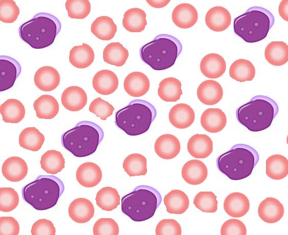
A rare, acute myeloid leukemia characterized by no significant myeloid maturation and more than 90% blast cells in the non-erythroid population. Various degrees of anemia, thrombocytopenia, or pancytopenia are present. Frequent clinical manifestations include fatigue, fever, bleeding disorders, and organomegaly, especially hepatosplenomegaly.