It has been reported in cases of extrapyramidal disorders, including Parkinson’s disease , parkinsonism , progressive supranuclear palsy , [14] hydrocephalus , [15] motor neuron disease , [16] Shy–Drager syndrome , [8] and various lesions in the brain. [17] [18] [19] [10] Medications that have been associated include Lithium , [20] sulpiride, [21] and a meperidine analog, 1-Methyl-4-phenyl-1,2,3,6-tetrahydropyridine. [22] Diagnosis [ edit ] ALO is often a benign and unilateral condition, and extensive evaluation may not be necessary. ... Clin Neuropharmacol. 1999 May-Jun. 22(3):176-9 ^ a b Tsuji S, Kikkawa S, Horiguchi J, Yamashita H, Kagaya A, Morinobu S, et al. Meige syndrome with apraxia of lid opening after the discontinuation of sulpiride treatment. ... Isolated eyelid-opening apraxia: report of a new levodopa-responsive syndrome. Neurology. 1994 Sep. 44(9):1752-4 ^ Lee KC, Finley R, Miller B.
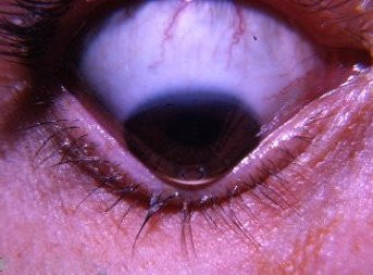
Defazio et al. (1998) described 10 new patients with 'so-called apraxia of eyelid opening.' They concluded that the term 'apraxia' may not be the correct descriptive term even when the eyelid disturbance occurs without any other central nervous system disease. Familial clustering of the isolated finding in 1 patient was consistent with a genetic contribution: 2 brothers, their father, and 2 paternal aunts were thought to be affected. Combining their 10 patients with 11 previously reported cases of isolated so-called apraxia of eyelid opening, Defazio et al. (1998) found that the peak age at onset was the sixth decade and that there was a female preponderance of 2 to 1. The characteristic inability to initiate lid elevation was frequently associated with failure to sustain lid elevation, thus suggesting that eyelid motor control may be abnormal.
Diagnosis [ edit ] Classification [ edit ] Prosopometamorphopsia is considered a face hallucination and is included under the umbrella of complex visual hallucinations. [5] Unlike other forms of hallucinations such as peduncular hallucinosis or Charles Bonnet syndrome , prosopometamorphopsia does not predominate at a particular time of day; it is a constant experience. [5] However, patients with Charles Bonnet syndrome have noted descriptions of prosopometamorphopsia. [4] This form of perceptual distortion along with others such as macropsia and micropsia (alteration of perceived object size) and palinopia (spatial and temporal varieties and polyopia) are classified under the category termed metamorphopsia. [6] These facial distortions can occur to either hallucinated perceptions or true (non-hallucinated) perceptions. [4] It is attributed to structural brain changes or functional disorders like epilepsy , migraine or eye diseases. [7] Treatment [ edit ] Antidepressants such as citalopram and the antipsychotic quetiapine have been recorded as unable to facilitate improvements for these symptoms. [7] Valproic acid was initially used to treat the woman who hallucinated the dragon-like faces and this alleviated her symptoms entirely, however, she went on to develop auditory hallucinations as a side effect. [7] She was subsequently prescribed rivastigmine instead which reduced the auditory hallucinations and reduced her visual symptoms. ... Neuropsychologia . 59 : 179–191. doi : 10.1016/j.neuropsychologia.2014.05.005 . ^ a b c Santhouse, A M; Howard, R J; ffytche, D H (2000). "Visual Hallucinatory Syndromes and the Anatomy of the Visual Brain" .
In addition to infections due to neutropenia, a patient with the Acute Radiation Syndrome will also be at risk for viral, fungal and parasitic infections. ... J Burn Care Res. 2011; 32:317-23. ^ Borden Institute. Chapter 2 Acute radiation syndrome ^ Borden Institute. Chapter 2 Acute radiation syndrome ^ Reeves GI.
However, the most common subcategories can be broken down as follows: 40% peripheral vestibular dysfunction, 10% central nervous system lesion, 15% psychiatric disorder, 25% presyncope/disequilibrium, and 10% nonspecific dizziness. [13] Some vestibular pathologies have symptoms that are comorbid with mental disorders. [14] While traditional medical teaching has focused on determining the cause of dizziness based on the category (e.g. vertigo vs presyncope), recent research suggests that this analysis is of limited clinical utility. [15] [16] Medical conditions that often have dizziness as a symptom include: [13] [17] [8] [18] Benign paroxysmal positional vertigo Meniere's disease Labyrinthitis Otitis media Brain tumor Acoustic neuroma Motion sickness Ramsay Hunt syndrome Migraine Multiple sclerosis Pregnancy Low blood pressure ( hypotension ) Low blood oxygen content ( hypoxemia ) Heart attack [19] Iron deficiency ( anemia ) Low blood sugar ( hypoglycemia ) Hormonal changes (e.g. thyroid disease, menstruation, pregnancy) Panic disorder Hyperventilation Anxiety Depression Age-diminished visual, balance, and perception of spatial orientation abilities a stroke is the cause of isolated dizziness in 0.7% of people who present to the emergency department . [7] Epidemiology [ edit ] About 20–30% of the population report to have experienced dizziness at some point in 2008. [7] Disequilibrium [ edit ] This section is about the loss of sense of balance. ... Many medications used to treat seizures, depression, anxiety, and pain affect the vestibular system and the central nervous system which can cause the symptom of disequilibrium. [20] See also [ edit ] Balance disorder Broken escalator phenomenon Chronic subjective dizziness Coriolis effect (perception) Equilibrioception Ideomotor phenomenon Illusions of self-motion Motion sickness Postural orthostatic tachycardia syndrome Proprioception Seasickness Spatial disorientation The spins , a state of dizziness and disorientation due to intoxication Vertigo References [ edit ] ^ " dizziness " at Dorland's Medical Dictionary ^ Dizziness at the US National Library of Medicine Medical Subject Headings (MeSH) ^ Reeves AG, Swenson RS (2008). ... (November 2010). "Part 10: acute coronary syndromes: 2010 American Heart Association Guidelines for Cardiopulmonary Resuscitation and Emergency Cardiovascular Care" .
Overview Dizziness is a term used to describe a range of sensations, such as feeling faint, woozy, weak or unsteady. Dizziness that creates the false sense that you or your surroundings are spinning or moving is called vertigo. Dizziness is one of the more common reasons adults visit their doctors. Frequent dizzy spells or constant dizziness can significantly affect your life. But dizziness rarely signals a life-threatening condition. Treatment of dizziness depends on the cause and your symptoms.
At an ultrastructural level the weakening of the corneal tissue is associated with a disruption of the regular arrangement of the collagen layers and collagen fibril orientation. [22] While keratoconus is considered a noninflammatory disorder, one study shows wearing rigid contact lenses by people leads to overexpression of proinflammatory cytokines , such as IL-6 , TNF-alpha , ICAM-1 , and VCAM-1 in the tear fluid. [23] A genetic predisposition to keratoconus has been observed, [24] with the disease running in certain families, [25] and incidences reported of concordance in identical twins. [26] The frequency of occurrence in close family members is not clearly defined, though it is known to be considerably higher than that in the general population, [14] and studies have obtained estimates ranging between 6% and 19%. [27] Two studies involving isolated, largely homogenetic communities have contrarily mapped putative gene locations to chromosomes 16q and 20q. [27] Most genetic studies agree on an autosomal dominant model of inheritance. [9] A rare, autosomal dominant form of severe keratoconus with anterior polar cataract is caused by a mutation in the seed region of mir-184 , a microRNA that is highly expressed in the cornea and anterior lens. [28] Keratoconus is diagnosed more often in people with Down's syndrome , though the reasons for this link have not yet been determined. [29] Keratoconus has been associated with atopic diseases , [30] which include asthma , allergies , and eczema , and it is not uncommon for several or all of these diseases to affect one person. Keratoconus is also associated with Alport syndrome , Down syndrome and Marfan syndrome . [30] A number of studies suggest vigorous eye rubbing contributes to the progression of keratoconus, and people should be discouraged from the practice. [31] [32] [33] [34] [35] [36] Keratoconus differs from ectasia which is caused by LASIK eye surgery. ... Keratoconus at Curlie Classification D ICD - 10 : H18.6 ICD - 9-CM : 371.6 OMIM : 148300 MeSH : D007640 DiseasesDB : 7158 External resources MedlinePlus : 001013 eMedicine : oph/104 NORD : Keratoconus GARD : Keratoconus Orphanet : 156071 v t e Diseases of the human eye Adnexa Eyelid Inflammation Stye Chalazion Blepharitis Entropion Ectropion Lagophthalmos Blepharochalasis Ptosis Blepharophimosis Xanthelasma Ankyloblepharon Eyelash Trichiasis Madarosis Lacrimal apparatus Dacryoadenitis Epiphora Dacryocystitis Xerophthalmia Orbit Exophthalmos Enophthalmos Orbital cellulitis Orbital lymphoma Periorbital cellulitis Conjunctiva Conjunctivitis allergic Pterygium Pseudopterygium Pinguecula Subconjunctival hemorrhage Globe Fibrous tunic Sclera Scleritis Episcleritis Cornea Keratitis herpetic acanthamoebic fungal Exposure Photokeratitis Corneal ulcer Thygeson's superficial punctate keratopathy Corneal dystrophy Fuchs' Meesmann Corneal ectasia Keratoconus Pellucid marginal degeneration Keratoglobus Terrien's marginal degeneration Post-LASIK ectasia Keratoconjunctivitis sicca Corneal opacity Corneal neovascularization Kayser–Fleischer ring Haab's striae Arcus senilis Band keratopathy Vascular tunic Iris Ciliary body Uveitis Intermediate uveitis Hyphema Rubeosis iridis Persistent pupillary membrane Iridodialysis Synechia Choroid Choroideremia Choroiditis Chorioretinitis Lens Cataract Congenital cataract Childhood cataract Aphakia Ectopia lentis Retina Retinitis Chorioretinitis Cytomegalovirus retinitis Retinal detachment Retinoschisis Ocular ischemic syndrome / Central retinal vein occlusion Central retinal artery occlusion Branch retinal artery occlusion Retinopathy diabetic hypertensive Purtscher's of prematurity Bietti's crystalline dystrophy Coats' disease Sickle cell Macular degeneration Retinitis pigmentosa Retinal haemorrhage Central serous retinopathy Macular edema Epiretinal membrane (Macular pucker) Vitelliform macular dystrophy Leber's congenital amaurosis Birdshot chorioretinopathy Other Glaucoma / Ocular hypertension / Primary juvenile glaucoma Floater Leber's hereditary optic neuropathy Red eye Globe rupture Keratomycosis Phthisis bulbi Persistent fetal vasculature / Persistent hyperplastic primary vitreous Persistent tunica vasculosa lentis Familial exudative vitreoretinopathy Pathways Optic nerve Optic disc Optic neuritis optic papillitis Papilledema Foster Kennedy syndrome Optic atrophy Optic disc drusen Optic neuropathy Ischemic anterior (AION) posterior (PION) Kjer's Leber's hereditary Toxic and nutritional Strabismus Extraocular muscles Binocular vision Accommodation Paralytic strabismus Ophthalmoparesis Chronic progressive external ophthalmoplegia Kearns–Sayre syndrome palsies Oculomotor (III) Fourth-nerve (IV) Sixth-nerve (VI) Other strabismus Esotropia / Exotropia Hypertropia Heterophoria Esophoria Exophoria Cyclotropia Brown's syndrome Duane syndrome Other binocular Conjugate gaze palsy Convergence insufficiency Internuclear ophthalmoplegia One and a half syndrome Refraction Refractive error Hyperopia Myopia Astigmatism Anisometropia / Aniseikonia Presbyopia Vision disorders Blindness Amblyopia Leber's congenital amaurosis Diplopia Scotoma Color blindness Achromatopsia Dichromacy Monochromacy Nyctalopia Oguchi disease Blindness / Vision loss / Visual impairment Anopsia Hemianopsia binasal
Keratoconus is the degeneration of the structure of the cornea , which is the clear tissue covering the front of the eye. In this condition, the shape of the cornea slowly changes from the normal round shape to a cone shape. Most people who develop keratoconus start out nearsighted, which tends to become worse over time. The earliest symptom is a slight blurring of vision that cannot be corrected with glasses. Over time, there may be eye halos, glare, or other night vision problems.The cause is unknown, but the tendency to develop keratoconus is probably present from birth.
Depending on the severity of disease, other signs or symptoms may include: Paralysis of muscles involved in breathing Difficulty swallowing Post-polio syndrome Post-polio syndrome is the appearance of new signs or symptoms or the progression of problems. ... Long-term complications for people who recover may include: Permanent paralysis Muscle shortening that causes deformed bones or joints Chronic pain Post-polio syndrome Prevention The most effective way to prevent polio is vaccination.
Poliomyelitis is a viral disease that can affect nerves and can lead to partial or full paralysis . It is caused by infection with the poliovirus. This virus can be spread by direct person-to-person contact, by contact with infected mucus or phlegm from the nose or mouth, or by contact with infected feces. There are three basic patterns of polio infection: subclinical infections, nonparalytic, and paralytic. Symptoms vary based on the pattern of infection and can range from asymptomatic with subclinical poliomyelitis to partial or full paralysis. Treatment is aimed at controlling symptoms while the infection runs its course.
Extended use of braces or wheelchairs may cause compression neuropathy , as well as a loss of proper function of the veins in the legs, due to pooling of blood in paralyzed lower limbs. [43] [73] Complications from prolonged immobility involving the lungs , kidneys and heart include pulmonary edema , aspiration pneumonia , urinary tract infections , kidney stones , paralytic ileus , myocarditis and cor pulmonale . [43] [73] Post-polio syndrome Main article: Post-polio syndrome Between 25 percent and 50 percent of individuals who have recovered from paralytic polio in childhood can develop additional symptoms decades after recovering from the acute infection, [74] notably new muscle weakness and extreme fatigue. This condition is known as post-polio syndrome (PPS) or post-polio sequelae. [75] The symptoms of PPS are thought to involve a failure of the oversized motor units created during the recovery phase of the paralytic disease. [76] [77] Contributing factors that increase the risk of PPS include aging with loss of neuron units, the presence of a permanent residual impairment after recovery from the acute illness, and both overuse and disuse of neurons. PPS is a slow, progressive disease, and there is no specific treatment for it. [75] Post-polio syndrome is not an infectious process, and persons experiencing the syndrome do not shed poliovirus. [1] Epidemiology This section needs to be updated . ... Please update this article to reflect recent events or newly available information. ( October 2020 ) The Poliovirus Antivirals Initiative was launched in 2007 with the aim of developing antiviral medications for polio, but while several promising candidates were identified, none have progressed beyond Phase II clinical trials. [155] [156] Pocapavir (a capsid inhibitor ) and V-7404 (a protease inhibitor ) may speed up viral clearance and are being studied for this purpose. [157] References ^ a b c d e f g h i j k l m n o p q r s t u v w x y z aa ab ac ad ae af ag Hamborsky J, Kroger A, Wolfe C, eds. (2015), "Poliomyelitis" , Epidemiology and Prevention of Vaccine-Preventable Diseases (The Pink Book) (13th ed.), Washington DC: Public Health Foundation, (chap. 18), archived from the original on 30 December 2016 . ^ a b "Post-Polio Syndrome Fact Sheet" . NIH. 16 April 2014. ... "Nonpoliovirus poliomyelitis simulating Guillain-Barré syndrome" . Archives of Neurology . 58 (9): 1460–4. doi : 10.1001/archneur.58.9.1460 .
Poliomyelitis is a viral infection caused by any of three serotypes of human poliovirus, which is part of the family of enteroviruses. Epidemiology Progress in global poliomyelitis eradication, since its beginning in 1988, has been remarkable. In 1988, 125 countries were endemic for poliomyelitis and an estimated 1000 children were being paralyzed every day by wild poliovirus. By the end of 2003, six polio-endemic countries remained (Afghanistan, Egypt, India, Niger, Nigeria, Pakistan), and less than 3 children per day were being paralyzed by the poliovirus. The Global Poliomyelitis Eradication Initiative is ongoing. There remain only 4 endemic countries (Pakistan, Afghanistan, India, and Nigeria) and just 2000 reported cases globally in 2006.
Overview Uterine prolapse occurs when pelvic floor muscles and ligaments stretch and weaken until they no longer provide enough support for the uterus. As a result, the uterus slips down into or protrudes out of the vagina. Uterine prolapse most often affects people after menopause who've had one or more vaginal deliveries. Mild uterine prolapse usually doesn't require treatment. But uterine prolapse that causes discomfort or disrupts daily life might benefit from treatment. Symptoms Mild uterine prolapse is common after childbirth. It generally doesn't cause symptoms.
Males are slightly more likely to develop ampullary cancer than are females. Inherited syndromes that increase cancer risk. Some gene mutations passed through generations of your family can increase your risk of ampullary cancer significantly. Only a small percentage of ampullary cancers are linked to inherited genes. The most common inherited syndromes that increase ampullary cancer risk are familial adenomatous polyposis and Lynch syndrome, which is also known as hereditary nonpolyposis colorectal cancer.
Carcinoma of the ampulla of Vater is a rare malignant tumor originating from the ampulla of Vater that can present with symptoms of general fatigue, loss of appetite, weight loss, nausea, vomiting, abdominal pain and, most commonly, painless obstructive jaundice. The tumor is believed to arise from duodenal, biliary or pancreatic epilthelium, resulting in the respective histological types. In general, carcinoma of the ampulla of Vater has a better prognosis (5-year survival rate of 45%) than cancers of the distal bile duct and pancreas.
External links [ edit ] Classification D ICD - 10 : D56.2 ICD - 9-CM : 282.45 MeSH : D055538 External resources Orphanet : 231237 Scholia has a topic profile for Delta-beta thalassemia . v t e Diseases of red blood cells ↑ Polycythemia Polycythemia vera ↓ Anemia Nutritional Micro- : Iron-deficiency anemia Plummer–Vinson syndrome Macro- : Megaloblastic anemia Pernicious anemia Hemolytic (mostly normo- ) Hereditary enzymopathy : Glucose-6-phosphate dehydrogenase deficiency glycolysis pyruvate kinase deficiency triosephosphate isomerase deficiency hexokinase deficiency hemoglobinopathy : Thalassemia alpha beta delta Sickle cell disease / trait Hereditary persistence of fetal hemoglobin membrane : Hereditary spherocytosis Minkowski–Chauffard syndrome Hereditary elliptocytosis Southeast Asian ovalocytosis Hereditary stomatocytosis Acquired AIHA Warm antibody autoimmune hemolytic anemia Cold agglutinin disease Donath–Landsteiner hemolytic anemia Paroxysmal cold hemoglobinuria Mixed autoimmune hemolytic anemia membrane paroxysmal nocturnal hemoglobinuria Microangiopathic hemolytic anemia Thrombotic microangiopathy Hemolytic–uremic syndrome Drug-induced autoimmune Drug-induced nonautoimmune Hemolytic disease of the newborn Aplastic (mostly normo- ) Hereditary : Fanconi anemia Diamond–Blackfan anemia Acquired: Pure red cell aplasia Sideroblastic anemia Myelophthisic Blood tests Mean corpuscular volume normocytic microcytic macrocytic Mean corpuscular hemoglobin concentration normochromic hypochromic Other Methemoglobinemia Sulfhemoglobinemia Reticulocytopenia v t e Medicine Specialties and subspecialties Surgery Cardiac surgery Cardiothoracic surgery Colorectal surgery Eye surgery General surgery Neurosurgery Oral and maxillofacial surgery Orthopedic surgery Hand surgery Otolaryngology ENT Pediatric surgery Plastic surgery Reproductive surgery Surgical oncology Transplant surgery Trauma surgery Urology Andrology Vascular surgery Internal medicine Allergy / Immunology Angiology Cardiology Endocrinology Gastroenterology Hepatology Geriatrics Hematology Hospital medicine Infectious disease Nephrology Oncology Pulmonology Rheumatology Obstetrics and gynaecology Gynaecology Gynecologic oncology Maternal–fetal medicine Obstetrics Reproductive endocrinology and infertility Urogynecology Diagnostic Radiology Interventional radiology Nuclear medicine Pathology Anatomical Clinical pathology Clinical chemistry Cytopathology Medical microbiology Transfusion medicine Other Addiction medicine Adolescent medicine Anesthesiology Dermatology Disaster medicine Diving medicine Emergency medicine Mass gathering medicine Family medicine General practice Hospital medicine Intensive care medicine Medical genetics Narcology Neurology Clinical neurophysiology Occupational medicine Ophthalmology Oral medicine Pain management Palliative care Pediatrics Neonatology Physical medicine and rehabilitation PM&R Preventive medicine Psychiatry Addiction psychiatry Radiation oncology Reproductive medicine Sexual medicine Sleep medicine Sports medicine Transplantation medicine Tropical medicine Travel medicine Venereology Medical education Medical school Bachelor of Medicine, Bachelor of Surgery Bachelor of Medical Sciences Master of Medicine Master of Surgery Doctor of Medicine Doctor of Osteopathic Medicine MD–PhD Related topics Alternative medicine Allied health Dentistry Podiatry Pharmacy Physiotherapy Molecular oncology Nanomedicine Personalized medicine Public health Rural health Therapy Traditional medicine Veterinary medicine Physician Chief physician History of medicine Book Category Commons Wikiproject Portal Outline
A number sign (#) is used with this entry because hereditary persistence of fetal hemoglobin (HPFH) can result from deletions within or encompassing the beta-globin gene cluster (see HBB, 141900) on chromosome 11p15, including deletions that also encompass the delta-globin gene (142000), or from point mutations in the promoter regions of either the HBG1 (142200) or the HBG2 (142250) gene. Other fetal hemoglobin quantitative trait loci (QTL) include HBFQTL2 (142470) on chromosome 6q23, HBFQTL3 (305435) on chromosome Xp22.2, and HBFQTL5 (142335) on chromosome 2p15, and HBFQTL6 (613566), caused by mutation in the KLF1 gene (600599) on chromosome 19p13. A QTL on chromosome 8q (HBFQTL4; 606789) is thought to interact with the common XmnI-G-gamma polymorphism in HBG2 (142250.0028) to influence the production of HbF. Description Classic hereditary persistence of fetal hemoglobin (HPFH) is characterized by a substantial elevation of fetal hemoglobin (HbF) in adult red blood cells. There are no other phenotypic or hematologic manifestations. Expression of the HBG1 and HBG2 genes, which encode the gamma isoforms of HbF, is normally suppressed shortly before birth and replaced by expression of the beta- (HBB; 141900) or delta- (HBD; 142000) chains, which form adult hemoglobin.
Delta-beta-thalassemia is a form of beta-thalassemia (see this term) characterized by decreased or absent synthesis of the delta- and beta-globin chains with a compensatory increase in expression of fetal gamma-chain synthesis. Epidemiology Prevalence of this form is not known. The condition is found in many ethnic groups but is most common in Greece and Italy. Clinical description The heterozygous form of the condition is clinically asymptomatic with mild microcytosis and no elevation of HbA2 whereas the few homozygous patients have a mild clinical presentation. When inherited with heterozygous classical beta-thalassemia, patients usually have the thalassemia intermedia phenotype, but the thalassemia major phenotype has been described in some cases. Etiology Delta-beta-thalassemia is commonly caused by deletions of the entire delta and beta gene sequences with production of only gamma-globin and formation of HbF.
For example, the differential for presentation with TIN includes: medications, autoimmune disease (e.g. Sjogren's syndrome, Sarcoid), malignancy (e.g. lymphoproliferative disease), and infection (e.g.
Monatsschrift für Psychiatrie und Neurologie, 1902, 12: 421-423. v t e Symptoms and signs relating to the nervous system Neurological examination · Cranial nerve examination Central nervous system Head Battle's sign Kernig's sign Macewen's sign Myerson's sign Stroop test Hirano body Other increased intracranial pressure Cushing's triad Lhermitte's sign Charcot's neurologic triad Peripheral nervous system Reflexes Combination Jendrassik maneuver Legs Plantar reflex Chaddock reflex Oppenheim's sign Westphal's sign Arms Hoffmann's sign Other Arms Froment's sign carpal tunnel syndrome Tinel sign Phalen maneuver Legs Gowers' sign Hoover's sign Lasègue's sign Trendelenburg's sign Torso Beevor's sign General Pain stimulus This medical sign article is a stub .
For a phenotypic description and discussion of genetic heterogeneity of migraine headaches, see MGR1 (157300). Mapping Since migraine is a syndrome instead of a clearly differentiated disease, Anttila et al. (2006) hypothesized that individual clinical components of migraine (i.e., traits such as pulsating pain and photophobia, among others) might represent reflections of specific rather than shared loci and thus independently contribute to susceptibility to migraine.