Influenza-like illness ( ILI ), also known as flu-like syndrome/symptoms , is a medical diagnosis of possible influenza or other illness causing a set of common symptoms . ... Other drugs associated with a flu-like syndrome include bisphosphonates , caspofungin , and levamisole . [14] [15] A flu-like syndrome can also be caused by an influenza vaccine or other vaccines, and by opioid withdrawal in physically dependent individuals.
Cardiac involvement in the acquired immunodeficiency syndrome: a multicentre clinical-pathological study. ... Cardiac involvement in the acquired immunodeficiency syndrome: a multicentre clinical-pathological study. ... Cardiac involvement in the acquired immunodeficiency syndrome: a multicentre clinical-pathological study.
Postmortem neuropathologic examination revealed no signs of Leigh syndrome (256000), but developmental and degenerative lesions were observed. ... Maesaka et al. (1976) described 2 sisters with pyruvate carboxylase deficiency, severe mental and motor retardation, and Leigh syndrome. The proband had lactic acidosis, low CSF glucose, hyperalaninemia, and increased urinary lactate, pyruvate, and alanine. ... INHERITANCE - Autosomal recessive ABDOMEN Liver - Hepatomegaly GENITOURINARY Kidneys - Renal tubular acidosis, proximal NEUROLOGIC Central Nervous System - Psychomotor retardation - Mental retardation - Developmental delay - Hypotonia - Seizures - Clonus - Cystic lesions consistent with Leigh syndrome ( 256000 ) - Neuronal loss in the cerebral cortex - Poor myelination - Periventricular leukomalacia - Subcortical leukodystrophy METABOLIC FEATURES - Lactic acidosis LABORATORY ABNORMALITIES - Decreased pyruvate carboxylase (PC) activity (less than 5%) - Increased serum lactate - Increased serum pyruvate - Increased serum alanine - Hypoglycemia - Increased serum ammonia (Group B) - Increased serum citrulline (Group B) - Increased serum lysine (Group B) - Intracellular redox disturbance (reduced cytoplasm and oxidized mitochondria (Group B)) - Increased lactate: pyruvate ratio (Group B) - Increased acetoacetate: beta-hydroxybutyrate ratio (Group B) - Immunoreactive PC protein - Presence of PC mRNA - A subset of Group B patients have absence of PC protein and mRNA MISCELLANEOUS - Onset at birth or in early infancy - Some patients may respond to thiamine treatment - Can be categorized into 3 groups - Group A, found in North American Indians, has lactic acidosis and psychomotor retardation - Group A patients die in the first years of life - Group B, found in France and United Kingdom, severe phenotype - Group B patients die by 3 months of age - Group C is relatively benign MOLECULAR BASIS - Caused by mutation in the pyruvate carboxylase gene (PC, 608786.0001 ) ▲ Close
Pyruvate carboxylase deficiency is an inherited disorder that causes lactic acid and other potentially toxic compounds to accumulate in the blood. High levels of these substances can damage the body's organs and tissues, particularly in the nervous system. Researchers have identified at least three types of pyruvate carboxylase deficiency, which are distinguished by the severity of their signs and symptoms. Type A, which has been identified mostly in people from North America, has severe symptoms that begin in infancy. Characteristic features include developmental delay and a buildup of lactic acid in the blood (lactic acidosis).
Pyruvate carboxylase (PC) deficiency is a rare neurometabolic disorder characterized by metabolic acidosis, failure to thrive, developmental delay, and recurrent seizures at an early age in severely affected patients. Epidemiology The overall prevalence of PC deficiency is not known and annual incidence has been reported to be 1/250,000 births. The disorder affects males and females equally. Clinical description Three clinical presentations of PC deficiency, probably constituting a continuum, have been described: infantile PC deficiency (type A); severe neonatal PC deficiency (type B); and intermittent/benign PC deficiency (type C). The only common feature is metabolic acidosis. Type A is characterized by infantile onset, generally with a severe course. Type B has a very severe course with a fatal outcome in early infancy, and Type C involves only episodic metabolic acidosis.
The diagnosis of PDHC deficiency is suspected in individuals with lactic acidemia who have a progressive or intermittent neurologic syndrome including: poor acquisition or loss of motor milestones, poor muscle tone, new-onset seizures, periods of incoordination (i.e., ataxia), abnormal eye movements, poor response to visual stimuli, and episodic dystonia. ... Brain MRI may reveal distinctive abnormalities, as described with Leigh syndrome or mitochondrial encephalomyopathy with lactic acidosis and stroke-like episodes (MELAS) [DiMauro & Schon 2008]. ... Two sisters with PC deficiency, severe intellectual disability and motor retardation, and Leigh syndrome improved clinically and biochemically after treatment with thiamine and lipoic acid.
Benign pyruvate carboxylase (PC) deficiency (Type C) is a rare, very mild form of PC deficiency characterized by episodic metabolic acidosis and normal or mildly delayed neurological development. Epidemiology Benign PC deficiency is a very rare form of PC deficiency and has been described in fewer than 10 patients to date. No ethnic predilection has been reported. Clinical description Onset typically occurs during the first year of life with episodic metabolic acidosis associated with lactic acidemia and occasionally with ketoacidosis during metabolic stress. Neurological development is normal or mildly impaired. Other signs include dystonia, episodic ataxia, dysarthria, transitory hemiparesis and seizures. Etiology Type C PC deficiency is caused by mutations in the PC gene (11q13.4-q13.5).
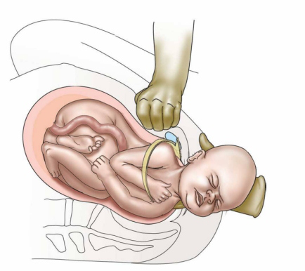
Severe neonatal pyruvate carboxylase (PC) deficiency (Type B) is a rare, extremely severe form of PC deficiency characterized by severe, early-onset metabolic acidosis, and a generally fatal outcome in early infancy. Epidemiology The exact prevalence of Type B pyruvate carboxylase deficiency is not known. The disorder has been reported to be more common in populations of Arab descent (Algerian, Egyptian, and Saudi Arabian). Higher incidence is also reported in France, Germany and the United Kingdom. Clinical description Patients develop clinical manifestations during the first 72 hours of life with severe truncal hypotonia and tachypnea.
Infantile pyruvate carboxylase (PC) deficiency (Type A) is a rare, severe form of PC deficiency characterized by infantile-onset, mild to moderate lactic acidemia, and a generally severe course. Epidemiology The specific prevalence of type A pyruvate carboxylase deficiency is not known but it has been reported most often in Native Americans from North American Algonquian-speaking groups including the Mi'kmaq, Cree, and Ojibwa tribes. In these groups, the carrier frequency may be as high as 1/10. Clinical description Patients with Type A PC deficiency usually first present with symptoms at the age of two to five months, often after normal early development. Clinical manifestations include mild to moderate metabolic acidosis with acute vomiting and tachypnea, failure to thrive, apathy, delayed intellectual and motor development, hypotonia, pyramidal dysfunction, ataxia, nystagmus and seizures. Renal tubular acidosis has also been reported. Etiology PC deficiency is caused by mutations in the PC gene (11q13.4-q13.5).
Pyruvate carboxylase deficiency is an inherited disorder that causes lactic acid and other potentially toxic compounds to accumulate in the blood. High levels of these substances can damage the body's organs and tissues, particularly in the nervous system. Researchers have identified at least three types of pyruvate carboxylase deficiency, types A, B, and C, which are distinguished by the severity of their signs and symptoms. This condition is caused by mutations in the PC gene and inherited in an autosomal recessive pattern.
Find sources: "Transfusion-related acute lung injury" – news · newspapers · books · scholar · JSTOR ( January 2017 ) ( Learn how and when to remove this template message ) Transfusion-related acute lung injury Other names TRALI Micrograph of diffuse alveolar damage , the histologic correlate of TRALI; H&E stain Specialty Pulmonology Transfusion-related acute lung injury ( TRALI ) is a serious blood transfusion complication characterized by the acute onset of non-cardiogenic pulmonary edema presenting with hypoxia following transfusion of blood products. [1] Although the incidence of TRALI has decreased with modified transfusion practices, it was the leading cause of transfusion-related deaths in the United States from fiscal year 2008 through fiscal year 2012. [2] Contents 1 Signs and symptoms 2 Cause 3 Pathophysiology 4 Diagnosis 5 Treatment 6 Epidemiology 7 See also 8 References 9 External links Signs and symptoms [ edit ] It is often impossible to distinguish TRALI from acute respiratory distress syndrome (ARDS). The typical presentation of TRALI is the sudden development of dyspnea , severe hypoxemia (O 2 saturation <90% in room air), hypotension , and fever that develop within 6 hours after transfusion and usually resolve with supportive care within 48 to 96 hours. ... In the remaining 20% of TRALI cases, non–antibody factors/biological response modifiers are suggested to contribute the second hit, and these may possibly include lipid mediators, extracellular vesicles, and aged blood cells. [12] Diagnosis [ edit ] Chest X-ray of transfusion-related acute lung injury (TRALI) compared to chest X-ray of the same person after treatment TRALI is defined as an acute lung injury that is temporally related to a blood transfusion; specifically, it occurs within the first six hours following a transfusion. [13] It is typically associated with plasma components such as platelets and fresh frozen plasma , though cases have been reported with packed red blood cells since there is some residual plasma in the packed cells. [10] Incidents have also been reported with other blood products including "cryoprecipitate, granulocytes, intravenous immune globulin, allogeneic and autologous stem cells". [14] It is a diagnosis upon examination of clinical manifestations that appear within 6 hours of transfusion, such as acute respiratory distress, tachypnea, hypotension, cyanosis, and dyspnea. TRALI is an uncommon syndrome, that is due to the presence of leukocyte antibodies in transfused plasma. ... In most cases leukoagglutination results in mild dyspnea and pulmonary infiltrates within about 6 hours of transfusion, and spontaneously resolves. [ citation needed ] Occasionally more severe lung injury occurs as a result of this phenomenon and acute respiratory distress syndrome (ARDS) results. Leukocyte filters may prevent TRALI for those patients whose lung injury is due to leukoagglutination of the donor white blood cells, but because most TRALI is due to donor antibodies to leukocytes, filters are not helpful in TRALI prevention.
Uterine cancer Other names Womb cancer Specialty Gynecology , oncology Symptoms Endometrial cancer : vaginal bleeding , pelvic pain [1] Uterine sarcoma : vaginal bleeding, mass in the vagina [2] Types Endometrial cancer , uterine sarcoma [3] Risk factors Endometrial cancer : obesity , metabolic syndrome , type 2 diabetes , family history of the condition [1] Uterine sarcoma : radiation therapy to the pelvis [2] Treatment Surgery , radiation therapy , chemotherapy , hormone therapy , targeted therapy [1] [2] Prognosis 81% 5 year survival (US) [4] Frequency 3.8 million (2015) [5] Deaths 90,000 (2015) [6] Uterine cancer , also known as womb cancer , are two types of cancer that develops from the tissues of the uterus . [3] Endometrial cancer forms from the lining of the uterus and uterine sarcoma forms from the muscles or support tissue of the uterus. [1] [2] Symptoms of endometrial cancer include unusual vaginal bleeding or pain in the pelvis . [1] Symptoms of uterine sarcoma include unusual vaginal bleeding or a mass in the vagina. [2] Risk factors for endometrial cancer include obesity , metabolic syndrome , type 2 diabetes , and a family history of the condition. [1] Risk factors for uterine sarcoma include prior radiation therapy to the pelvis. [2] Diagnosis of endometrial cancer is typically based on an endometrial biopsy . [1] A diagnosis of uterine sarcoma may be suspected based on symptoms, a pelvic exam , and medical imaging . [2] Endometrial cancer can often be cured while uterine sarcoma typically is harder to treat. [3] Treatment may include a combination of surgery , radiation therapy , chemotherapy , hormone therapy , and targeted therapy . [1] [2] Just over 80% of people survive more than 5 years following diagnosis. [4] In 2015 about 3.8 million people were affected globally and it resulted in 90,000 deaths. [5] [6] Endometrial cancer is relatively common while uterine sarcoma is rare. [3] In the United States they represent 3.6% of new cancer cases. [4] They most commonly occur in women between the ages of 55 and 74. [4] Contents 1 Causes 2 Types 3 Epidemiology 3.1 United Kingdom 3.2 United States 4 References 5 External links Causes [ edit ] It is not known with certainty what the causes for uterine cancer may be, though hormone imbalance is speculated as a risk factor. ... External links [ edit ] Classification D ICD - 10 : Xxx.x ICD - 9-CM : xxx Clinically reviewed uterine cancer information for patients UK uterine cancer statistics v t e Tumors of the female urogenital system Adnexa Ovaries Glandular and epithelial / surface epithelial- stromal tumor CMS: Ovarian serous cystadenoma Mucinous cystadenoma Cystadenocarcinoma Papillary serous cystadenocarcinoma Krukenberg tumor Endometrioid tumor Clear-cell ovarian carcinoma Brenner tumour Sex cord–gonadal stromal Leydig cell tumour Sertoli cell tumour Sertoli–Leydig cell tumour Thecoma Granulosa cell tumour Luteoma Sex cord tumour with annular tubules Germ cell Dysgerminoma Nongerminomatous Embryonal carcinoma Endodermal sinus tumor Gonadoblastoma Teratoma / Struma ovarii Choriocarcinoma Fibroma Meigs' syndrome Fallopian tube Adenomatoid tumor Uterus Myometrium Uterine fibroids/leiomyoma Leiomyosarcoma Adenomyoma Endometrium Endometrioid tumor Uterine papillary serous carcinoma Endometrial intraepithelial neoplasia Uterine clear-cell carcinoma Cervix Cervical intraepithelial neoplasia Clear-cell carcinoma SCC Glassy cell carcinoma Villoglandular adenocarcinoma Placenta Choriocarcinoma Gestational trophoblastic disease General Uterine sarcoma Mixed Müllerian tumor Vagina Squamous-cell carcinoma of the vagina Botryoid rhabdomyosarcoma Clear-cell adenocarcinoma of the vagina Vaginal intraepithelial neoplasia Vaginal cysts Vulva SCC Melanoma Papillary hidradenoma Extramammary Paget's disease Vulvar intraepithelial neoplasia Bartholin gland carcinoma v t e Women's health Reproductive & Sexual health Reproductive health Reproductive tract External female genitalia (vulva) Clitoris Clitoral hood Labia minora Labia majora Vagina Cervix Uterus Fallopian tube Ovary Reproductive system disease Maternal health Pregnancy Unintended pregnancy Gravidity and parity Obstetrics Antenatal care Adolescent pregnancy Complications of pregnancy Hyperemesis gravidarum Ectopic pregnancy Miscarriage Obstetrical bleeding Gestational diabetes Hypertension Preeclampsia Eclampsia Childbirth Midwifery Preterm birth Multiple births Oxytocin Obstructed labor Cesarian section Retained placenta Obstetrical fistulae Vesicovaginal fistula Rectovaginal fistula Episiotomy husband stitch Postpartum care Postpartum confinement Maternal deaths Perinatal mortality Stillbirths Abortion Mother-to-child transmission Sterilization Compulsory sterilization Breastfeeding and mental health Reproductive life plan Infertility Childlessness Assisted reproductive technology In vitro fertilization Parenting Adoption Fostering Contraception & Family planning Unsafe sex Intrauterine devices Oral contraceptives Condoms Contraceptive prevalence Contraceptive security Planned parenthood Fertility awareness Menstruation Culture and menstruation Feminine hygiene Menarche Menstrual cycle Menstrual aids Cloth menstrual pad Menstrual cup Tampon Sanitary pad Dysmenorrhea Menorrhagia Amenorrhoea Menopause Hormone replacement therapy Sexual health Sexually transmitted infections HIV Human papilloma virus HPV vaccine Pelvic inflammatory disease Female genital mutilation Clitoridectomy Infibulation Breast ironing Child marriage Forced marriage Leblouh Polygamy Sexual intercourse Orgasm Dyspareunia Sex differences Sex education Puberty Breast health Gynaecological disorders Vaginitis Non-reproductive health Violence against women Abuse during childbirth Domestic violence Intimate partner violence Misogyny Sexual harassment Sexual assault Rape Femicide Gender discrimination Non-communicable diseases Cancer Lung cancer Breast cancer Uterine cancer Endometrial cancer Cervical cancer Papanicolaou test Ovarian cancer Cardiovascular disease Dementia Alzheimer's disease Bone health Osteoporosis Hip fracture Anaemia Mental health Anxiety Depression Major depressive disorder Urinary tract Urethra Urinary tract infection Urinary incontinence Sociocultural factors Poverty Disadvantaged Gender equality Healthcare inequality Gender disparities in health Social determinants of health Reproductive justice Women's empowerment Politics, Research & Advocacy United Nations The Convention on the Elimination of All Forms of Discrimination against Women Declaration on the elimination of violence against women International Day of the Girl Child Commission on the Status of Women UN Women United States Office of Research on Women's Health Women's Health Initiative International Center for Research on Women Nurses' Health Study Black Women's Health Study Cartwright Inquiry Society for Women's Health Research Women's health by country Women's health in China Women's health in Ethiopia Women's health in India Family planning Birth control in the United States Category Commons WikiProject
Excessive alcohol consumption Excessive consumption of antidiuretics or inadequate levels of antidiuretic hormone Intestinal malabsorption caused by short bowel syndrome Ketogenic diet Biochemistry [ edit ] Biotin is a coenzyme for five carboxylases in the human body (propionyl-CoA carboxylase, methylcrotonyl-CoA carboxylase, pyruvate carboxylase, and 2 forms of acetyl-CoA carboxylase.) ... External links [ edit ] Classification D ICD - 10 : E53.8 ICD - 9-CM : 266.2 MeSH : C531633 C531633, C531633 External resources eMedicine : ped/238 GeneReviews/NCBI/NIH/UW entry on Biotinidase deficiency OMIM entries on Biotinidasa deficiency v t e Malnutrition Protein-energy malnutrition Kwashiorkor Marasmus Catabolysis Vitamin deficiency B vitamins B 1 Beriberi Wernicke–Korsakoff syndrome Wernicke's encephalopathy Korsakoff's syndrome B 2 Riboflavin deficiency B 3 Pellagra B 6 Pyridoxine deficiency B 7 Biotin deficiency B 9 Folate deficiency B 12 Vitamin B 12 deficiency Other A: Vitamin A deficiency Bitot's spots C: Scurvy D: Vitamin D deficiency Rickets Osteomalacia Harrison's groove E: Vitamin E deficiency K: Vitamin K deficiency Mineral deficiency Sodium Potassium Magnesium Calcium Iron Zinc Manganese Copper Iodine Chromium Molybdenum Selenium Keshan disease Growth Delayed milestone Failure to thrive Short stature Idiopathic General Anorexia Weight loss Cachexia Underweight
Pyruvate kinase deficiency is a genetic blood disorder characterized by low levels of an enzyme called pyruvate kinase , which is used by red blood cells. Without pyruvate kinase, red blood cells break down too easily, resulting in low levels of these cells (hemolytic anemia). The signs and symptoms of the disease may vary greatly from person to person. However, they usually include jaundice, enlargement of the spleen , and mild or severe hemolysis (red cell breakdown), leading to anemia. In some cases, the problems may first appear while in utero, causing a condition in which abnormal amounts of fluid build up in two or more body areas of the fetus (hydrops fetalis).
A number sign (#) is used with this entry because red cell pyruvate kinase (PK) deficiency is caused by homozygous or compound heterozygous mutation in the gene encoding pyruvate kinase (PKLR; 609712) on chromosome 1q22. Description Red cell pyruvate kinase deficiency is the most common cause of hereditary nonspherocytic hemolytic anemia. PK deficiency is also the most frequent enzyme abnormality of the glycolytic pathway (Zanella et al., 2005). Clinical Features Valentine et al. (1961) first reported pyruvate kinase deficiency in 3 patients with congenital nonspherocytic hemolytic anemia. Tanaka et al. (1962) observed a compensated hemolytic anemia in young adults who had been relatively little incapacitated.
Pyruvate kinase deficiency is an inherited disorder that affects red blood cells, which carry oxygen to the body's tissues. People with this disorder have a condition known as chronic hemolytic anemia, in which red blood cells are broken down (undergo hemolysis) prematurely, resulting in a shortage of red blood cells (anemia). Specifically, pyruvate kinase deficiency is a common cause of a type of inherited hemolytic anemia called hereditary nonspherocytic hemolytic anemia. In hereditary nonspherocytic hemolytic anemia, the red blood cells do not assume a spherical shape as they do in some other forms of hemolytic anemia. Chronic hemolytic anemia can lead to unusually pale skin (pallor), yellowing of the eyes and skin (jaundice), extreme tiredness (fatigue), shortness of breath (dyspnea), and a rapid heart rate (tachycardia).
Differential diagnosis Secondary PK deficiency has also been reported, occurring in the context of hematological diseases (acute/chronic leukemia, myelodysplastic syndromes and sideroblastic anemia). In case of persistent normocytic hemolytic anemia in which hemoglobin abnormalities and antiglobulin reactions have been excluded, spherocytes are absent, and osmotic fragility is normal, the diagnosis of hereditary nonspherocytic hemolytic anemia should be considered.
If you had diabetes while pregnant (gestational diabetes), you and your child are at higher risk of developing prediabetes. Polycystic ovary syndrome. Women with this common condition — characterized by irregular menstrual periods, excess hair growth and obesity — have a higher risk of prediabetes. ... Other conditions associated with an increased risk of prediabetes include: High blood pressure Low levels of high-density lipoprotein (HDL) cholesterol, the "good" cholesterol High levels of triglycerides — a type of fat in your blood Metabolic syndrome When certain conditions occur with obesity, they are associated with insulin resistance, and can increase your risk for diabetes — and heart disease and stroke. A combination of three or more of these conditions is often called metabolic syndrome: High blood pressure Low levels of HDL High triglycerides High blood sugar levels Large waist size Complications Prediabetes has been linked with long-term damage, including to your heart, blood vessels and kidneys, even if you haven't progressed to type 2 diabetes.
A predisease state of hyperglycemia with high risk for diabetes Prediabetes Specialty Endocrinology Prediabetes is a component of the metabolic syndrome and is characterized by elevated blood sugar levels that fall below the threshold to diagnose diabetes mellitus . ... In 2014, 29.1 million people or 9% of the US population had diabetes. [44] In 2011–2012, the prevalence of diabetes in the U.S. using hemoglobin A1C, fasting plasma glucose or the two-hour plasma glucose definition was 14% for total diabetes, 9% for diagnosed diabetes, 5% for undiagnosed diabetes and 38% for prediabetes. [45] See also [ edit ] Metabolic syndrome Diabetes Insulin resistance Cardiovascular disease References [ edit ] ^ American Diabetes Association (January 2017). "2.
Dermatophagia – extreme nail biting / biting of skin to point of an obsessive compulsive disorder (OCD) or other condition leading to self mutilating behaviour such as autistic spectrum disorders (as is the case in this example) or Lesch-Nyhan Syndrome . [ citation needed ] Compulsive behavior is defined as performing an action persistently and repetitively without it necessarily leading to an actual reward or pleasure. [ citation needed ] Compulsive behaviors could be an attempt to make obsessions go away. [1] The act is usually a small, restricted and repetitive behavior, yet not disturbing in a pathological way. [ citation needed ] Compulsive behaviors are a need to reduce apprehension caused by internal feelings' a person wants to abstain from or control. [2] A major cause of the compulsive behaviors is said to be obsessive–compulsive disorder (OCD). [1] [3] "The main idea of compulsive behavior is that the likely excessive activity is not connected to the purpose to which it appears directed." [ citation needed ] Furthermore, there are many different types of compulsive behaviors including shopping , hoarding , eating , gambling , trichotillomania and picking skin , itching , checking , counting , washing, sex , and more. ... "Advances in the behavior analytic treatment of trichotillomania and Tourette's Syndrome" . Journal of Early and Intensive Behavior Intervention . 1 (1): 57–64. doi : 10.1037/h0100282 . ... Schwartz Susan Swedo Emily Colas Vic Meyer Popular culture Literature/Comics Fictional Matchstick Men Plyushkin Xenocide Nonfiction Everything in Its Place Just Checking Media As Good as It Gets The Aviator Matchstick Men Adrian Monk " $pringfield " Straight Up Related Obsessive–compulsive personality disorder Obsessional jealousy PANDAS Primarily Obsessional OCD Relationship obsessive–compulsive disorder Social anxiety disorder Tourette syndrome ^ Moritz, Steffen; Rufer, Michael (2011).
Malformations often lead to cardiac failure, cranial bruits (pattern 1), hydrocephaly , and subarachnoid hemorrhage in neonates. [4] The heart failure is due to the size of the arteriovenous shunt that can steal 80% or more of the cardiac output, with large volumes of blood under high pressure returning to the right heart and pulmonary circulation and sinus venosus atrial septal defects . [4] [5] It is also the most common cause of death in such patients. [8] Associated conditions [ edit ] Non-developmental syndromes also directly or indirectly affect the Great Cerebral Vein of Galen, although they are extremely rare. These include superior vena cava syndrome (SVCS), and thrombosis of the lateral sinus, superior sagittal sinus, internal jugular vein, or of the Great Cerebral Vein of Galen itself. ... External links [ edit ] Classification D ICD - 10 : Q28.2 MeSH : C536535 C536535, C536535 External resources eMedicine : article/1179888 Orphanet : 1053 v t e Congenital vascular defects / Vascular malformation Great arteries / other arteries Aorta Patent ductus arteriosus Coarctation of the aorta Interrupted aortic arch Double aortic arch Right-sided aortic arch Overriding aorta Aneurysm of sinus of Valsalva Vascular ring Pulmonary artery Pulmonary atresia Stenosis of pulmonary artery Subclavian artery Aberrant subclavian artery Umbilical artery Single umbilical artery Great veins Superior / inferior vena cava Congenital stenosis of vena cava Persistent left superior vena cava Pulmonary vein Anomalous pulmonary venous connection ( Total , Partial ) Scimitar syndrome Arteriovenous malformation Cerebral arteriovenous malformation
A number sign (#) is used with this entry because of evidence that capillary malformation-arteriovenous malformation-2 (CMAVM2) is caused by heterozygous mutation in the EPHB4 gene (600011) on chromosome 7q22. Description Capillary malformation-arteriovenous malformation-2 (CMAVM2) is an autosomal dominant disorder with variable expressivity. Patients have small multifocal cutaneous capillary malformations (CMs) on the head, neck, trunk, and/or extremities, sometimes in association with arteriovenous malformations (AVMs), which are typically located in the brain, face, or extremities. Some affected individuals also exhibit Parkes Weber lesions of the extremities, and vein of Galen aneurysmal malformations (VGAMs) are present in some patients (Amyere et al., 2017). For a discussion of genetic heterogeneity of CMAVM, see 608354. Clinical Features Yu et al. (2017) reported an 8-year-old white boy who had multiple capillary malformations since infancy.
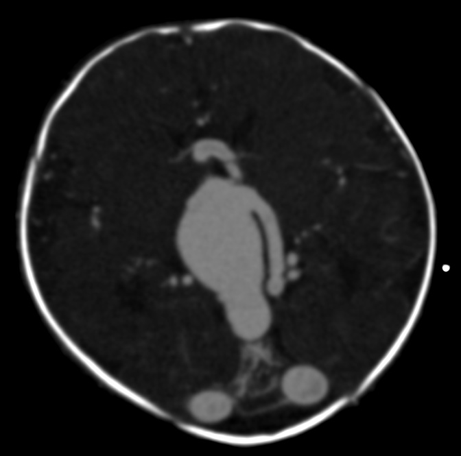
A congenital vascular malformation characterized by dilation of the embryonic precursor of the vein of Galen. It is a sporadic lesion that occurs during embryogenesis. Epidemiology The lesion is rare, with less than 800 cases (representing less than 10% of all cerebral arteriovenous malformations) reported so far. Vein of Galen aneurysmal malformation (VGAM) is more frequent in males than in females. Clinical description Cardiac insufficiency of variable severity is the principle manifestation that leads to detection of the malformation in newborns. It is sometimes associated with respiratory problems and/or hepatic, renal, and/or cerebral (encephalomalacia) manifestations, the main concern for therapeutic management.
Vein of Galen aneurysm is a rare form of arteriovenous malformation in which the embryonic precursor to the vein of Galen, a vein at the base of the brain, dilates causing too much blood to rush to the heart. This can lead to rapid heart failure. Other features may include increased head circumference resulting from hydrocephalus, unusually prominent veins on the face and scalp, developmental delay, persistent headache, and other neurological findings. Vein of Galen aneurysm is often recognized on an ultrasound late in pregnancy. In other cases, it is diagnosed after birth. Although the exact cause remains unknown, this condition appears to result from a defect in early fetal development. Treatment is aimed at decreasing the blood flow through the malformation while maximizing the blood supply to the brain.
A number sign (#) is used with this entry because of evidence that syndromic or nonsyndromic congenital generalized hypertrichosis can be caused by palindrome-mediated interchromosomal insertion at chromosome Xq27.1. ... Tadin-Strapps et al. (2003) described a Mexican kindred with X-linked recessive inheritance of a syndrome in which affected males exhibited generalized hypertrichosis, dental anomalies, and deafness, whereas carrier females exhibited only mild hypertrichosis.
Hypertrichosis lanuginosa congenita is a congenital (present from birth) skin disease characterized by excessive lanugo (very fine, soft, unpigmented) hair covering the entire body, with the exception of the palms, soles, and mucous membranes. The hair can grow to be 3 to 5 cm in length. This condition appears to follow an autosomal dominant pattern of inheritance.
Description Congenital hypertrichosis lanuginosa is a rare disorder characterized by excessive lanugo hair present at birth covering the entire body surface except the mucosae, palms, and soles (summary by De Raeve and Keymolen, 2011). Clinical Features Beighton (1970) observed a 6-year-old boy with extreme generalized hypertrichosis. He was born with double eyebrows. The father, grandfather, and great-grandfather had excessive hair over the entire body until about age 4. Other males in each generation escaped the excessive hairiness, and the trait may have been transmitted through an unaffected female. There was no gingival fibromatosis (135400) in this family. De Raeve and Keymolen (2011) reported a 15-month-old boy who presented with a history of excessive hairiness since birth that increased progressively during infancy.
Hypertrichosis lanuginosa congenita is a rare congenital skin disease characterized by the presence of 3 to 5cm long lanugo-type hair on the entire body, with the exception of palms, soles, and mucous membranes.
Losartan was added as an alternative to beta adrenergic-blocking agents in 2014 after studies showed its efficacy in children and young adults with Marfan syndrome who were randomly assigned to losartan or atenolol [Lacro et al 2014]. ... TGFBR1 or TGFBR2 or Loeys-Dietz syndrome. Surgical management is more aggressive with aortic root repair at 4.0 cm [Williams et al 2007, MacCarrick et al 2014].
Conditions that lower testosterone production, such as Klinefelter syndrome or pituitary insufficiency, can be associated with gynecomastia. ... Risk factors Risk factors for gynecomastia include: Adolescence Older age Use of anabolic steroids to enhance athletic performance Certain health conditions, including liver and kidney disease, thyroid disease, hormonally active tumors, and Klinefelter syndrome Complications Gynecomastia has few physical complications, but it can cause psychological or emotional problems caused by appearance.
A number sign (#) is used with this entry because posterior amorphous corneal dystrophy (PACD) is a chromosome 12q21.33 contiguous gene deletion syndrome. Clinical Features Carpel et al. (1977) observed a posterior corneal dystrophy characterized by irregular sheetlike areas of opacification with involvement of the Descemet membrane and, in some instances, alterations of the normal endothelial mosaic. ... INHERITANCE - Autosomal dominant HEAD & NECK Eyes - Posterior amorphous corneal dystrophy extending to the corneal limbus - Disorganization of posterior stromal lamellae by electron microscopy - Hyperopia - Corneal thinning - Flattened corneal topography - Corneal scleralization, superior - Anterior iris surface and stromal abnormalities - Fine iris processes extending to Schwalbe line for 360 degrees - Corectopia (in some patients) - Iris coloboma (in some patients) MISCELLANEOUS - Onset in infancy - Nonprogressive - Variable clinical presentation of corneal findings - Association with other ocular anomalies - Contiguous gene deletion syndrome MOLECULAR BASIS - Contiguous gene deletion of 700-1,670kb on chromosome 21q21.33 ▲ Close
Posterior amorphous corneal dystrophy (PACD) is a very rare form of stromal corneal dystrophy (see this term) characterized by irregular amorphous sheet-like opacities in the posterior corneal stroma and in Descemet membrane and mildly impaired vision. Epidemiology Prevalence of this form of corneal dystrophy is not known. To date cases have been reported primarily in the USA. Clinical description Patients usually develop corneal abnormalities in infancy or childhood. The condition is non-progressive or slowly progressive. Visual acuity is usually only minimally affected but in some more severe cases, penetrating keratoplasty (PK) may be warranted. Unlike other corneal dystrophies, non-corneal manifestations have been observed and include abnormalities of the iris (iridocorneal adhesions, corectopia, and pseudopolycoria).
Human disease Posterior amorphous corneal dystrophy Specialty Ophthalmology Posterior amorphous corneal dystrophy (PACD) is a rare form of corneal dystrophy . It is not yet linked to any chromosomal locus. The first report describing this dystrophy dates back to 1977. [1] References [ edit ] ^ Carpel EF, Sigelman RJ, Doughman DJ (May 1977). "Posterior amorphous corneal dystrophy". Am. J. Ophthalmol. 83 (5): 629–32. doi : 10.1016/0002-9394(77)90127-1 . PMID 301356 . External links [ edit ] Classification D OMIM : 612868 MeSH : C567546 C567546, C567546 v t e Types of corneal dystrophy Epithelial and subepithelial Epithelial basement membrane dystrophy Gelatinous drop-like corneal dystrophy Lisch epithelial corneal dystrophy Meesmann corneal dystrophy Subepithelial mucinous corneal dystrophy Bowman's membrane Reis–Bucklers corneal dystrophy Thiel-Behnke dystrophy Stroma Congenital stromal corneal dystrophy Fleck corneal dystrophy Granular corneal dystrophy Lattice corneal dystrophy Macular corneal dystrophy Posterior amorphous corneal dystrophy Schnyder crystalline corneal dystrophy Descemet's membrane and endothelial Congenital hereditary endothelial dystrophy Fuchs' dystrophy Posterior polymorphous corneal dystrophy X-linked endothelial corneal dystrophy This article about the eye is a stub . You can help Wikipedia by expanding it . v t e
Thus, the disorder may be considered a tumor predisposition syndrome (summary by Camacho-Vanegas et al., 2012). ... Mapping Martignetti et al. (1997, 1999) used microsatellite markers in a genome screen for the gene locus in diaphyseal medullary stenosis with malignant fibrous histiocytoma in 3 unrelated families. They linked the syndrome to a region of approximately 3 cM on 9p22-p21, with a maximum 2-point lod score of 5.49 with marker D9S171 at recombination fraction (theta) 0.05.
It can be divided into cancer-associated retinopathy (CAR) and melanoma-associated retinopathy (MAR). [2] The condition is associated with retinal degeneration caused by autoimmune antibodies recognizing retinal proteins as antigens and targeting them. [3] AIR's prevalence is extremely rare, with CAR being more common than MAR. [2] It is more commonly diagnosed in females (approximately 60% of diagnosed patients are females) in the age range of 50-60. [2] Contents 1 Types 1.1 Cancer-associated retinopathy 1.2 Melanoma-associated retinopathy 2 Signs and symptoms 3 Diagnosis 4 Treatment 4.1 Immunoglobulin 4.2 Plasmapheresis 4.3 Corticosteroids 5 References Types [ edit ] Cancer-associated retinopathy [ edit ] A division of AIR, cancer-associated retinopathy is a paraneoplastic syndrome , which is a disorder caused by an immune system response to an abnormality. ... This is because they are both paraneoplastic syndromes. AIR symptoms are numerous and shared by many other diseases. [2] Symptoms Painless Vision Loss Blind Spots in Vision Photopsia Nyctalopia Scotomas Dislike/avoidance of light Loss of contrast sensitivity Incomplete colour blindness Decreased night vision [2] [6] [7] Diagnosis [ edit ] Diagnosis of AIR can be difficult due to the overlap of symptoms with other disorders. [2] Examination of the fundus (inner surface of eye) can show no results or it can show narrowing of the blood vessels, abnormal colouration of the optic disc , and retinal atrophy. [2] [5] Fundus examination results are not indicative of autoimmune retinopathy but they are used to initiate the diagnostic process.
Autoimmune retinopathy represents a spectrum of rare autoimmune diseases that primarily affect retinal photoreceptor function and lead to progressive vision loss. Included in this spectrum are cancer-associated retinopathy (CAR), melanoma-associated retinopathy (MAR) and presumed non-paraneoplastic autoimmune retinopathy (npAIR). Autoimmune retinopathy typically presents in the fifth and sixth decades with rapidly progressive, bilateral, painless visual deterioration. Examination of the fundus (the back inner part of the eye) is usually normal at presentation. The underlying reason for the autoimmune attack on retinal cells remains unknown.
At age 4 years, he developed a progressive 3-lineage hypoplasia and myelodysplastic syndrome and underwent successful hematopoietic stem cell transplantation. ... Acquired somatic mutations in the GATA1 gene that result in synthesis of GATA1s have been found in individuals with Down syndrome with both transient myeloproliferative disorder and acute megakaryoblastic leukemia (see 190685).
X-linked dyserythropoietic anemia with abnormal platelets and neutropenia is a rare, genetic, constitutional dyserythropoietic anemia disorder characterized by moderate to severe anemia without thrombocytopenia, variable degrees of neutropenia, and bone marrow biopsy findings of trilineage dysplasia and hypocellularity of erythroid and granulocytic lineages. Peripheral blood findings include anisocytosis, macrocytosis, poikilocytosis, elliptocytes, and fragmented erythrocytes.
Serologically, diagnostic markers can be tested; specifically, the streptozyme test is used and measures multiple streptococcal antibodies: antistreptolysin , antihyaluronidase, antistreptokinase, antinicotinamide-adenine dinucleotidase, and anti- DNAse B antibodies. [2] Differential diagnosis [ edit ] The differential diagnosis of acute proliferative glomerulonephritisis is based on the following: [ citation needed ] Causes of acute glomerulonephritis: IgA Nephropathy Lupus nephritis Type 1 membranoproliferative glomerulonephritis Bacterial endocarditis Shunt nephritis Cryoglobulinemia Nephrotic syndrome Causes of generalized edema: Malnutrition Malabsorption Renal affection Liver cell failure Right side heart failure Angioedema Prevention [ edit ] Antibiotic type It is unclear whether or not acute proliferative glomerulonephritis (i.e., poststreptococcal glomerulonephritis) can be prevented with early prophylactic antibiotic therapy, with some authorities arguing that antibiotics can prevent development of acute proliferative glomerulonephritis [14] Treatment [ edit ] Acute management of acute proliferative glomerulonephritis mainly consists of blood pressure (BP) control. ... External links [ edit ] Classification D ICD - 10 : N00.8 ICD - 9-CM : 580.0 DiseasesDB : 29306 External resources MedlinePlus : 000503 eMedicine : med/889 Scholia has a topic profile for Acute proliferative glomerulonephritis . v t e Disease of the kidney glomerules Primarily nephrotic Non-proliferative Minimal change Focal segmental Membranous Proliferative Mesangial proliferative Endocapillary proliferative Membranoproliferative/mesangiocapillary By condition Diabetic Amyloidosis Primarily nephritic , RPG Type I RPG / Type II hypersensitivity Goodpasture syndrome Type II RPG / Type III hypersensitivity Post-streptococcal Lupus diffuse proliferative IgA Type III RPG / Pauci-immune Granulomatosis with polyangiitis Microscopic polyangiitis Eosinophilic granulomatosis with polyangiitis General glomerulonephritis glomerulonephrosis