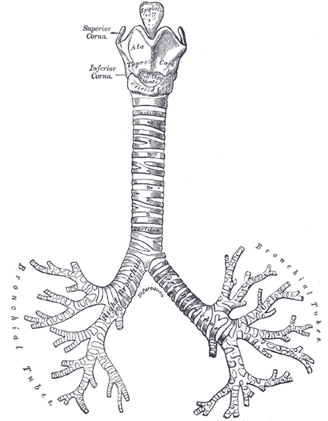
Secondary airway malacia was defined as airway malacia secondary to esophageal atresia, VATER/VACTERL association (condition with vertebral anomalies, anal atresia, congenital heart disease, tracheoesophageal fistula or esophageal atresia, renourinary anomalies, or radial limb defects), vascular or other external compression of the airways, or specific syndromes. Treatment [ edit ] Time Minimally Invasive, usually in conjunction with Continuous Positive Airflow Pressure.
Idiopathic pure sudomotor failure Specialty Dermatology Idiopathic pure sudomotor failure (IPSF) is the most common cause of a rare disorder known as acquired idiopathic generalized anhidrosis (AIGA), a clinical syndrome characterized by generalized decrease or absence of sweating without other autonomic and somatic nervous dysfunctions and without persistent organic cutaneous lesions . [1] The term IPSF was first introduced in 1994 after researchers at Saitama Medical School speculated the primary lesion sites in patients were within cholinergic receptors of the sweat glands.
Dependence Concepts Physical dependence Psychological dependence Withdrawal Disorders Drugs Alcoholism Amphetamine Barbiturate Benzodiazepine Caffeine Cannabis Cocaine Nicotine Opioid Non-drug stimuli Tanning dependence Treatment and management Detoxification Alcohol detoxification Drug detoxification Behavioral therapies Cognitive behavioral therapy Relapse prevention Contingency management Community reinforcement approach and family training Motivational enhancement therapy Motivational interviewing Motivational therapy Physical exercise Treatment programs Drug rehab Residential treatment center Heroin-assisted treatment Intensive outpatient program Methadone maintenance Smoking cessation Nicotine replacement therapy Tobacco cessation clinics in India Twelve-step program Support groups Addiction recovery groups List of twelve-step groups Harm reduction Category:Harm reduction Drug checking Reagent testing Low-threshold treatment programs Managed alcohol program Moderation Management Needle exchange program Responsible drug use Stimulant maintenance Supervised injection site Tobacco harm reduction See also Addiction medicine Allen Carr Category:Addiction Discrimination against drug addicts Dopamine dysregulation syndrome Cognitive control Inhibitory control Motivational salience Incentive salience Sober companion Category Television portal v t e Digital media use and mental health Proposed or recognised diagnostic categories Gaming disorder (Video game addiction) Problematic social media use Internet addiction disorder Problematic smartphone use Nomophobia Computer addiction Television addiction Internet sex addiction Disciplines involved Digital sociology Digital anthropology Psychiatry Psychology Neuroscience Associated psychiatric conditions Depression Anxiety Attention deficit hyperactivity disorder Bipolar disorder Related concepts Electronic media and sleep Screen time Digital detox Smartphone zombie Cyberbullying Media multitasking Online problem gambling Technostress Computer rage
She was the child of consanguineous parents who had lost 4 other children due to alleged sudden infant death syndrome. No cardiac malformation was documented in the 5 patients with 3 SMN2 copies.
Spinal muscular atrophy 1 (SMA1) , also known as Werdnig Hoffmann disease, is a genetic neuromuscular disorder that affects the nerve cells that control voluntary muscles (motor neurons). Without treatment, symptoms of SMA1 become apparent before 6 months of age and include worsening muscle weakness and poor muscle tone (hypotonia) due to loss of the lower motor neurons in the spinal cord and brain stem. Feeding and breathing problems are also present. SMA1 is caused by changes (pathogenic variants also called mutations) in the SMN1 gene and is typically inherited in an autosomal recessive manner. Diagnosis of SMA1 is suspected by symptoms and confirmed by genetic testing. SMA has been added to the list of recommended newborn screening tests in the United States, so that it can be detected prior to symptoms developing.
Spinal muscular atrophy is a genetic disorder characterized by weakness and wasting (atrophy ) in muscles used for movement (skeletal muscles). It is caused by a loss of specialized nerve cells, called motor neurons that control muscle movement. The weakness tends to be more severe in the muscles that are close to the center of the body (proximal) compared to muscles away from the body's center (distal). The muscle weakness usually worsens with age. There are many types of spinal muscular atrophy that are caused by changes in the same genes. The types differ in age of onset and severity of muscle weakness; however, there is overlap between the types.
Proximal spinal muscular atrophies are a group of neuromuscular disorders characterized by progressive muscle weakness resulting from the degeneration and loss of the lower motor neurons in the spinal cord and the brain stem nuclei. Epidemiology Prevalence is estimated at around 1/30,000. Clinical description Four subtypes have been defined according to the age of onset and severity of the disease: type 1 (SMA1), the most severe form, with onset before six months of age; type 2 (SMA2), with onset between 6 and 18 months of age, type 3 (SMA3), with onset between childhood and adolescence, and type 4 (SMA4), the least severe form, with adult onset (see these terms). All types are characterized by muscle weakness and atrophy of varying severity, particularly affecting the lower limbs and respiratory muscles. The weakness is almost always symmetric and progressive. Scoliosis, muscle retractions, and joint contractures may occur. Constipation and gastroesophageal reflux are frequent. Etiology Around 95% of cases of SMA are caused by homozygous deletions (either of exon 7, or of exons 7 and 8) in the SMN1 gene (5q12.2-q13.3) encoding the SMN (survival motor neuron) protein.
Pott's fracture , also known as Pott's syndrome I and Dupuytren fracture , is an archaic term loosely applied to a variety of bimalleolar ankle fractures. [1] The injury is caused by a combined abduction external rotation from an eversion force.
Pityriasis lichenoides chronica (PLC) is a skin disease that causes the development of small, scaling, raised spots ( papules ) on the skin. PLC is the relatively mild form of the disease pityriasis lichenoides . A person with PLC tends to have multiple episodes of papules on the skin lasting for months or a few years, meaning the disease is chronic. The papules develop gradually. They first appear pink and scaly, and they gradually flatten and become brown in color over a period of weeks or months. Papules at various stages may be present at any one time. The cause of PLC is unknown, but it is not contagious.
Wartenberg's sign is not a feature of, and should not be confused with, Wartenberg's syndrome . The latter involves compression at the wrist of the superficial sensory branch of the radial nerve which does not innervate hand muscles.
Instead, in adult athletes a more common injury is to the ulnar collateral ligament of the elbow, an injury that often requires Tommy John surgery in order for the athlete to resume high-level competitive throwing. [ citation needed ] See also [ edit ] Tennis elbow Golfer's elbow Repetitive strain injury Radial tunnel syndrome Panner disease References [ edit ] ^ Awh, M.D., Mark H.
Some individuals with muscle GSD 0 have a disruption of the heart's normal rhythm (arrhythmia) known as long QT syndrome. In all affected individuals, muscle GSD 0 impairs the heart's ability to effectively pump blood and increases the risk of cardiac arrest and sudden death, particularly after physical activity.
A number sign (#) is used with this entry because of evidence that liver glycogen storage disease-0 (GSD0A) is caused by homozygous or compound heterozygous mutation in the GYS2 gene (138571), which encodes glycogen synthase-2, on chromosome 12p12. See 611556 for a description of muscle glycogen storage disease caused by mutation in the GYS1 gene (138570). Clinical Features In a well-studied family, Lewis et al. (1963) demonstrated that infantile hypoglycemia was due to a deficiency of glycogen synthetase in the liver. The cases were probably of the same type as those reported by Broberger and Zetterstrom (1961) because urinary excretion of catecholamines was not influenced by hypoglycemia. The observations of Lewis et al. (1963) are particularly convincing evidence for autosomal recessive inheritance of this one form, although iron-clad proof awaits demonstration of a partial deficiency in both parents.
A genetically inherited anomaly of glycogen metabolism and a form of glycogen storage disease (GSD) characterized by fasting hypoglycemia. This is not a glycogenosis, strictly speaking, as the enzyme deficiency decreases glycogen reserves. Epidemiology It is an extremely rare disease; about 20 cases have been reported in the literature so far. Clinical description It commonly appears in infancy or in early childhood. Patients present with morning fatigue and fasting hypoglycemia (without hepatomegaly) associated with hyperketonemia but without hyperalaninemia or hyperlactacidemia.
Glycogen storage disease type 0, liver (liver GSD 0), a form of glycogen storage disease (GSD), is a rare abnormality of glycogen metabolism (how the body uses and stores glycogen, the storage form of glucose). Unlike other types of GSD, liver GSD 0 does not involve excessive or abnormal glycogen storage, and causes moderately decreased glycogen stores in the liver. Symptoms typically begin in infancy or in early childhood and may include drowsiness, sweating, lack of attention, fasting hypoglycemia associated with hyperketonemia , seizures, and other findings. It is caused by a deficiency of the enzyme glycogen synthetase in the liver, due to mutations in the GYS2 gene. It is inherited in an autosomal recessive manner. Treatment involves a specific diet that includes frequent meals with high protein intake during the day, and uncooked starch in the evening.
Also, it may be associated with hyponatremia (hypervolemic hyponatremia). [1] Causes [ edit ] Excessive sodium and/or fluid intake : IV therapy containing sodium [2] As a transfusion reaction to a rapid blood transfusion . [2] [3] High intake of sodium [2] Sodium and water retention : Heart failure [2] Liver cirrhosis [2] Nephrotic syndrome [2] Corticosteroid therapy [2] Hyperaldosteronism [2] Low protein intake [2] Fluid shift into the intravascular space : Fluid remobilization after burn treatment [2] Administration of hypertonic fluids , e.g. mannitol [2] or hypertonic saline solution Administration of plasma proteins , such as albumin [2] Treatment [ edit ] This section has no medical references for verification or relies exclusively on non-medical sources .
Overview Moles (nevi) are a common type of skin growth. They often appear as small, dark brown spots and are caused by clusters of pigment-forming cells (melanocytes). Most people have 10 to 40 moles that appear during childhood and adolescence and may change in appearance or fade over time. Most moles are harmless. Rarely, they become cancerous. Being aware of changes in your moles and other pigmented patches is important to detecting skin cancer, especially malignant melanoma. Symptoms The typical mole is a small brown spot. But moles come in different colors, shapes and sizes: Color and texture. Moles can be brown, tan, black, blue, red or pink. They can be smooth, wrinkled, flat or raised.
In a follow-up of one of these families (DEM4154), reported by Santos et al. (2006) to be negative for limb deformities or any other syndromic features, Santos-Cortez et al. (2016) found that 6 of the 7 affected individuals had severe asymmetric lower limb anomalies, including short and/or bent tibiae, bent or absent fibulae, and absent knee joint.
Some researchers consider Dent disease 2 to be a mild variant of a similar disorder called Lowe syndrome. Frequency Dent disease is a rare condition, with about 250 affected families reported.