-
Low Back Pain
Wikipedia
Red flags Red flags are warning signs that may indicate a more serious problem Red flag [36] Possible cause [1] Previous history of cancer Cancer Unintentional weight loss Loss of bladder or bowel control Cauda equina syndrome Significant motor weakness or sensory problems Loss of sensation in the buttocks ( saddle anesthesia ) Significant trauma related to age Fracture Chronic corticosteroid use Osteoporosis Severe pain after lumbar surgery in past year Infection Fever Urinary tract infection Immunosuppression Intravenous drug use The presence of certain signs, termed red flags , indicate the need for further testing to look for more serious underlying problems, which may require immediate or specific treatment. [5] [37] The presence of a red flag does not mean that there is a significant problem. ... When warranted, imaging such as MRI can provide clear detail about disc related causes of back pain (L4–L5 disc herniation shown) Imaging is indicated when there are red flags, ongoing neurological symptoms that do not resolve, or ongoing or worsening pain. [5] In particular, early use of imaging (either MRI or CT) is recommended for suspected cancer, infection, or cauda equina syndrome . [5] MRI is slightly better than CT for identifying disc disease; the two technologies are equally useful for diagnosing spinal stenosis. [5] Only a few physical diagnostic tests are helpful. [5] The straight leg raise test is almost always positive in those with disc herniation. [5] Lumbar provocative discography may be useful to identify a specific disc causing pain in those with chronic high levels of low back pain. [42] Similarly, therapeutic procedures such as nerve blocks can be used to determine a specific source of pain. [5] Some evidence supports the use of facet joint injections , transforminal epidural injections and sacroilliac injections as diagnostic tests. [5] Most other physical tests, such as evaluating for scoliosis , muscle weakness or wasting, and impaired reflexes, are of little use. [5] Complaints of low back pain are one of the most common reasons people visit doctors. [9] [43] For pain that has lasted only a few weeks, the pain is likely to subside on its own. [44] Thus, if a person's medical history and physical examination do not suggest a specific disease as the cause, medical societies advise against imaging tests such as X-rays , CT scans , and MRIs . [43] Individuals may want such tests but, unless red flags are present, [10] [45] they are unnecessary health care . [9] [44] Routine imaging increases costs, is associated with higher rates of surgery with no overall benefit, [46] [47] and the radiation used may be harmful to one's health. [46] Fewer than 1% of imaging tests identify the cause of the problem. [9] Imaging may also detect harmless abnormalities, encouraging people to request further unnecessary testing or to worry. [9] Even so, MRI scans of the lumbar region increased by more than 300% among United States Medicare beneficiaries from 1994 to 2006. [11] Prevention Exercise appears to be useful for preventing low back pain. [48] Exercise is also probably effective in preventing recurrences in those with pain that has lasted more than six weeks. [1] [49] Medium-firm mattresses are more beneficial for chronic pain than firm mattresses. [50] There is little to no evidence that back belts are any more helpful in preventing low back pain than education about proper lifting techniques. [48] [51] There is no quality data that supports medium firm mattresses over firm mattresses. ... For acute or chronic radicular pain syndromes massage therapy is recommended only if low back pain is considered a symptom.PLAT, SPARC, NAB2, SPAST, HSPB8, HTRA1, ALDH18A1, STAT6, FAS, TBX6, ABCD1, NGF, IL1B, LBP, TNF, IL6, CALCA, CXCL8, COMT, IL1A, LEP, CCL2, NUBP1, EMSLR, TP63, CASP9, COASY, BDNF, CRP, CISD3, SEMA3A, SLCO6A1, COPD, RGS6, GDF15, NLRP3, MIR204, ADAMTS3, ADAMTS4, ZFYVE9, MSC, FST, ITLN1, ADAMTS5, RTN4, MCF2L2, SIRT1, PES1, SPECC1, TSKU, LCS1, F11R, DNAJC5, PER2, MARCHF1, PACC1, SPEN, ACR, GDF5, ZNF35, ACAN, AMH, BMP3, TSPO, CASP1, CAT, CD6, DNMT3B, NR3C1, IL1RN, IL6ST, MMP1, MMP9, OPRM1, FURIN, PPARG, PTGS2, RARRES2, RASA1, RHAG, RTN1, CCL4, ACP5, TAC1, TLR4, TP53, VDR, SMS

-
Otitis Media
Wikipedia
Inflammation of the middle ear Otitis media Other names Otitis media with effusion : serous otitis media, secretory otitis media A bulging tympanic membrane which is typical in a case of acute otitis media Specialty Otorhinolaryngology Symptoms Ear pain, fever, hearing loss [1] [2] Types Acute otitis media, otitis media with effusion, chronic suppurative otitis media [3] [4] Causes Viral, bacterial [4] Risk factors Smoke exposure, daycare [4] Prevention Vaccination, breastfeeding [1] Medication Paracetamol (acetaminophen), ibuprofen , benzocaine ear drops [1] Frequency 471 million (2015) [5] Deaths 3,200 (2015) [6] Otitis media is a group of inflammatory diseases of the middle ear . [2] One of the two main types is acute otitis media ( AOM ), [3] an infection of rapid onset that usually presents with ear pain. [1] In young children this may result in pulling at the ear, increased crying, and poor sleep. [1] Decreased eating and a fever may also be present. [1] The other main type is otitis media with effusion ( OME ), typically not associated with symptoms, [1] although occasionally a feeling of fullness is described; [4] it is defined as the presence of non-infectious fluid in the middle ear for more than three months. [4] Chronic suppurative otitis media ( CSOM ) is middle ear inflammation that results in discharge from the ear for more than three months. [7] It may be a complication of acute otitis media. [4] Pain is rarely present. [4] All three types of otitis media may be associated with hearing loss . [2] [3] The hearing loss in OME, due to its chronic nature, may affect a child's ability to learn. [4] The cause of AOM is related to childhood anatomy and immune function . [4] Either bacteria or viruses may be involved. [4] Risk factors include exposure to smoke, use of pacifiers , and attending daycare . [4] It occurs more commonly among indigenous peoples and those who have cleft lip and palate or Down syndrome . [4] [8] OME frequently occurs following AOM and may be related to viral upper respiratory infections , irritants such as smoke, or allergies . [3] [4] Looking at the eardrum is important for making the correct diagnosis. [9] Signs of AOM include bulging or a lack of movement of the tympanic membrane from a puff of air. [1] [10] New discharge not related to otitis externa also indicates the diagnosis. [1] A number of measures decrease the risk of otitis media including pneumococcal and influenza vaccination , breastfeeding , and avoiding tobacco smoke. [1] The use of pain medications for AOM is important. [1] This may include paracetamol (acetaminophen), ibuprofen , benzocaine ear drops, or opioids . [1] In AOM, antibiotics may speed recovery but may result in side effects. [11] Antibiotics are often recommended in those with severe disease or under two years old. [10] In those with less severe disease they may only be recommended in those who do not improve after two or three days. [10] The initial antibiotic of choice is typically amoxicillin . [1] In those with frequent infections tympanostomy tubes may decrease recurrence. [1] In children with otitis media with effusion antibiotics may increase resolution of symptoms, but may cause diarrhoea, vomiting and skin rash. [12] Worldwide AOM affects about 11% of people a year (about 325 to 710 million cases). [13] [14] Half the cases involve children less than five years of age and it is more common among males. [4] [13] Of those affected about 4.8% or 31 million develop chronic suppurative otitis media. [13] The total number of people with CSOM is estimated at 65–330 million people. [15] Before the age of ten OME affects about 80% of children at some point. [4] Otitis media resulted in 3,200 deaths in 2015 – down from 4,900 deaths in 1990. [6] [16] Contents 1 Signs and symptoms 2 Causes 3 Diagnosis 3.1 Acute otitis media 3.2 Otitis media with effusion 3.3 Chronic suppurative otitis media 3.4 Adhesive otitis media 4 Prevention 5 Management 5.1 Antibiotics 5.2 Tympanostomy tube 5.3 Otitis media with effusion 5.4 Chronic suppurative otitis media 5.5 Alternative medicine 6 Outcomes 6.1 Membrane rupture 6.2 Hearing loss 7 Epidemiology 8 Etymology 9 References 10 External links Signs and symptoms [ edit ] Otitis media. ... Those with craniofacial malformations such as cleft lip and palate , Down syndrome , and microcephaly are at higher risk. [ citation needed ] Worldwide approximately 11% of the human population is affected by AOM every year, or 709 million cases. [13] [14] About 4.4% of the population develop CSOM. [14] According to the World Health Organization , CSOM is a primary cause of hearing loss in children. [29] Adults with recurrent episodes of CSOM have a higher risk of developing permanent conductive and sensorineural hearing loss. ... Secretory otitis media (Ear disorder) at the Encyclopædia Britannica Otitis media (Pathology) at the Encyclopædia Britannica Classification D ICD - 10 : H65 - H67 ICD - 9-CM : 017.40 , 055.2 , 381.0 , 381.1 , 381.2 , 381.3 , 381.4 , 382 MeSH : D010033 DiseasesDB : 29620 External resources MedlinePlus : 000638 eMedicine : emerg/351 ent/426 complications, ent/209 with effusion, ent/212 Medical treat., ent/211 Surgical treat. ped/1689 Wikimedia Commons has media related to Otitis media . v t e Diseases of the outer and middle ear Outer ear Otitis externa Otomycosis Middle ear and mastoid Otitis media Mastoiditis Bezold's abscess Gradenigo's syndrome Tympanosclerosis Cholesteatoma Perforated eardrum Symptoms Ear pain Hearing loss Tests Otoscope pneumatic tympanometry v t e Common cold Viruses Adenovirus Coronavirus Enterovirus Rhinovirus Symptoms Cough Fatigue Fever Headache Loss of appetite Malaise Muscle aches Nasal congestion Rhinorrhea Sneezing Sore throat Weakness Complications Acute bronchitis Bronchiolitis Croup Otitis media Pharyngitis Pneumonia Sinusitis Strep throat Drugs Antiviral drugs Pleconaril (experimental) Authority control NDL : 00573957RSPH4A, TNF, A2ML1, IL6, MUC5AC, IL1B, IL10, FBXO11, BPIFA1, RPL38, SH3PXD2B, MUC4, TGIF1, MUC1, IL1A, FGFR1, MECOM, EYA4, NF2, E2F4, NAGLU, SCGB1A1, DNAH5, TP73, BTC, CTSL, TBX1, CTSK, CTSB, CBY1, SCN2B, EDARADD, IDUA, LMNA, PHEX, ENPP1, CAT, SALL4, NOS2, CXCR4, PNP, TNFSF11, CD3D, CYBC1, CCDC47, DCLRE1C, NSD1, SLC2A1, NCF1, PRX, TCIRG1, BTK, WAS, NBN, NCF2, NCF4, ALMS1, CD3E, CD247, IKBKB, TLK2, FGFR3, DNAAF3, RPGR, IL2RG, RAG2, NIPBL, RAG1, IL7R, RNF168, CYBB, CYBA, CLCN7, PTPRC, SNX10, STX17-AS1, CCDC39, MBL2, TLR2, TLR4, CD14, TLR9, CASP3, DUSP1, COPD, PYCARD, MBL3P, DEFB4A, STS, MCPH1, DHRS2, DEFB4B, MPO, MUC5B, CCR2, CAPN14, SFTPA2, TRAP, SFTPA1, KAT5, MIR146B, SMC2, POLD4, ADAMTS13, NISCH, MIR146A, CFAP97, FLVCR1, TIPRL, CRYGEP, SLC25A21, ITFG1, METTL8, PACC1, PPP2R2D, GALNT14, POLE4, PORCN, ABO, PIK3CD, VEGFA, ELANE, KRT5, ITGB2, ITGAM, ITGAL, ITGA5, CXCL8, IL6R, IL5, IL2, IFNG, FUT2, ATN1, CD46, CRYGC, COL2A1, CD44, CASP9, CASP1, VPS51, C7, BPI, AQP1, AQP8, AKT1, LIMK1, MEFV, TLR5, PIK3CG, SLC11A1, CCL3, CCL2, S100B, S100A12, S100A9, S100A8, S100A1, RAC1, PTEN, PIP, JAG1, MIF, PIK3CB, PIK3CA, PAX9, PAK1, SERPINE1, OPHN1, NT5E, MUC2, MMP9, MMP8, MMP2, H3P5

-
Pulmonary Embolism
Wikipedia
Alterations in blood flow : immobilization (after surgery, long-haul flight), injury , pregnancy (also procoagulant), obesity (also procoagulant), cancer (also procoagulant) Factors in the vessel wall : surgery, catheterizations causing direct injury ("endothelial injury") Factors affecting the properties of the blood (procoagulant state): Estrogen -containing hormonal contraception [ citation needed ] Genetic thrombophilia ( factor V Leiden , prothrombin mutation G20210A , protein C deficiency , protein S deficiency , antithrombin deficiency, hyperhomocysteinemia and plasminogen / fibrinolysis disorders) Acquired thrombophilia ( antiphospholipid syndrome , nephrotic syndrome , paroxysmal nocturnal hemoglobinuria ) Cancer (due to secretion of pro-coagulants) Although most pulmonary embolisms are the result of proximal leg deep vein thrombosis (DVTs), there are still many other risk factors that can also result in a pulmonary embolism.FGA, MTHFR, EPO, PLAU, THBD, XDH, MERTK, TBXA2R, KLKB1, CAT, DAB2IP, PLAT, FGB, HP, MMP2, MMP9, CX3CL1, FGG, VCAM1, NOS3, CCL2, EDN1, TBXAS1, CX3CR1, CCN2, F13A1, F5, STAT4, TRNW, PLP1, PROC, PIGA, PTEN, PROS1, ABO, COX7A2L, TLR4, KLRC4, TRNS1, PROCR, TSPAN15, ERAP1, TET2, AGGF1, SLC44A2, SELENON, FUNDC2, TRIM8, IL23R, F11-AS1, UBAC2, TRNS2, ND5, TRNQ, ENG, TRNL1, HLA-B, HABP2, GNAQ, GDF2, F2, CD55, JAK2, CCR1, CBS, C4A, SERPINC1, FAS, AKT1, IL12A, IL10, KCNJ5, COX3, TRNH, TRNF, ND6, ACVRL1, ND4, ND1, IL12A-AS1, COX2, COX1, MPL, MEFV, SMAD4, CPB2, NPPB, TNNI3, SERPINF2, P2RY12, HGF, ALB, CRP
-
Hiv/aids In Africa
Wikipedia
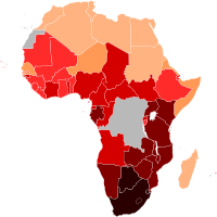
By the 1960s, about 2,000 people in Africa may have had HIV, [15] including people in Kinshasa whose tissue samples from 1959 and 1960 have been preserved and studied retrospectively. [19] The first epidemic of HIV/AIDS is believed to have occurred in Kinshasa in the 1970s, signalled by a surge in opportunistic infections such as cryptococcal meningitis , Kaposi's sarcoma , tuberculosis , and pneumonia . [20] [21] History [ edit ] Acquired immunodeficiency syndrome (AIDS) is a fatal disease caused by the slow-acting human immunodeficiency virus (HIV). The virus multiplies in the body until it causes immune system damage, leading to diseases of the AIDS syndrome. HIV emerged in Africa in the 1960s and traveled to the United States and Europe the following decade.
-
Health Effects Of Wine
Wikipedia
The health effects of wine are mainly determined by its active ingredient alcohol . [1] [2] Some studies found that, when comparing people who consume alcohol, drinking small quantities of alcohol (up to one standard drink per day for women and one to two drinks per day for men) is associated with a decreased risk of heart disease , stroke , diabetes mellitus , metabolic syndrome and early death. [2] [3] However, other studies found no such effect. [4] Drinking more than the standard drink amount increases the risk of heart disease, high blood pressure , atrial fibrillation , stroke [3] and cancer . [5] Mixed results are also observed in light drinking and cancer mortality. [5] [6] [7] [8] Risk is greater in young people due to binge drinking which may result in violence or accidents. [3] About 88,000 deaths in the US are estimated to be due to alcohol each year. [9] Alcoholism reduces a person's life expectancy by around ten years [10] and excessive alcohol use is the third leading cause of early death in the United States. [3] According to systematic reviews and medical associations, people who are nondrinkers should not start drinking wine. [3] [7] [11] Wine has a long history of use as an early form of medication , being recommended variously as a safe alternative to drinking water , an antiseptic for treating wounds, a digestive aid , and as a cure for a wide range of ailments including lethargy , diarrhea and pain from child birth . [12] Ancient Egyptian papyri and Sumerian tablets dating back to 2200 BC detail the medicinal role of wine, making it the world's oldest documented human-made medicine. [13] : 433 Wine continued to play a major role in medicine until the late 19th and early 20th century, when changing opinions and medical research on alcohol and alcoholism cast doubt on its role as part of a healthy lifestyle. ... Studies showed a connection between alcohol consumption among pregnant mothers and an increased risk of mental retardation and physical abnormalities in what became known as fetal alcohol syndrome , prompting the use of alcohol packaging warning messages in several countries. [13] : 341–2 French paradox and the benefits of consumption [ edit ] Main article: French paradox The French have a diet that is high in full-fat dairy products such as cheeses , and also have low rates of heart disease . ... Sulfonic acids : Acamprosate Religion and alcohol Christian views on alcohol alcohol in the Bible Islam and alcohol History Bratt System Related Index of alcohol-related articles Austrian syndrome Ban on caffeinated alcoholic beverages Brief intervention Gateway drug effect Last call Mood disorder Non-alcoholic fatty liver disease Self-medication Spins Sober companion Sober living houses Sobering center Town drunk Category v t e Health effects of food, drink, and use of natural substances Food Chocolate Honey Sugar Drink Alcohol Coffee Tea Wine Phytochemicals Caffeine Natural phenols and polyphenols (including tannins) Other Cannabis Noise pollution Pesticide exposure Sun exposure Tobacco Drink portal

-
Cystocele
Wikipedia
Up to one third of women with Marfan syndrome have a history of vaginal wall collapse. Ehlers-Danlos syndrome in women is associated with a rate of 3 out of 4. [6] Risk factors [ edit ] Risk factors for developing a cystocele are: an occupation involving or history of heavy lifting pregnancy and childbirth chronic lung disease /smoking family history of cystocele [20] [21] exercising incorrectly [15] ethnicity (risk is greater for Hispanic and whites) [22] hypoestrogenism pelvic floor trauma connective tissue disorders spina bifida [6] hysterectomy [23] cancer treatment of pelvic organs [24] childbirth ; correlates to the number of births forceps delivery age chronically high intra-abdominal pressures chronic obstructive pulmonary disease constipation [12] obesity [6] Connective tissue disorders predispose women to developing cystocele and other pelvic organ prolapse. ... Further reading [ edit ] Using splinting to support and diminish pain while coughing , Craven and Hirnle's Fundamentals of Nursing: Human Health and Function, 6th edition External links [ edit ] Cystocele, Pelvic Organ Prolapse Classification D ICD - 10 : N81.1 ICD - 9-CM : 618.01 - 618.02 MeSH : D052858 DiseasesDB : 3391 SNOMED CT : 40421008 v t e Female diseases of the pelvis and genitals Internal Adnexa Ovary Endometriosis of ovary Female infertility Anovulation Poor ovarian reserve Mittelschmerz Oophoritis Ovarian apoplexy Ovarian cyst Corpus luteum cyst Follicular cyst of ovary Theca lutein cyst Ovarian hyperstimulation syndrome Ovarian torsion Fallopian tube Female infertility Fallopian tube obstruction Hematosalpinx Hydrosalpinx Salpingitis Uterus Endometrium Asherman's syndrome Dysfunctional uterine bleeding Endometrial hyperplasia Endometrial polyp Endometriosis Endometritis Menstruation Flow Amenorrhoea Hypomenorrhea Oligomenorrhea Pain Dysmenorrhea PMS Timing Menometrorrhagia Menorrhagia Metrorrhagia Female infertility Recurrent miscarriage Myometrium Adenomyosis Parametrium Parametritis Cervix Cervical dysplasia Cervical incompetence Cervical polyp Cervicitis Female infertility Cervical stenosis Nabothian cyst General Hematometra / Pyometra Retroverted uterus Vagina Hematocolpos / Hydrocolpos Leukorrhea / Vaginal discharge Vaginitis Atrophic vaginitis Bacterial vaginosis Candidal vulvovaginitis Hydrocolpos Sexual dysfunction Dyspareunia Hypoactive sexual desire disorder Sexual arousal disorder Vaginismus Urogenital fistulas Ureterovaginal Vesicovaginal Obstetric fistula Rectovaginal fistula Prolapse Cystocele Enterocele Rectocele Sigmoidocele Urethrocele Vaginal bleeding Postcoital bleeding Other / general Pelvic congestion syndrome Pelvic inflammatory disease External Vulva Bartholin's cyst Kraurosis vulvae Vestibular papillomatosis Vulvitis Vulvodynia Clitoral hood or clitoris Persistent genital arousal disorder

-
Giant-Cell Carcinoma Of The Lung
Wikipedia
Contents 1 Classification 2 Cytology 3 Tissue architectural features 4 Macroscopic features 5 Staining and immunohistochemistry 6 Differential diagnosis 7 Sites of metastasis 8 Pathogenesis 9 Combined/multiphasic tumors containing giant cells 10 Imaging characteristics 11 Positron emission tomography scanning 12 Metabolic pathways 13 Paraneoplastic syndromes 14 Treatment 15 Prognosis 16 Epidemiology 17 History 18 References 19 External links Classification [ edit ] For several decades, primary lung cancers were consistently dichotomously classified for treatment and research purposes into small-cell lung carcinomas (SCLCs) and non-small-cell lung carcinomas (NSCLCs), based on an oversimplified approach that is now clearly outmoded. ... In a number of cases of severe cavitation, the resected tumor remnant consists of only a thin rim of proliferating cells. [ citation needed ] Positron emission tomography scanning [ edit ] On positron emission tomography (PET) scanning, GCCL has been found to have exceedingly high standardized uptake values (SUV) for radioactive glucose, values that are statistically significantly higher than in other histological variants of lung cancer. [35] Metabolic pathways [ edit ] PET scanning suggests that GCCL are tumors with particularly rapid metabolism, and that the metabolic pathways of GCCL may be unusually dependent on, or interlinked to, glycolysis . [35] Paraneoplastic syndromes [ edit ] GCCL have been long known [36] for secretion of the beta subunit of human chorionic gonadotropin ( beta -HCG), often in large amounts, which can lead to very high levels of estrogen and painful gynecomastia (breast enlargement) in males as paraneoplastic signs. [37] Giant-cell lung cancers are well known for their paraneoplastic production and secretion of granulopoietic colony stimulating factor (G-CSF) [29] [38] GCCL has also been reported to produce plasminogen activator as a paraneoplastic phenomenon. [9] Treatment [ edit ] Because of its rarity, there have been no randomized clinical trials of treatment of GCCL, and all information available derives from small retrospective institutional series or multicenter metadata. [39] Prognosis [ edit ] Giant-cell lung cancers have long been considered to be exceptionally aggressive malignancies [40] [15] [41] that grow very rapidly [29] and have a very poor prognosis. [42] Many small series have suggested that the prognosis of lung tumors with giant cells is worse than that of most other forms of non-small-cell lung cancer (NSCLC), [11] including squamous cell carcinoma, [42] and spindle cell carcinoma. [42] The overall five-year survival rate in GCCL varies between studies but is generally considered to be very low.

-
Beta Thalassemia
Wikipedia
Less often, abnormal splice variants are believed to contribute to the disease. [13] Deletion forms: Deletions of different sizes involving the β globin gene produce different syndromes such as (β o ) or hereditary persistence of fetal hemoglobin syndromes. [14] Alleles without a mutation that reduces function are characterized as (β). ... External links [ edit ] Classification D ICD - 10 : D56.1 ICD - 9-CM : 282.44 OMIM : 141900 MeSH : D017086 DiseasesDB : 3087 External resources eMedicine : article/199534 Orphanet : 848 Scholia has a topic profile for Beta thalassemia . v t e Diseases of red blood cells ↑ Polycythemia Polycythemia vera ↓ Anemia Nutritional Micro- : Iron-deficiency anemia Plummer–Vinson syndrome Macro- : Megaloblastic anemia Pernicious anemia Hemolytic (mostly normo- ) Hereditary enzymopathy : Glucose-6-phosphate dehydrogenase deficiency glycolysis pyruvate kinase deficiency triosephosphate isomerase deficiency hexokinase deficiency hemoglobinopathy : Thalassemia alpha beta delta Sickle cell disease / trait Hereditary persistence of fetal hemoglobin membrane : Hereditary spherocytosis Minkowski–Chauffard syndrome Hereditary elliptocytosis Southeast Asian ovalocytosis Hereditary stomatocytosis Acquired AIHA Warm antibody autoimmune hemolytic anemia Cold agglutinin disease Donath–Landsteiner hemolytic anemia Paroxysmal cold hemoglobinuria Mixed autoimmune hemolytic anemia membrane paroxysmal nocturnal hemoglobinuria Microangiopathic hemolytic anemia Thrombotic microangiopathy Hemolytic–uremic syndrome Drug-induced autoimmune Drug-induced nonautoimmune Hemolytic disease of the newborn Aplastic (mostly normo- ) Hereditary : Fanconi anemia Diamond–Blackfan anemia Acquired: Pure red cell aplasia Sideroblastic anemia Myelophthisic Blood tests Mean corpuscular volume normocytic microcytic macrocytic Mean corpuscular hemoglobin concentration normochromic hypochromic Other Methemoglobinemia Sulfhemoglobinemia ReticulocytopeniaHBB, HBA2, HAMP, EPO, LCN2, TFR2, CACNA1H, DHODH, TFRC, UMPS, CAD, HBB-LCR, HBG2, HBG1, HBA1, VDR, HFE, CD34, GH1, APOE, GSTM1, PROS1, BCL11A, UGT1A1, F2, UGT1A6, GATA1, IFNL3, UGT1A, PTH, CD38, CD19, UGT1A10, UGT1A8, HSPA4, KLF1, ALB, HPGDS, MTHFR, CSF3, UGT1A7, UGT1A5, PGD, KLF10, SLC35A2, UGGT1, F5, PC, UGT1A3, GPT, UGT1A4, UGT1A9, IQCB1, HBFQTL2, H6PD, TNFSF11, OGA, HCST, FHL5, TNFRSF11A, ACOT7, ZHX2, PPRC1, LPCAT3, SERPINA3, CUL9, CADM1, RN7SL263P, PSC, CD24, POTEM, POTEKP, MIR223, LINC01194, GSTK1, ACTBL2, ERFE, HJV, SLCO6A1, CHPT1, SOX6, XPO1, PRRX2, AHSP, CRYL1, SOST, DLL1, PRDX5, YY1, PSMB6, TRPV1, CYP2E1, GYPB, GYPA, GSTT1, GSTA1, GLUD1, GHRH, FOXO3, ESR1, ELANE, CD55, CYP3A4, CRP, VEGFA, CR1, CPT2, CPT1A, CPB2, CP, CHIT1, CD59, ENTPD1, CAPS, FAS, ACTG2, GYPE, HNRNPA1, HPT, IFNA1, UGDH, TNF, TGFB1, TERT, PRDX2, CCL18, SCD, ACTG1, PRS, PRH2, PRH1, POMC, PRRX1, PMCH, PRDX1, TNFRSF11B, NOS3, LPA, LMO2, IL7, IL6R, IL3, IFNA13, LINC02605

-
Postpartum Infections
Wikipedia
External links [ edit ] Classification D ICD - 10 : O85 ICD - 9-CM : 672 MeSH : D011645 External resources eMedicine : article/796892 v t e Pathology of pregnancy , childbirth and the puerperium Pregnancy Pregnancy with abortive outcome Abortion Ectopic pregnancy Abdominal Cervical Interstitial Ovarian Heterotopic Embryo loss Fetal resorption Molar pregnancy Miscarriage Stillbirth Oedema , proteinuria and hypertensive disorders Gestational hypertension Pre-eclampsia HELLP syndrome Eclampsia Other, predominantly related to pregnancy Digestive system Acute fatty liver of pregnancy Gestational diabetes Hepatitis E Hyperemesis gravidarum Intrahepatic cholestasis of pregnancy Integumentary system / dermatoses of pregnancy Gestational pemphigoid Impetigo herpetiformis Intrahepatic cholestasis of pregnancy Linea nigra Prurigo gestationis Pruritic folliculitis of pregnancy Pruritic urticarial papules and plaques of pregnancy (PUPPP) Striae gravidarum Nervous system Chorea gravidarum Blood Gestational thrombocytopenia Pregnancy-induced hypercoagulability Maternal care related to the fetus and amniotic cavity amniotic fluid Oligohydramnios Polyhydramnios Braxton Hicks contractions chorion / amnion Amniotic band syndrome Chorioamnionitis Chorionic hematoma Monoamniotic twins Premature rupture of membranes Obstetrical bleeding Antepartum placenta Circumvallate placenta Monochorionic twins Placenta accreta Placenta praevia Placental abruption Twin-to-twin transfusion syndrome Labor Amniotic fluid embolism Cephalopelvic disproportion Dystocia Shoulder dystocia Fetal distress Locked twins Nuchal cord Obstetrical bleeding Postpartum Pain management during childbirth placenta Placenta accreta Preterm birth Postmature birth Umbilical cord prolapse Uterine inversion Uterine rupture Vasa praevia Puerperal Breastfeeding difficulties Low milk supply Cracked nipples Breast engorgement Childbirth-related posttraumatic stress disorder Diastasis symphysis pubis Postpartum bleeding Peripartum cardiomyopathy Postpartum depression Postpartum psychosis Postpartum thyroiditis Puerperal fever Puerperal mastitis Other Concomitant conditions Diabetes mellitus Systemic lupus erythematosus Thyroid disorders Maternal death Sexual activity during pregnancy Category

-
Tennis Elbow
Wikipedia
Major, described as "lawn-tennis elbow". [71] [72] See also [ edit ] Tennis portal Olecranon bursitis Repetitive strain injury Radial tunnel syndrome References [ edit ] ^ a b c d e f g h i j k l Hubbard, MJ; Hildebrand, BA; Battafarano, MM; Battafarano, DF (June 2018). ... Tennis elbow: Lateral elbow pain syndrome. Scand j. work environ. & health 5 (1979): suppl. 3, 15–18. ... Classification D ICD - 10 : M77.1 ICD - 9-CM : 726.32 MeSH : D013716 DiseasesDB : 12950 External resources MedlinePlus : 000449 eMedicine : orthoped/510 pmr/64 sports/59 Patient UK : Tennis elbow v t e Soft tissue disorders Capsular joint Synoviopathy Synovitis / Tenosynovitis Calcific tendinitis Stenosing tenosynovitis Trigger finger De Quervain syndrome Transient synovitis Ganglion cyst osteochondromatosis Synovial osteochondromatosis Plica syndrome villonodular synovitis Giant-cell tumor of the tendon sheath Bursopathy Bursitis Olecranon Prepatellar Trochanteric Subacromial Achilles Retrocalcaneal Ischial Iliopsoas Synovial cyst Baker's cyst Calcific bursitis Noncapsular joint Symptoms Ligamentous laxity Hypermobility Enthesopathy / Enthesitis / Tendinopathy upper limb Adhesive capsulitis of shoulder Impingement syndrome Rotator cuff tear Golfer's elbow Tennis elbow lower limb Iliotibial band syndrome Patellar tendinitis Achilles tendinitis Calcaneal spur Metatarsalgia Bone spur other/general: Tendinitis / Tendinosis Nonjoint Fasciopathy Fasciitis : Plantar Nodular Necrotizing Eosinophilic Fibromatosis / contracture Dupuytren's contracture Plantar fibromatosis Aggressive fibromatosis Knuckle pads v t e Inflammation Symptoms Flushing (Rubor) Fever (Calor) Swelling (Tumor) Pain (Dolor) Malaise Mechanism Acute Plasma-derived mediators Bradykinin complement C3 C5a MAC coagulation Factor XII Plasmin Thrombin Cell-derived mediators preformed: Lysosome granules biogenic amines Histamine Serotonin synthesized on demand: cytokines IFN-γ IL-8 TNF-α IL-1 eicosanoids Leukotriene B4 Prostaglandins Nitric oxide Kinins Chronic Macrophage Epithelioid cell Giant cell Granuloma Other Acute-phase reaction Vasodilation Increased vascular permeability Exudate Leukocyte extravasation Chemotaxis Tests Full blood count Leukocytosis C-reactive protein Erythrocyte sedimentation rate General Lymphadenopathy List of inflammed body part states

-
Chronic Wasting Disease
Wikipedia
TSEs are a family of diseases thought to be caused by misfolded proteins called prions and includes similar diseases such as BSE (mad cow disease) in cattle, Creutzfeldt-Jakob disease (CJD) in humans and scrapie in sheep. [1] In the US, CWD affects mule deer , white-tailed deer , red deer , sika deer , elk , caribou , and moose . [2] Natural infection causing CWD affects members of the deer family . [2] Experimental transmission of CWD to other species such as squirrel monkeys, and genetically modified mice has been shown. [3] In 1967, CWD was first identified in mule deer at a government research facility in northern Colorado , United States . [2] It was initially recognized as a clinical "wasting" syndrome and then in 1978, it was identified more specifically as a TSE disease. ... Williams performed necropsies on deer and elk that had died of an unknown syndrome. She recognized that the brain lesions in these animals were consistent with transmissible spongiform encephalopathy (TSE). [7] In 1978, she and neuropathologist Stuart Young cowrote the first scientific paper that named the disease and described it as a TSE. [8] Signs and symptoms [ edit ] Most cases of CWD occur in adult animals; the youngest animal to exhibit clinical symptoms of the disease was 15 months. [9] The disease is progressive and always fatal. ... Animal and Plant Health Inspection Service Chronic Wasting Disease Alliance Chronic wasting disease Centers for Disease Control and Prevention Chronic wasting disease (CWD) of deer and elk , Canadian Food Inspection Agency Chronic wasting disease (CWD) USGS National Wildlife Health Center CWD information & testing Colorado Parks and Wildlife [ https://www.dnr.illinois.gov/programs/CWD/Pages/default.aspx Chronic wasting diseas Illinois Department of Natural Resources Chronic wasting disease management Minnesota Department of Natural Resources Chronic wasting disease (CWD) Nebraska Game and Parks Commission Chronic wasting disease New York State Department of Environmental Conservation Chronic wasting disease Program Pennsylvania Department of Agriculture Chronic wasting disease Pennsylvania Game Commission Chronic wasting disease Wisconsin Department of Natural Resources Chronic wasting disease (CWD) Wyoming Wildlife, Wyoming Game and Fish v t e Prion diseases and transmissible spongiform encephalopathy Prion diseases in humans inherited/ PRNP : fCJD Gerstmann–Sträussler–Scheinker syndrome Fatal familial insomnia sporadic: sCJD Sporadic fatal insomnia Variably protease-sensitive prionopathy acquired/ transmissible: iCJD vCJD Kuru Prion diseases in other animals Bovine spongiform encephalopathy Camel spongiform encephalopathy Scrapie Chronic wasting disease Transmissible mink encephalopathy Feline spongiform encephalopathy Exotic ungulate encephalopathy

-
Side Effects Of Bicalutamide
Wikipedia
Contents 1 Central nervous system 1.1 Hot flashes 1.2 Sexual dysfunction 1.3 Psychiatric conditions 2 Breasts and reproductive system 2.1 Breast changes 2.1.1 Management of breast changes 2.1.2 Male breast cancer 2.2 Lower reproductive system 2.2.1 Male birth defects 3 Skin, fat, and bone 3.1 Skin changes 3.1.1 Sensitivity to light 3.2 Fat distribution 3.3 Bone density and fractures 4 Gastrointestinal system 5 Heart, liver, kidneys, and lungs 5.1 Cardiovascular system 5.1.1 Coagulation 5.2 Kidney function 5.2.1 Anemia 5.3 Liver toxicity 5.4 Lung toxicity 6 Modification of side effects by castration 7 References Central nervous system [ edit ] Hot flashes [ edit ] In the EPC trial, at 7.4 years follow-up, the rate of hot flashes was 9.2% for bicalutamide monotherapy relative to 5.4% for placebo, which was regarded as relatively low. [8] In the LAPC subgroup of the EPC trial, the rate of hot flashes with bicalutamide monotherapy was 13.1% (relative to 50.0% for castration). [8] [9] Sexual dysfunction [ edit ] Bicalutamide may cause sexual dysfunction, including decreased sex drive and erectile dysfunction. [8] However, the rates of these side effects with bicalutamide monotherapy are very low. [8] In the EPC trial, at 7.4 years follow-up, the rates of decreased libido and impotence were only 3.6% and 9.3% in the 150 mg/day bicalutamide monotherapy group relative to 1.2% and 6.5% for placebo, respectively. [8] Similarly, in the trials of 150 mg/day bicalutamide monotherapy for advanced prostate cancer, fewer than 10% of men reported decreased sex drive or reduced erectile function as a side effect. [9] About two-thirds of men in these trials, who had advanced prostate cancer and were of almost invariably advanced age, [10] maintained sexual interest, while sexual function was slightly reduced by 18%. [9] Most men experience sexual dysfunction only moderately or not at all with bicalutamide monotherapy, and the same is true during monotherapy with other NSAAs . [11] Bicalutamide monotherapy at a dosage of 50 mg/day had no effect on nocturnal erections in men with prostate cancer. [12] [13] Similarly to in men, bicalutamide has been associated with minimal or no sexual dysfunction in women. [14] A phase III clinical study of 50 mg/day bicalutamide in conjunction with a combined oral contraceptive in women with severe hirsutism due to polycystic ovary syndrome (PCOS) carefully assessed the side effect of decreased libido and found that the incidence with bicalutamide did not differ from the control group. [14] Minimal rates of reduced sex drive have also been associated with the related NSAA flutamide . [15] [16] These findings are in accordance with the fact that women with complete androgen insensitivity syndrome (CAIS) show normal sexual function in spite of complete loss of androgen receptor (AR) signaling. [17] They are also in accordance with a variety of findings concerning testosterone levels and sexual function in premenopausal women, in which no change in parameters of sexual function, including libido, have been observed in association with increases or decreases in testosterone levels. [17] It appears that testosterone levels within the normal physiological range are not importantly involved in sexual desire or function in women. [18] Psychiatric conditions [ edit ] At 5.3 years follow-up, the incidence of depression was 5.5% for bicalutamide monotherapy relative to 3.0% for placebo in the EPC trial, and the incidence of asthenia (weakness or fatigue) was 10.2% for bicalutamide monotherapy relative to 5.1% for placebo. [19] Rarely, bicalutamide has been associated with hallucinations . [20] This is thought to be secondary to AR antagonism. [20] Breasts and reproductive system [ edit ] Bicalutamide monotherapy and breast side effects in dose-ranging studies in men Study N Dosage Gynecomastia Breast tenderness Ref Tyrrell et al. (1998) a 386 10 mg/day 9% 11% [21] 30 mg/day 26% 42% 50 mg/day 36% 48% 100 mg/day 79% 86% 150 mg/day 78% 89% 200 mg/day 79% 79% Kennealey & Furr (1991) b 210 10 mg/day 29% 38% [22] 30 mg/day 60% 64% 50 mg/day 52% 60% Zanardi et al. (2006) c 66 0 mg/week (controls) 0% 0% [23] [24] [25] 50 mg/week (~7 mg/day) 44% 32% 100 mg/week (~14 mg/day) 50% 64% Footnotes: a = Testosterone levels increased to ~460–610 ng/dL and estradiol levels to ~32–51 pg/mL. b = Testosterone levels increased to ~505–715 ng/dL and estradiol levels to ~32–53 pg/mL. c = Testosterone levels increased to ~540–600 ng/dL and estradiol levels to ~29–34 pg/mL. ... "Combined Oral Contraception and Bicalutamide in Polycystic Ovary Syndrome and Severe Hirsutism: A Double-Blind Randomized Controlled Trial" .
-
Diabetic Retinopathy
Wikipedia
NIH recommends [13] that all pregnant women with diabetes have dilated eye examinations each trimester. People with Down syndrome , who have extra chromosome 21 material, almost never acquire diabetic retinopathy. ... "Role of endogenous angiogenesis inhibitors in Down syndrome". The Journal of Craniofacial Surgery . 20 Suppl 1 (Suppl 1): 595–6. doi : 10.1097/SCS.0b013e3181927f47 . ... External links [ edit ] Diabetic retinopathy resource guide courtesy of National Eye Institute, National Institutes of Health (NEI/NIH) Diabetic Eye Disease National Institute of Diabetes and Digestive and Kidney Diseases, National Institutes of Health (NIDDK/NIH) NHS Diabetic Eye Screening Programme Classification D ICD - 10 : H36 ( E10.3 E11.3 E12.3 E13.3 E14.3 ) ICD - 9-CM : 250.5 MeSH : D003930 DiseasesDB : 29372 External resources MedlinePlus : 000494 eMedicine : oph/414 oph/415 Wikimedia Commons has media related to Diabetic retinopathy . v t e Diseases of the human eye Adnexa Eyelid Inflammation Stye Chalazion Blepharitis Entropion Ectropion Lagophthalmos Blepharochalasis Ptosis Blepharophimosis Xanthelasma Ankyloblepharon Eyelash Trichiasis Madarosis Lacrimal apparatus Dacryoadenitis Epiphora Dacryocystitis Xerophthalmia Orbit Exophthalmos Enophthalmos Orbital cellulitis Orbital lymphoma Periorbital cellulitis Conjunctiva Conjunctivitis allergic Pterygium Pseudopterygium Pinguecula Subconjunctival hemorrhage Globe Fibrous tunic Sclera Scleritis Episcleritis Cornea Keratitis herpetic acanthamoebic fungal Exposure Photokeratitis Corneal ulcer Thygeson's superficial punctate keratopathy Corneal dystrophy Fuchs' Meesmann Corneal ectasia Keratoconus Pellucid marginal degeneration Keratoglobus Terrien's marginal degeneration Post-LASIK ectasia Keratoconjunctivitis sicca Corneal opacity Corneal neovascularization Kayser–Fleischer ring Haab's striae Arcus senilis Band keratopathy Vascular tunic Iris Ciliary body Uveitis Intermediate uveitis Hyphema Rubeosis iridis Persistent pupillary membrane Iridodialysis Synechia Choroid Choroideremia Choroiditis Chorioretinitis Lens Cataract Congenital cataract Childhood cataract Aphakia Ectopia lentis Retina Retinitis Chorioretinitis Cytomegalovirus retinitis Retinal detachment Retinoschisis Ocular ischemic syndrome / Central retinal vein occlusion Central retinal artery occlusion Branch retinal artery occlusion Retinopathy diabetic hypertensive Purtscher's of prematurity Bietti's crystalline dystrophy Coats' disease Sickle cell Macular degeneration Retinitis pigmentosa Retinal haemorrhage Central serous retinopathy Macular edema Epiretinal membrane (Macular pucker) Vitelliform macular dystrophy Leber's congenital amaurosis Birdshot chorioretinopathy Other Glaucoma / Ocular hypertension / Primary juvenile glaucoma Floater Leber's hereditary optic neuropathy Red eye Globe rupture Keratomycosis Phthisis bulbi Persistent fetal vasculature / Persistent hyperplastic primary vitreous Persistent tunica vasculosa lentis Familial exudative vitreoretinopathy Pathways Optic nerve Optic disc Optic neuritis optic papillitis Papilledema Foster Kennedy syndrome Optic atrophy Optic disc drusen Optic neuropathy Ischemic anterior (AION) posterior (PION) Kjer's Leber's hereditary Toxic and nutritional Strabismus Extraocular muscles Binocular vision Accommodation Paralytic strabismus Ophthalmoparesis Chronic progressive external ophthalmoplegia Kearns–Sayre syndrome palsies Oculomotor (III) Fourth-nerve (IV) Sixth-nerve (VI) Other strabismus Esotropia / Exotropia Hypertropia Heterophoria Esophoria Exophoria Cyclotropia Brown's syndrome Duane syndrome Other binocular Conjugate gaze palsy Convergence insufficiency Internuclear ophthalmoplegia One and a half syndrome Refraction Refractive error Hyperopia Myopia Astigmatism Anisometropia / Aniseikonia Presbyopia Vision disorders Blindness Amblyopia Leber's congenital amaurosis Diplopia Scotoma Color blindness Achromatopsia Dichromacy Monochromacy Nyctalopia Oguchi disease Blindness / Vision loss / Visual impairment Anopsia Hemianopsia binasal bitemporal homonymous Quadrantanopia subjective Asthenopia Hemeralopia Photophobia Scintillating scotoma Pupil Anisocoria Argyll Robertson pupil Marcus Gunn pupil Adie syndrome Miosis Mydriasis Cycloplegia Parinaud's syndrome Other Nystagmus Childhood blindness Infections Trachoma Onchocerciasis v t e Diabetes Types Type 1 Type 2 LADA Gestational diabetes Diabetes and pregnancy Prediabetes Impaired fasting glucose Impaired glucose tolerance Insulin resistance KPD MODY Neonatal Transient Permanent Type 3c (pancreatogenic) Type 3 Blood tests Blood sugar level Glycosylated hemoglobin Glucose tolerance test Postprandial glucose test Fructosamine Glucose test C-peptide Noninvasive glucose monitor Insulin tolerance test Management Diabetic diet Anti-diabetic drugs Insulin therapy intensive conventional pulsatile Cure Embryonic stem cells Artificial pancreas Other Gastric bypass surgery Complications Diabetic comas Hypoglycemia Ketoacidosis Hyperosmolar hyperglycemic state Diabetic foot ulcer Neuropathic arthropathy Organs in diabetes Blood vessels Muscle Kidney Nerves Retina Heart Diabetic skin disease Diabetic dermopathy Diabetic bulla Diabetic cheiroarthropathy Neuropathic ulcer Hyperglycemia Hypoglycemia Other Glossary of diabetes History of diabetes Notable people with type 1 diabetes Authority control NDL : 00935390ICAM1, AGT, CASP3, THBS1, VEGFA, SIRT1, PON1, CRP, AGTR1, SOD1, AGER, GAD2, NOS3, CCL2, SERPINF1, AKR1B1, HIF1A, PGF, NFE2L2, EDN1, ANGPT2, CAT, BDNF, INS, CCN2, FGF2, PPARGC1A, FLT1, OCLN, CLDN1, GAPDH, CD59, SERPINA3, NUTF2, KDM1A, MFN2, BDKRB2, HRAS, ITGA4, KCNJ10, BDKRB1, NOS1, FUCA1, TIMM44, NPR3, IGF1R, AQP4, CYBA, EP300, VTN, KEAP1, ARHGAP22, NOX4, PLXDC2, MYSM1, HS6ST3, NLRP3, KIAA0825, EPO, LINC01611, IGF1, LMO7-AS1, LINC02774, MAN2A1, IL1B, IL6, LMO7, MMP9, RBFOX1, MTHFR, SERPINE1, PDR, PLXNA2, TBC1D4, ACE, UCHL3, TNF, PPARG, LINC02196, ALB, MALAT1, KDR, REN, PPARA, SOD2, TXNIP, IL10, TLR4, MOK, GLP1R, EHMT1, GCG, MIR126, TGFB1, TTR, UCP2, DECR1, ADIPOQ, AKT1, AKR1A1, FN1, PIK3CG, PIK3CD, PIK3CB, MAPK3, PIK3CA, GFER, LOX, GABPA, SLC2A1, GFAP, KMT2D, VDR, ZGLP1, ROBO4, RBP4, VCAM1, TXN, MMP2, MIR200B, TCF7L2, LGALS1, HMGB1, SST, CD34, GOLGA6A, MIAT, RCBTB1, ITGA2, PNPLA2, RHO, ALOX15, SORD, HP, LTA, APOE, AIF1, IL27, APOA1, KNG1, NFKB1, IL17A, LILRB1, HEMGN, STK38, P2RX7, MYDGF, SUCNR1, RPE, RENBP, SP1, RAC1, PTX3, PSMD9, PRL, STAT3, MAPK1, PRKCB, TEK, MEG3, VEGFC, IL17D, POSTN, GH1, EGFR, MIR21, MIR15A, GSTT1, PARP1, CYBB, DPP4, MIR29A, RMC1, WNK1, PRKCA, MIR29B1, PRKCZ, SLC19A3, GORASP1, MIR29B2, ENG, PTGS2, SCG3, MAP1LC3B, CXCR4, ANGPTL4, PLG, ANGPTL3, GSTM1, FGF21, CASP1, PECAM1, PDGFRB, POU2F3, OPN4, PRRT2, NRP1, SRR, MIR30B, GJA1, EPHB2, CTNNB1, SYT1, FOXO1, SMPD1, HNF1A, SLC5A2, NME1-NME2, SELP, SELE, TIMP3, ADA2, TLR2, CTSD, SIRT6, MIR20B, APLN, CTNNBL1, MZB1, MIR383, GP6, ROS1, BCL2, MIR133B, UCP1, MIR150, RELA, RBP3, ALDH2, ACE2, GHRH, LRG1, SPARC, NPY, CAPN10, IL1A, CLDN7, NTN1, MBL2, IL4, LRP6, IL18, LPA, CXCL10, HCAR2, CNR1, INSR, ADCYAP1, LCN1, IRS1, MIR15B, HMGA1, KLKB1, TNFSF15, KCNQ1OT1, KCNJ11, CHN2, ATP6AP2, HOXB3, LIPG, HDAC6, HFE, CORT, IGFBP3, MIR145, MIR146A, CD40, SIRT3, NME2, CLDN19, NGF, CD36, NOS2, CCN1, TNC, CFH, CDKN2B-AS1, TUBD1, UTS2, NFAT5, SMOX, MIR1470, NES, HOTAIR, MTCO2P12, MIR2116, MIR543, ANXA2, TLR7, ISYNA1, RN7SL263P, EMCN, FOXO6, TLR9, ARPP21, HPSE, RBPMS, HOTTIP, ING4, IGHD1-14, SIRT5, INTU, NOX1, SIRT4, PABPC1, CCR2, ANGPT1, POLDIP2, RNF19A, TARDBP, HEY1, TXN2, IL17RA, CASP14, ADNP, REM1, RGCC, BANCR, ACP1, SDS, ATP6V0A2, MIR3197, RMDN1, SCAF8, NLRP1, ANPEP, TVP23B, MBL3P, MMRN1, NOX3, ATF6, ANG, FBXO8, MIR152, CPVL, PPARGC1B, MIR203A, MIR204, TMEM217, CNKSR3, RMDN2, GPBAR1, ROMO1, UPRT, MAGEC3, ACVR1C, TAS1R3, KLHDC7A, C1QTNF3, MIR211, UCN3, AZIN2, GORAB, SNHG7, IGSF21, ZNRF1, AHSG, H19, MIR195, OR10A4, MIR148A, MIR144, MIR122, FENDRR, LIN28B, LEKR1, HES5, MIR155, ARMS2, SUMO4, ENHO, MALRD1, MIR17, GADL1, ADRB3, C1QTNF9, MIR183, JAG1, MIR192, MIR217, MIR219A1, MIR590, STAP2, AKR1B10, FAM20C, MIR384, SLC50A1, MYO5C, ANGPTL8, ADCY10, MCTP2, SYBU, MIR451A, MIR221, MIR146B, TMEM63B, FBXW7, RMDN3, ENOX1, CDKAL1, MIR411, MIR449B, CCHCR1, CCNL1, MIR377, MIR93, PLXDC1, MIR223, MIR23B, PLVAP, COLEC12, COL18A1, MIR27A, SCUBE1, ARMC9, MIR27B, RTN4R, MIR28, PINK1, MRPL14, FBRS, VSIR, MIR30A, PRDM16, TRPV4, MIR320A, SESN2, CYP2C19, AHSA1, CXCL8, CDH13, LGALS3, AFF3, KIF11, KLK1, KCNQ1, JUND, JUNB, JUN, ITGB3, CCR5, CNTF, ABCB1, IL1RN, COL11A2, IKBKB, IGL, COMP, IFNG, IDUA, IDE, ID2, MAP3K8, HSPB2, LRP1, CDH5, MEFV, MME, CASP8, PCSK2, SERPINB2, RUNX2, PEBP1, TNFRSF11B, OGG1, NVL, NUCB2, NT5E, CD14, PNP, NOVA1, NOTCH2, NID1, NGFR, NFKBIA, NF1, CD40LG, COX2, MMP10, CD74, MMP3, HSPB1, HSPA5, HNF4A, CRX, MAPK14, FOSB, FOS, FLT4, CST3, FGF13, FGF3, PTK2B, FABP4, FAAH, F2, ERCC4, ERCC1, ERBB3, EPHB1, CTSH, ELK3, ELAVL2, ELANE, EGF, DUSP1, DSPP, SARDH, GAD1, CRK, HLA-DQB1, GCK, HLA-A, HK2, ATF2, HHEX, HGF, H1-2, GUCA1A, GSTP1, GSR, GRM5, NR3C1, GRIK2, GRB2, GPX4, GNAT1, GLUL, GLO1, GLI1, GCLC, GIP, GH2, GGT1, GDNF, CA1, SERPINA1, TXNRD2, XBP1, B4GALT2, AOC3, API5, OGT, LOH19CR1, HMGA2, AIMP2, PLA2G7, ST8SIA4, SLMAP, CNBP, VWF, PLAU, AVP, VPREB1, VLDLR, VIP, VIM, BAX, UPP1, UMOD, SUMO1, UBE2I, TYRP1, CTSF, FGF16, PROM1, TSC22D1, CARM1, YAP1, RAPGEF3, SEMA3A, SIGMAR1, APOB, AKT3, ARF6, ATF4, HDAC9, CLOCK, VPS26A, TBPL1, ROCK2, ABCG2, GRAP2, KL, XPR1, ATM, SOCS3, SELENBP1, HSPB3, PER2, PHLDA2, TRPC1, CFB, RPS19, RPGR, ROCK1, RNASE3, RDH5, RARRES2, RAF1, PTPN1, PTN, PTEN, HTRA1, PRNP, MAPK13, MAPK7, PRKCD, PRKAB1, PRKAA2, PRKAA1, SRGN, C3, PPARD, PON2, C5AR1, VPS51, RPE65, RS1, TM7SF2, RXRG, TLE1, TIMP1, BMP4, SULT1A1, SPP1, SOD3, BSG, SLC12A3, SLC6A2, ACR, ST3GAL4, SHBG, SELL, SDHB, CXCL12, SDC1, CX3CL1, CCL21, CCL15, CCL8, CCL3, SERPING1, S100A12, VAV2

-
Osteoarthritis
Wikipedia
Causes [ edit ] Damage from mechanical stress with insufficient self repair by joints is believed to be the primary cause of osteoarthritis. [18] Sources of this stress may include misalignments of bones caused by congenital or pathogenic causes; mechanical injury; excess body weight; loss of strength in the muscles supporting a joint; and impairment of peripheral nerves, leading to sudden or uncoordinated movements. [18] However exercise , including running in the absence of injury, has not been found to increase the risk of knee osteoarthritis. [19] Nor has cracking one's knuckles been found to play a role. [20] Primary [ edit ] The development of osteoarthritis is correlated with a history of previous joint injury and with obesity, especially with respect to knees. [21] Changes in sex hormone levels may play a role in the development of osteoarthritis, as it is more prevalent among post-menopausal women than among men of the same age. [1] [22] Conflicting evidence exists for the differences in hip and knee osteoarthritis in African American and Caucasians. [23] Occupational [ edit ] See also: Occupational disease and Occupational injury Increased risk of developing knee and hip osteoarthritis was found among those who work with manual handling (e.g. lifting), have physically demanding work, walk at work, and have climbing tasks at work (e.g. climb stairs or ladders). [6] With hip osteoarthritis in particular, increased risk of development over time was found among those who work in bent or twisted positions. [6] For knee osteoarthritis in particular, increased risk was found among those who work in a kneeling or squatting position , experience heavy lifting in combination with a kneeling or squatting posture, and work standing up. [6] Women and men have similar occupational risks for the development of osteoarthritis. [6] Secondary [ edit ] Lateral Frontal Secondary osteoarthritis of the ankle (due to an old bone fracture ) in an 82-year-old woman This type of osteoarthritis is caused by other factors but the resulting pathology is the same as for primary osteoarthritis: Alkaptonuria Congenital disorders of joints Diabetes doubles the risk of having a joint replacement due to osteoarthritis and people with diabetes have joint replacements at a younger age than those without diabetes. [24] Ehlers-Danlos syndrome Hemochromatosis and Wilson's disease Inflammatory diseases (such as Perthes' disease ), ( Lyme disease ), and all chronic forms of arthritis (e.g., costochondritis , gout , and rheumatoid arthritis ). ... Ligamentous deterioration or instability may be a factor. Marfan syndrome Obesity Joint infection Pathophysiology [ edit ] Healthy hip joint Hip joint with osteoarthritis [25] While osteoarthritis is a degenerative joint disease that may cause gross cartilage loss and morphological damage to other joint tissues, more subtle biochemical changes occur in the earliest stages of osteoarthritis progression.TGFB1, COMP, COL2A1, MATN3, ALDH1A2, DPEP1, GDF5, FRZB, PPARG, RUNX2, SMAD3, ASPN, IL1B, AGER, CTSK, SOD2, FGF18, CALCA, TGM2, CHI3L1, TRPV4, S100A4, CXCL2, CLU, EDIL3, SLC2A1, BMP4, CXCL6, VIM, ACTB, PLOD2, ENO1, LTBP3, AKR1C1, CTSD, TRIM2, BMP6, COL1A2, GSTK1, TFPI2, ETFA, ISOC2, SCRN1, SGCG, MSN, SDHA, CLIC1, TXNRD1, SDCBP, VDAC2, HADHA, TNFSF15, NDUFV1, NDUFS8, EZR, NME2, MYH13, IDH2, IDH3A, MVP, COL6A2, ESD, DPYSL2, OPA1, CLIC4, IMMT, HIBCH, EEF2, DAPP1, YWHAQ, FTL, P3H3, PDCD6IP, AKR1C2, GAPDH, SEC23A, PLCD1, ADGRG2, GLS, RCN3, TRAP1, ACAA2, DDX3X, RAN, PSMB1, PLS3, POU3F3, ACO1, PPP2R1A, AK4, PDHA1, ATP1A3, ATP6V1B2, ACAN, MMP13, COL1A1, SLC39A8, DOT1L, COL11A1, COL11A2, ANKH, FGFR3, GNL3, MEFV, COL9A1, F9, TGFA, MCF2L, CCN6, COL9A3, COG5, GNAS, GHR, CLCN7, DUS4L, LTBP1, LMNA, CHADL, UQCC1, SUPT3H, MAPT, SLC26A2, UNC5C, CAMK2B, COL9A2, GLIS3, SLC40A1, MYOF, PDE1C, SPP1, LMX1B, MMP1, AVL9, DKK1, HECW1, F8, MMP3, CTNNB1, ADAMTS5, CCN2, STMN2, KIF26B, IL4, TBC1D1, STAT3, F2RL1, EXT2, EXT1, SOST, SYT1, DYNC1I1, ESR1, SIRT1, ANAPC4, EPHB2, RNF19A, EPAS1, TSKU, MAPK8, MAPK1, MIR140, CPSF6, CXCR4, PIK3CG, AIP, HGD, HIF1A, HLA-DPA1, HLA-DPB1, HMGB1, IL18, IL17A, HPGD, CRADD, IL10, AIMP2, VEGFA, ACLY, LINC02319, CXCL8, TNFSF11, MTCO2P12, IFNG, IGF1, SORBS2, H2BC4, IL6, IL1A, IL1RN, MSC, MFHAS1, AKT1, PRG4, LEP, TIMP1, TLR4, FN1, UFSP2, AHSA1, MTOR, GABPA, GBA, KIF22, ZMPSTE24, TRPS1, MPHOSPH9, ADIPOQ, RBM6, PHEX, AEBP1, PIK3CA, PIK3CB, PTGES, ITGA4, ADAMTS3, ADAMTS4, PIK3CD, VDR, GRAP2, TNF, POLDIP2, MAPK14, TMEM241, BGLAP, NLRP3, COL3A1, BCL2, BMP2, PTGS2, MIR146A, CASP3, KIRREL3, ZNF687, BTF3P11, FAM53A, OPCML, COL10A1, GPR101, NFE2L2, SCARB2, RELA, COL5A2, COL5A1, PTHLH, SLC44A2, KIF7, NAV3, CPNE4, TNFRSF11B, CRP, CANT1, GLT8D1, CRK, WNK1, NGF, SOX9, CXCL12, SCN3A, ATP7B, RETN, SCUBE1, COX2, GORASP1, CCL2, CMC1, RAPH1, RYR3, SFRP1, MMP9, FGF2, POSTN, HTRA1, GJA1, CCN4, MIR27A, CD44, PTH, FOS, MIR145, CALM1, HFE, HMOX1, NOTCH1, ACE, ADAM12, MAPK3, VIP, S100A8, VCAM1, TIMP3, TLR2, JUN, DIO2, CCR2, MMP2, ALOX5, WNT5A, BECN1, MIR223, EBI3, MIR34A, MIR21, ENPP1, DNMT3A, TNC, TP53, TLR3, CCL5, CXCL10, TGFBR1, LEPR, LGALS3, TGFBR2, NR1I2, CDKN2A, GRN, TIMP2, SLC52A1, IL23A, TTR, CD14, TAC1, BMP5, COL6A4P1, IL37, SMAD2, HOTAIR, PRKAA1, TNFRSF1B, UCMA, IL13, PRKAA2, P2RX7, DDR2, NFKB1, FOXO3, CLOCK, PPARA, CD68, PDCD5, GNAQ, LRRC32, MIR149, NOS2, TNFAIP6, CD40, OSM, HDAC4, MAP1LC3B, CNGB1, MIR130A, ELF3, ELAVL2, LGALS1, GREM1, EDN1, LPAR1, ICAM1, ADAM17, CCND1, BGN, BDNF, WNT16, RARRES2, PTEN, PLG, CSF2, IL11, MIR204, TGFB3, IL18R1, MMRN1, ALCAM, EZH2, PRKAB1, FTO, IL16, ALOX15, FGFR1, IL2, SQSTM1, TRPV1, IL34, IL1R1, HMGB2, NCOA3, MIA, TKTL1, MALAT1, CAT, MIR127, SPAG9, NM, CALM2, APLN, CASP1, KL, BMP7, SOCS1, PLAAT4, C4BPA, TNFRSF11A, S100A9, XIST, ROS1, POMC, MMP8, HAVCR2, TXN, ESR2, IL22, MIR373, PANK2, MFAP1, SERPINE1, CX3CL1, CNR1, FSTL1, FGF1, FCGR3B, THBS1, SGSH, SDC4, MIR17, PAEP, PRDX2, MIR93, ARNTL, CCR5, ADM, MIR98, S100B, TNFRSF1A, MIR483, YAP1, ABCB6, CXCR3, MEG3, MATN1, APRT, FAS, MIR206, TTN, GLI1, EGR1, MIR455, MIP, IL17F, CD86, IL33, SIRT6, SMAD1, GFER, SIRT3, CD46, MCL1, SOD1, AURKA, SLPI, SNRPN, IL7, ST14, MRC1, IL6ST, SRY, MGP, SREBF2, PLA2G4A, FOXO4, MMP7, IL5, TACR1, CCN3, MAP3K7, KNG1, TWIST1, NGFR, S100A12, JUNB, S100A11, JUND, TSC1, IRF5, KDR, IRF1, PECAM1, PRNP, MYD88, BEST1, SDC1, OPRK1, TGFB2, PITX1, MOK, LRP1, TCF4, ZFP36, FGF23, A2M, FOSB, CNMD, CDH11, MIR203A, AQP1, FABP4, PTK2B, PADI2, EFEMP1, FCGR3A, DANCR, MIR210, WWP2, MIR20A, ADAMTS16, FOXM1, HPSE, TNFSF13B, CALM3, FLT4, FMOD, CX3CR1, CXCL13, CALR, SULF1, ETS1, SEMA3A, DNMT3B, GDE1, DCN, TLR9, BAX, ATF3, MIR155, MIR15A, CHST11, DMD, NOX4, DPP4, TARDBP, EFEMP2, DUSP1, PYCARD, MIR195, HBP1, SULF2, TRPV5, ADAMTS9, PADI4, CEMIP, MIR23B, BSG, IL21, MIR590, CISH, ASPSCR1, MIR671, MIR27B, CARD14, WNT3A, HLA-DRB1, ACVRL1, CCN5, TNFRSF6B, VIM2P, ADAM15, RIPK1, HSPA4, HSPA5, ZC3H12A, TP63, CILP, IGF2, IGFBP7, CCN1, IL32, SOCS3, JAG1, GPR22, GLB1, MIR193B, LRPPRC, CASP8, ASPRV1, GPI, GLP1R, ADAMTS14, CCR6, CAPG, CXCL1, MIR29A, RENBP, CCL11, SYVN1, PAQR7, REN, S100A10, PTRH1, TRIM63, FOXD2-AS1, ROM1, CLEC4D, NEAT1, COPD, S100A1, OSCAR, PANX3, CTHRC1, HS6ST2, HMGA2, PTH1R, ARMH1, MIR29B2, MIR320A, PRKCZ, MIR337, MIR449A, MIR488, MIR146B, PRELP, MIR495, MIR634, PON1, ACTD, MIR675, TRAP, DIP, LINC01672, H3P13, H3P28, MIR30A, MIR29B1, REG1A, MIR26B, RSPO2, MIR126, MIR144, MIR181A2, MIR186, RAC1, PVT1, PTPN1, PTN, MIR19B1, PTK2, PTGS1, PTGER4, MIR211, PTCH1, PSMD7, MIR22, CCL17, BTNL2, ADAMTS12, CCL20, RABEPK, SIGMAR1, TRAF6, TRAF3, LANCL1, KLF2, TNNC1, RACK1, GPNMB, KHDRBS1, TIMP4, EBNA1BP2, ADAMTS7, THY1, THBS4, KDM6B, TGFBI, LRCH1, PHLPP1, CLEC3A, NAMPT, PIEZO1, SNURF, ULK1, TNKS, HYAL2, TNFSF14, TNFSF10, VTN, ARHGEF7, BUD31, PAPSS2, NR1D1, ARHGEF2, VEGFC, CRLF1, CD163, NTN1, ATG5, ADAMTS1, TYROBP, BRD4, PART1, PPP1R11, SFRP4, SMN2, SMN1, SEMA6A, CFAP97, GAS5, PCGEM1, SFTPD, XYLT1, SFRP2, ANP32A, SEMA4A, SELE, FBRS, SMURF2, ADIPOR2, XCL1, KLF3-AS1, CCL22, SUMO2, LGR4, OPTC, STAT1, FBXO8, BBC3, TBX5, DKK2, HPGDS, FOXP3, ASCC1, FIS1, ADIPOR1, PGPEP1, SSRP1, ANGPTL4, LEF1, NME8, HDAC7, SOX11, TREM1, EGLN1, PLCG1, H3P40, CRYAB, GGCX, NHS, CYBB, CYBA, NFATC2, CCR3, MSTN, HDAC2, CIRBP, ALPL, RPSA, STMN1, LCN2, MYOG, HDAC1, NODAL, C5AR1, CD82, CALCR, NQO1, CHAT, DECR1, OLR1, OMD, AGTR1, NTRK1, CHUK, FLT1, CHI3L2, NR4A1, FUT1, CYP19A1, CREB1, ANGPT1, MYC, MTRR, GZMA, STS, MMP12, GRIN2B, B2M, LUM, CXCL9, CXCR5, GLA, NR3C1, GRP, CRH, BDKRB2, CPB1, CRAT, MMP19, MPO, LRP5, HAS1, RHOB, LOXL2, GPR39, AR, CSF1R, CSF3, APOE, APOA1, LIF, ANXA6, MTF1, CMKLR1, ANK1, P2RX4, MRPL49, JAK2, PAX5, CCNB1, DNMT1, ETS2, ELANE, ESRRG, PCNA, CXCR1, HYAL1, CEBPB, IL15, CD34, IRF4, INHBA, ERN1, CDH5, IL9, ITGA6, CD38, IL6R, PRKN, PFKFB3, HOXD9, CXCR2, ITGB2, FHL2, CD19, DMP1, FGFR2, EGFR, CDKN2D, ITGB1, IL3, CASR, CDK2, MINDY4, ROPN1L, CNR2, COL8A2, SENP2, CXCL16, ZNF410, JAM3, KLHL42, TRIB3, SFMBT2, CDA, CPB2, VANGL2, CDK1, KDM2B, PBXIP1, CORT, AICDA, CDC42, SPZ1, ZNF469, CDH2, MAVS, RPTOR, SESN2, MICAL3, COMT, COL15A1, COL13A1, LGR6, SLC38A1, FAM3A, CENPE, RUBCNL, PINK1, CEBPD, SCD5, HDAC11, CDK15, SECISBP2, CHRM3, CAMKMT, CHGA, TPP1, NLRX1, C9orf16, MAPKAP1, PHF23, MMP28, IRX1, BHLHE41, TUT7, MUL1, CLC, CLEC7A, CDK9, PNPLA3, SLC5A7, CNC2, TLR10, LTB4R, CDKN1A, TXNDC5, CETP, NOD2, DCSTAMP, CEP70, HHIP, PPP1R2C, TET1, CCR7, CDSN, CCR4, TM2D3, ROBO3, HIF3A, MMP25, CPA6, PLAUR, SLC12A9, DYNC1LI1, DLL1, SPCS1, KLF15, SETD2, DVL2, MYLIP, CD274, ICOS, DSP, IL19, A1CF, RHOD, DSC3, ERVW-1, KCNIP3, RRM2B, MINK1, CD207, DLX5, IL20, IL21R, DLG4, TAS2R13, DIO3, DEFB4A, EXOSC3, NDUFA13, DHX9, RMDN1, SGSM3, E2F1, APEX2, SLC17A5, EPHA4, POT1, EPHA3, ENG, PCDHGA12, ELN, PRPF31, KIFBP, ELK1, ELAVL1, FGF21, EGR2, NUPR1, IL36RN, IL17B, EGF, RNU1-1, EFNB2, SIGLEC7, EEF1A1, FOXP1, B3GAT1, EDNRB, DKK3, EDNRA, AGO2, NAAA, E2F2, DCC, DCTN4, FAM20C, DBP, CSNK1E, CRTAC1, PIWIL2, RMDN3, CHDH, CSF1, CRYGD, TRPV6, SOX6, TENT5A, SYBU, SELENOS, HSH2D, CENPJ, HDAC8, PBK, ZC4H2, HAPLN1, BCAP29, MYDGF, CRHR2, CRHR1, METTL3, LTB4R2, CCL28, SAR1A, CREBBP, SPHK2, MEPE, CSPG4, MOCOS, TUG1, IL20RA, HSPA14, TLR7, SLC25A37, PHF21A, HOOK1, ANGPT4, CYP2D6, ISYNA1, UFC1, SIRT7, CYP1B1, CUX1, ADA2, RIPK4, CASZ1, CTSS, CTSL, CYTL1, WNT4, SIAE, DGCR8, SMOX, CPVL, DDIT4, CTSB, FBLIM1, CSTA, SLC25A10, ORAI1, LRG1, UBASH3B, CD53, MIR324, MIR335, ABCD2, MIR338, MIR340, MIR376C, MIR370, AKT2, MIR377, MIR384, MIR4435-2HG, MIR448, AIF1, MIR451A, MIR410, MIR485, AHR, AHCY, MIR202, APLNR, AGTR2, MIR502, MIR486-1, AGT, COL6A4P2, UCA1, NCF1, SPAG11A, GGTLC5P, MIR302D, MIR302B, MIR133B, ANPEP, MIR200B, ARG2, AREG, AQP9, FASLG, APP, MIR216A, MIR221, MIR23A, XIAP, MIR24-1, BIRC2, ANXA5, ANGPT2, GPR166P, MIR301A, MIR30B, MIR31, ANG, MIR33A, ALOX5AP, MIR95, ALDOA, MIR17HG, CCL3L3, MIAT, VN1R17P, ZFAS1, MIR33B, MIR558, MIR576, LOC102724197, ACR, HOTAIRM1, MELTF-AS1, SNHG16, COMMD3-BMI1, MIR4454, MIR4784, DNM3OS, ERVK-18, LINC-ROR, PRNCR1, LINC01534, THRIL, SIK1B, HOTTIP, CDR1-AS, PACERR, GACAT3, LINC02210-CRHR1, ABO, THRA1/BTR, CST12P, AOC1, LOC107987479, LOC112694756, LINC02605, ABCA1, ABAT, MIR4262, FAS-AS1, MIR577, GGTLC4P, MIR582, ADRB2, MIR602, MIR608, MIR615, PARP1, MIR641, GGTLC3, GGT2, HULC, ZBED3-AS1, ADCYAP1, PMS2P6, ADCY6, MIR1227, MIR454, ADAM10, ADAM8, CDKN2B-AS1, ADA, MIR216B, MIR940, ACVR1, ACTN1, ACP3, DEFB4B, MIR1246, MIR1277, RHOA, MIR19A, MIR199A2, CBLL2, RUNX1, DUSP19, LAYN, CASP10, CILP2, SIK1, CASP7, PLB1, GPBAR1, RMDN2, CAST, DDR1, AMOT, SYNE3, GPR151, ZNF780B, IFNLR1, OXER1, CHST13, VPS51, ADAMTS15, RHOV, NLRP6, PTCRA, PAOX, ADCY4, LCTL, NLRC3, SHROOM1, PPARGC1B, UNC5B, SAAL1, TMEM60, ABCC11, DCLK3, TSLP, CD47, CD36, UCN2, GGTLC1, MTDH, NAF1, DNER, MUC16, CD27, UCN3, SLCO6A1, TIRAP, TNFRSF13C, CD247, FOXP4, PRDX5, TADA1, MRGPRX3, MRGPRX4, KRIT1, CCK, RBM45, CCAL1, LRRC15, PRSS55, MARCHF8, MIR199A1, MIR141, MIRLET7E, MIR100, MIR106A, MIR107, MIR10A, MIR122, BDKRB1, BCS1L, TNFRSF17, MIR132, MIR137, MIR139, BCL9, MIR142, HES5, MIR143, BCL2L1, BAD, MIR148A, ATR, MIR150, SERPINC1, ASPA, ASIP, MIR181C, ARSA, MIR191, RHOH, SNX19, SNHG5, C4B, HNF1A-AS1, GPRC6A, SEMA3D, RICTOR, EBF3, TAC4, CASC2, PLA2G4F, MRGPRX1, ASPM, C3, IFNL1, C1R, B4GALNT3, PRTG, CA13, NUDT7, TSPO, FFAR4, BST2, AGBL3, ZFP36L1, BMPR1A, H3P44, ZACN, PADI6, BMI1, ATP9B, ENHO, EPHB4, ZNF423, SNHG1, NUP62, MAP3K3, STC1, MECP2, ABCC8, MDM4, MDM2, SMCP, MCAM, MAZ, TAT, TBX1, TAZ, SMAD4, MAD2L1, TCF3, TCF7L2, TRB, LRPAP1, TEK, TERF1, TFAM, TFAP2A, TFDP1, TFF1, TFF2, TFF3, LPP, TGFB1I1, LOX, MAP3K4, MAP3K5, MET, FSCN1, SHMT2, SHOX, MPST, SLC8A1, SLC14A1, SLC20A1, SLC20A2, SLC25A1, MPP1, SMARCA4, CD200, SUMO3, MOS, SOAT1, TRIM21, MMP14, SOD3, SOX2, SOX4, SOX5, MMP10, MAP3K11, SP1, SERPINA3, SREBF1, MIF, SRM, MFAP4, LNPEP, LMNB1, LIMS1, KDM6A, KIR3DS1, HSP90B2P, TRH, TRIO, KCNMA1, KCNA4, TUFT1, ITIH1, ITGB3, TYRP1, UCN, COL14A1, UQCRFS1, UVRAG, TRAF2, ITGAM, ITGA2, IRS1, IRAK1, INSRR, INPP5D, VIPR2, IDO1, VWF, WNT3, IL13RA2, WNT7A, WNT10B, KLK1, TRAF1, TGFBR3, TMSB4X, LIG4, LGALS9, THRA, LGALS8, TIA1, TIE1, LDLR, TLR1, LAIR2, LAIR1, LAG3, TLR5, TM7SF2, CLEC3B, HSP90B1, KRT13, TNFAIP3, KRT5, TNPO1, KPNA2, KIF11, KLRB1, TPH1, TPI1, TPM1, TPSAB1, CRISP2, NR2C2, SHH, SHBG, MSD, PSMD11, PDPK1, PDK2, PDK1, PDGFRA, MAPK10, MAP2K1, MAP2K7, EIF2AK2, PDE4A, PDE3A, PDCD1, PSMB3, CDK16, PSMD12, PRKCD, PCSK1, PTGDS, PTGER2, PTGER3, PAX7, PAM, PAK2, SERPINB2, PTH2R, PRDX1, PCSK6, P4HB, PTPN7, PRKD1, PRKAR2B, PTPN14, SERPINA4, PLEC, PLAGL1, SERPINF2, PLK1, FXYD1, PLA2G1B, PKM, PLXNB1, PMS2P2, PRRX1, PIK3R1, SERPINE2, POU1F1, PI3, PRKAR1A, PPARD, PGF, PPM1A, PPP1R1A, PPP2CA, PGAM1, PPP3R2, SRGN, PF4, SERPINF1, PDYN, PRKACB, PRKACG, PTPN11, PTPRK, SGK1, CCL21, SCN9A, NFATC1, CCL3, CCL3L1, CCL4, NEK2, CCL7, CCL8, NELL1, CCL13, NEDD9, CCL18, NCAM1, GADD45B, SAA3P, CXCL5, MUTYH, RNR2, ND2, ND1, MTHFR, MAP2K4, SET, COX1, MSX2, MSX1, MSH3, MSH2, SCN8A, SAA2, PTX3, NT5E, P4HA1, OPRM1, OPRD1, RARA, RARB, OCRL, OAT, ROR2, NTRK2, RHAG, RHEB, RLBP1, RORA, RPE65, SAA1, RPL29, RPLP0, RPS3, RPS6KA1, RPS6KA3, RRAS, NPPA, NPAS2, NOTCH4, NOTCH3, NOTCH2, NFKBIA, NFATC4, WNT11, WRN, XBP1, IPO8, GDF2, NOD1, GCK, SPAG11B, GCG, SPON1, C1D, GC, GATA4, GATA1, FST, GAS6, SEMA3C, FUT2, GGT1, BATF, ANP32B, PRDX4, SLC35A1, FPR2, PDPN, FOLH1, CXCR6, GNA13, FLG, YME1L1, MAP3K2, FOXO1, GFAP, SPRY2, ADAM28, CCR10, GPX4, KEAP1, TLK1, USP3, GPX1, SLC23A2, GRK5, MED12, CASP8AP2, AKT3, HDAC6, GJC1, GPR17, UST, GH1, KIF20A, GNAI3, GNAI1, GMDS, GLRX, G3BP1, GLO1, GPC3, CHST4, GIP, CALCRL, CBLIF, RASGRP2, LILRB1, PPARGC1A, HDAC9, ESRRB, FASN, FAP, ACSL4, PLAU, MCF2L2, FAAH, F11, GPD1L, GANAB, F5, F2R, ACSL6, SATB2, SIK3, ATF6, ESRRA, ERG, HARS2, ANGPTL2, EREG, LPAR3, ERCC1, SMUG1, DDX58, FAM215A, PATZ1, CLEC5A, CD2AP, SIRT2, FAT1, CPSF4, CDC42EP1, RALBP1, FOXD1, CKAP4, FOXC1, FKBP5, KIF2C, LILRB3, FH, FGF13, CNTRL, FGF8, FGF6, CD160, FGF5, MLXIP, PSIP1, FGF4, FZD10, DDX20, POLG2, FBN2, CHP1, TREX1, B4GALT7, GABARAP, CASC3, CILK1, NLRP1, GRIA1, IKBKE, IL13RA1, HSD11B1, CBX4, APOL1, LGR5, KHSRP, IFNA13, SLC25A12, PSMG1, IFNA1, IRF8, ICAM3, HSPD1, HSP90AA1, HSPA1A, HES1, PIK3R3, PRMT1, TNFSF13, HPRT1, HPN, RIPK2, HP, FADD, HOXD13, IL1RL2, HOXA1, NRP1, SOCS2, CFLAR, ITGA8, MAP4K3, HNRNPD, IL2RG, YWHAB, YWHAZ, ZAP70, CNBP, ZNF143, ZNF185, SCG2, BAG6, TFEB, GHS, IL7R, NR4A3, IL4R, IL2RB, IFNB1, IL2RA, IKBKB, IHH, AXIN1, FZD1, FZD4, IGFBP6, SPARCL1, IGFBP5, BBOX1, RECK, IGFBP3, IGFBP1, HNRNPK, HNF4A, NCOR2, GSTT1, HABP2, H3-3B, PDLIM7, MAPKAPK2, KLF4, H3-3A, LHX2, PPIG, GTF2H1, ZFYVE9, MSH6, PEX16, CYP7B1, EIF2AK3, NOG, ITM2A, GGPS1, GSTM1, SLC25A27, GSTA4, GSM1, PDIA3, GAL3ST1, LITAF, GDF15, TBPL1, VPS4B, GRINA, HARS1, XPR1, HDAC3, P4HA2, KAT2B, FOXA1, HMX1, PER2, IER3, HMMR, SPHK1, HLA-G, DDX18, HK2, AP1S2, HK1, SKAP2, KALRN, HCLS1, MAP3K14, HGF, CH25H, PKD2L1, HCRTR1, USP14, SLC16A4, SLC16A3, PDLIM1, PRPF3, ATG12, HCRT, LPAR2, SPTBN1

-
Friedreich's Ataxia
Wikipedia
Glutathione controls oxidative stress. [55] It is being used in a number of related mitochondrial diseases clinical trials such as Leigh syndrome [56] and is planned for a clinical trial for FRDA in 2019. [57] Flavonoids (food additives) [ edit ] Epicatechin is a natural flavonoid being developed by Cardero Therapeutics. ... "Treatment for speech disorder in Friedreich ataxia and other hereditary ataxia syndromes" (PDF) . The Cochrane Database of Systematic Reviews . 10 (10): CD008953. doi : 10.1002/14651858.CD008953.pub2 . ... "EPI-743 reverses the progression of the pediatric mitochondrial disease--genetically defined Leigh Syndrome". Molecular Genetics and Metabolism . 107 (3): 383–8. doi : 10.1016/j.ymgme.2012.09.007 . ... Sanjay Bidichandani Classification D ICD - 10 : G11.1 ICD - 9-CM : 334.0 OMIM : 229300 MeSH : D005621 DiseasesDB : 4980 External resources MedlinePlus : 001411 eMedicine : article/1150420 Patient UK : Friedreich's ataxia GeneReviews : Friedreich Ataxia Orphanet : 95 v t e Diseases of the nervous system , primarily CNS Inflammation Brain Encephalitis Viral encephalitis Herpesviral encephalitis Limbic encephalitis Encephalitis lethargica Cavernous sinus thrombosis Brain abscess Amoebic Brain and spinal cord Encephalomyelitis Acute disseminated Meningitis Meningoencephalitis Brain / encephalopathy Degenerative Extrapyramidal and movement disorders Basal ganglia disease Parkinsonism PD Postencephalitic NMS PKAN Tauopathy PSP Striatonigral degeneration Hemiballismus HD OA Dyskinesia Dystonia Status dystonicus Spasmodic torticollis Meige's Blepharospasm Athetosis Chorea Choreoathetosis Myoclonus Myoclonic epilepsy Akathisia Tremor Essential tremor Intention tremor Restless legs Stiff-person Dementia Tauopathy Alzheimer's Early-onset Primary progressive aphasia Frontotemporal dementia / Frontotemporal lobar degeneration Pick's Dementia with Lewy bodies Posterior cortical atrophy Vascular dementia Mitochondrial disease Leigh syndrome Demyelinating Autoimmune Inflammatory Multiple sclerosis For more detailed coverage, see Template:Demyelinating diseases of CNS Episodic/ paroxysmal Seizures and epilepsy Focal Generalised Status epilepticus For more detailed coverage, see Template:Epilepsy Headache Migraine Cluster Tension For more detailed coverage, see Template:Headache Cerebrovascular TIA Stroke For more detailed coverage, see Template:Cerebrovascular diseases Other Sleep disorders For more detailed coverage, see Template:Sleep CSF Intracranial hypertension Hydrocephalus Normal pressure hydrocephalus Choroid plexus papilloma Idiopathic intracranial hypertension Cerebral edema Intracranial hypotension Other Brain herniation Reye syndrome Hepatic encephalopathy Toxic encephalopathy Hashimoto's encephalopathy Both/either Degenerative SA Friedreich's ataxia Ataxia–telangiectasia MND UMN only: Primary lateral sclerosis Pseudobulbar palsy Hereditary spastic paraplegia LMN only: Distal hereditary motor neuronopathies Spinal muscular atrophies SMA SMAX1 SMAX2 DSMA1 Congenital DSMA Spinal muscular atrophy with lower extremity predominance (SMALED) SMALED1 SMALED2A SMALED2B SMA-PCH SMA-PME Progressive muscular atrophy Progressive bulbar palsy Fazio–Londe Infantile progressive bulbar palsy both: Amyotrophic lateral sclerosis v t e Mitochondrial diseases Carbohydrate metabolism PCD PDHA Primarily nervous system Leigh disease LHON NARP Myopathies KSS Mitochondrial encephalomyopathy MELAS MERRF PEO No primary system DAD MNGIE Pearson syndrome Chromosomal OPA1 Kjer's optic neuropathy SARS2 HUPRA syndrome TIMM8A Mohr–Tranebjærg syndrome see also mitochondrial proteins v t e Non-Mendelian inheritance : anticipation Trinucleotide Polyglutamine (PolyQ), CAG Dentatorubral-pallidoluysian atrophy Huntington's disease Kennedy disease Spinocerebellar ataxia 1, 2, 3, 6, 7, 17 ( Machado-Joseph disease ) Non-polyglutamine CGG ( Fragile X syndrome ) GAA ( Friedreich's ataxia ) CTG ( Myotonic dystrophy type 1 ) CTG ( Spinocerebellar ataxia 8 ) CAG ( Spinocerebellar ataxia 12 ) Tetranucleotide CCTG ( Myotonic dystrophy type 2 ) Pentanucleotide ATTCT ( Spinocerebellar ataxia 10 ) Authority control LCCN : sh85051980FXN, NFE2L2, GABPA, CASP3, GPAA1, PPARG, GAA, FANCC, PIP5K1B, CTCF, PPARGC1A, SIRT3, GFAP, MSH2, EPO, IGF1, HDAC3, IL1B, SOD2, BRCA2, GRAP2, SLC1A3, RACK1, DNM1L, PCLAF, TJP2, TAT, TBX1, AIMP2, XBP1, FOXH1, TRAF2, CPNE3, CCT3, UCHL1, VCAM1, EZR, TP53, SYNE1, AHSA1, GABARAP, VTRNA2-1, POU5F1P4, POU5F1P3, MIR323A, MIR124-1, CTCFL, CHCHD4, FTMT, BCL2L12, ACE2, HAMP, LYRM4, SLC17A7, COQ8A, RETN, HPGDS, MLH3, POLDIP2, RNF19A, ISCU, CRTC1, SCT, SETX, SELP, ACO2, ATXN1, HSPA5, HFE, GPX1, GH1, GABRB1, FTH1, FMR1, ERBB2, DNMT1, NQO1, CYP2A6, MAPK14, CRK, COX8A, CD68, CCK, RUNX1, CAT, CAPG, BAX, ATF4, AR, AGTR1, ACTC1, HIF1A, HSPA9, ROS1, ICAM1, RORC, RAD51, MAPK1, PRKAA2, ACTB, POU5F1, PMS2, PHF1, PDK1, PDGFRB, OPA1, NPPB, MYH4, MYH2, MTHFR, MLH1, MIP, MEF2C, CD46, LPL, LAMP1, IL10, IFNG, LINC01672

-
Muscular Dystrophy, Limb-Girdle, Autosomal Recessive 1
Omim
., 256030), central core disease (117000), thyrotoxic myopathy, various scapuloperoneal syndromes (e.g., 181430), chronic polymyositis, and, spinal muscular atrophy (e.g., 253300). ... They referred to 3 different iduronidase (252800) mutations causing Hurler syndrome (607014) in 4 different non-Jewish families in the Galilee region of Israel (Bach et al., 1993) and 5 different mutations in the arylsulfatase A gene (607574) causing metachromatic leukodystrophy (250100), occurring on at least 3 different haplotypes in the same region (Heinisch et al., 1995).CAPN3, COG3, DYSF, BRAF, ANO5, TTN, FRZB, SGCD, FKRP, POMT1, ACTB, PLEC, FLNC, FKTN, DNAH8, CTNNB1, CAPN5, GH1
-
Chronic Kidney Disease
Wikipedia
If the cause is unknown, it is called idiopathic . [34] By anatomical location [ edit ] Vascular disease includes large vessel disease such as bilateral kidney artery stenosis and small vessel disease such as ischemic nephropathy, hemolytic-uremic syndrome , and vasculitis . Glomerular disease comprises a diverse group and is classified into: Primary glomerular disease such as focal segmental glomerulosclerosis and IgA nephropathy (or nephritis) Secondary glomerular disease such as diabetic nephropathy and lupus nephritis Tubulointerstitial disease includes drug- and toxin-induced chronic tubulointerstitial nephritis, and reflux nephropathy . ... Measurement of kidney length on the US image is illustrated by '+' and a dashed line. [44] Nephrotic syndrome . Hyperechoic kidney without demarcation of cortex and medulla. [44] Chronic pyelonephritis with reduced kidney size and focal cortical thinning. ... External links [ edit ] Classification D ICD - 10 : N18 ICD - 9-CM : 585.9 585.1-585.5 403 MeSH : D007676 DiseasesDB : 11288 External resources MedlinePlus : 000471 eMedicine : article/238798 Patient UK : Chronic kidney disease Chronic kidney disease at Wikipedia's sister projects Definitions from Wiktionary Media from Wikimedia Commons News from Wikinews Quotations from Wikiquote Texts from Wikisource Textbooks from Wikibooks Resources from Wikiversity Dialysis Complications of Chronic Renal Failure at eMedicine Chronic Renal Failure Information from Great Ormond Street Hospital v t e Kidney disease Glomerular disease See Template:Glomerular disease Tubules Renal tubular acidosis proximal distal Acute tubular necrosis Genetic Fanconi syndrome Bartter syndrome Gitelman syndrome Liddle's syndrome Interstitium Interstitial nephritis Pyelonephritis Balkan endemic nephropathy Vascular Renal artery stenosis Renal ischemia Hypertensive nephropathy Renovascular hypertension Renal cortical necrosis General syndromes Nephritis Nephrosis Renal failure Acute renal failure Chronic kidney disease Uremia Other Analgesic nephropathy Renal osteodystrophy Nephroptosis Abderhalden–Kaufmann–Lignac syndrome Diabetes insipidus Nephrogenic Renal papilla Renal papillary necrosis Major calyx / pelvis Hydronephrosis Pyonephrosis Reflux nephropathy v t e Organ failure General Heart failure Respiratory failure Liver failure Acute Chronic Renal failure Acute Chronic Encephalopathy Multiple Multiple organ dysfunction syndrome Authority control GND : 4127098-8TGFB1, ADIPOQ, CCL2, ACE, HAMP, CYBA, ADIPOR1, SERPINE1, CYBB, RELA, HMGCR, ACAT1, CBR1, CAT, SREBF2, TNF, ALB, EPO, IL6, MYH9, CRP, IGF1, NOX4, RETN, PTGS2, PPARA, PAX2, CYP2B6, HMOX1, NFKB1, NGF, APRT, GUCA2B, LRP2, CYP2C19, CPT1A, DNAJB11, CD68, FABP1, ABCG1, GTPBP4, MLXIPL, MSR1, NOS3, HBEGF, FASN, SREBF1, SLC22A2, AGTR1, SLC34A1, FGF23, SCARB1, NCF1, ABCA1, KL, PTGS1, PTH, IL1B, CCR5, EDN1, HAVCR1, NFE2L2, MTOR, NOS1, HP, EPOR, BCL2, LCAT, CCL5, ICAM1, ADIPOR2, OPA1, RNLS, PLAU, NR3C1, ABCB1, CLU, MFN1, BGLAP, SLC19A3, PCSK2, NR4A1, PGF, OLR1, CYP1A1, INSR, IRS1, LPIN1, POSTN, LPL, APOL1, PTGER4, SLC19A2, DNM1L, BECN1, DGAT1, ABCC3, F2, XDH, VCAM1, DRD1, UMOD, TERT, NQO1, SLC19A1, LIPC, SHH, KEAP1, SOAT2, NAT2, DDC, IL20, ABCC2, FIS1, ARG2, SLC46A1, C6, AGTR2, DGAT2, BNIP3, CST3, NPHS1, AFF3, SDCCAG8, RREB1, GCKR, RPN1, REN, CFH, ALMS1P1, TMEM229B, SLC30A8, SLC16A9, PTPRN2, PDILT, ATXN2, THEM4, AQP4-AS1, SLC6A13, NTAN1, PRODH, GATM, SLC25A26, ZNF618, UBE2Q2, IDI2, TFDP2, TLL1, LRSAM1, SLC22A16, PLXDC2, THSD7A, PKD2, WDR72, LINC01723, LPA, AOC1, LPP, LINC02188, LINC02284, ACADL, MTHFR, ACADS, KCNQ1, NFATC1, LINC01804, NFIC, HOXD-AS2, LINC01006, ADCY8, TRIM49B, INHBC, MIR1908, IL10, LINC00624, ZAR1L, CCSER1, LINC01721, IGF2R, C12orf75, AGT, ASPG, PKD1, VEGFA, OTOGL, VDR, SLC22A1, FADS1, SIPA1L3, PDZRN3, WDR37, SLC7A9, MMP20, MYO19, DSCAM, NFAT5, CERS4, DPEP1, PRKAG2, FTO, SFMBT1, LUC7L3, FADS2, ABCG2, SPATA5L1, SLC13A3, BCAS3, ELOVL2, RGS14, SLC47A1, SLC2A9, IGAN1, SHROOM3, DAB2, DACH1, PDXDC1, TMEM258, LARP4B, GJA10, NPHS2, PIP5K1B, LRIG1, L3MBTL3, DIANPH, KIRREL2, C9, MYRF, DNAH17, RAPGEF5, CDON, CALM2, DLGAP2, CPS1, SYNE2, PKD2L1, NAT8, CRH, MPO, GLA, COL4A3, ESR1, IL4, IL1RN, B2M, IL1A, MMP9, HLA-DRB1, SELE, CXCL8, GSTT1, KLK1, APOE, IFNG, ACTN4, ENPP1, GSTM1, HGF, LCN2, HLA-A, ERAL1, GLP1R, PPARG, SLC17A5, COPD, GCG, RBM45, SOST, SLC5A2, PON1, MUC1, EGFR, SLCO6A1, FN1, MMP2, CD46, FGF21, COL4A4, SPP1, CASR, CNDP1, AGER, EHMT1, OR10A4, DPP4, CX3CR1, GSTK1, HIF1A, CCN2, PTHLH, VWF, MAPK1, MCTP2, TRPC6, TLR4, PRTN3, NOS2, INF2, CD2AP, PVT1, ZGLP1, CABIN1, CFHR5, SLC12A3, NPHP1, NPY, MIR499A, LINC02210-CRHR1, APOB, APOA1, AVP, CD40, FCGR3A, DECR1, ADD1, FCGR3B, BMP7, F3, COL4A5, KNG1, CYP3A4, ABO, GSTP1, LEP, AHSG, CRHR1, GIP, BTBD9, NLRP3, SLC9A3, SOD2, APOL3, BMP2, NT5C1A, CAPG, GABPA, BDKRB2, ARSA, SLC4A1, SLC3A1, SELP, CXCL12, TFPI, GNB3, S100A10, CLDN19, GPT, GPX1, KLK3, GSTM2, PTX3, GUCA2A, PCSK9, HNF1B, UCP2, CD14, NPHP3, FOXP3, COL1A1, DIO1, MTRF1, KLK4, INVS, COX8A, TLR2, CYP2D6, CTNNB1, C1QL1, SMUG1, ADAMTS13, CRHR2, EXOSC3, EFNB2, EPHB2, WNT4, ETF1, HBHR, CUBN, CHGA, CHDH, XRCC1, RGMA, PDXP, HLA-B, CXCL16, TP53, TNNI3, CD28, FRMD3, CSF2, PAPPA, PIK3CA, HNP1, MBL2, IL2RA, KLKB1, PIK3CG, PIK3CD, PIK3CB, ADA, ADM, IL17A, MEN1, CCR2, IFNA1, IFNA13, MIR122, IL2, MGP, TNFRSF11B, CXCR2, MIR146A, LDLR, LGALS3, AGXT, MIR29B2, ISG20, MIR29B1, HLA-G, KIR3DL1, MIR133B, MIR302B, PARP1, CLCNKB, SIRT6, CHPT1, CD40LG, DCDC2, GHRL, ARID4B, CLTC, CLCN5, TLR9, VUR, KTWS, CDH13, ANKH, CCR1, PLF, RCBTB1, MOCOS, CD151, ADRA1A, CHI3L1, ADRA1B, WDR11, ZC4H2, EPB41L4B, ADRA1D, MIR4453HG, CCHCR1, ATF7IP, CD59, PPP6R3, ERCC8, SLC9C1, TRPV2, PLCE1, CPD, RNF19A, MGAT4C, POU2F3, PRDX5, MICA, KLRC4-KLRK1, CPOX, IL17RA, MIR499B, TMEM245, CPT2, PGR-AS1, CR1, CREB1, MCF2L, ACHE, ACACA, CRK, TBC1D9, MAPK14, POLDIP2, COL11A2, ACP5, SMARCAL1, CCR3, CCR4, CCR6, CCR7, ZMIZ1, MIR885, ACKR2, KIR2DS2, COL4A1, CD24, COL4A2, NXPH1, HILPDA, KLF15, HPGDS, TINAG, LINC00963, B3GAT1, ACTB, FOXD3, MDD1, TNFRSF8, KIDINS220, ARNT, CREBRF, ERFE, SKOR1, PTX4, PLB1, SIK1, AFP, PTPRVP, LYPD4, NRG4, C1QL3, STS, ASS1, MTFMT, ADRB2, ATM, ATP12A, ATP4A, FBXO32, MIRLET7B, AQP2, RHOV, DBA2, MCIDAS, NEK8, SLC24A5, ALDH2, LINC00667, ALK, ALOX5, AMBP, GOLGA6A, ANXA5, CASC2, RLS1, FNDC5, SPESP1, APC, ENHO, MALAT1, PRSS54, APEX1, IL31, JAG1, MIRLET7C, ATP5PF, MIR30C2, P2RY12, RUNX3, MIR214, CORO7, CD19, CD27, VKORC1, GRAMD2B, WNK1, IL25, MIR29A, RUNX2, GORASP1, CD80, ABCG8, BTBD8, ACE2, ZNF410, CD36, MIR30C1, ENTPD1, TTC21B, COQ8B, HOGA1, BRCA2, ATR, MIR142, BDKRB1, KNSTRN, CSH1, BDNF, BMP4, MIR149, MIR192, SARNP, SEMA6D, C3, ESPN, MIR196A2, MIR19B1, APOL4, CASP3, MIR21, PPP1R2C, CAV1, LIMK2, CYP24A1, KLRK1, PRKAA1, RNASE3, RNASE2, RET, GRN, GSN, GSTA1, RBBP4, RARRES2, MOK, RAC1, PTPRF, PTH1R, GYPA, HDAC1, HDAC2, HFE, PTGDS, PSMA6, PSEN1, CFHR1, MAP2K5, HLA-DQA1, PRKD1, PRKCB, PRKAB1, RTN1, S100A4, S100A9, CBLIF, SOD1, SMARCB1, SMARCA4, SMARCA2, GALNT3, GEM, GFRA3, GGT1, GH1, SLC9A5, SLC9A1, SLC6A3, SAA1, GLB1, SHC1, SHBG, SGK1, SELL, GNAO1, CCL15, CCL14, GNG7, SCT, GOT2, PRKAA2, HLA-DQB1, GAD1, HLA-DQB2, MUTYH, MTR, MTM1, KIR3DL2, KIR3DS1, KTN1, MMP1, NR3C2, MIF, MFAP1, MET, MEST, MEIS1, MEFV, DNAJB9, SMCP, LBP, MCAM, SMAD3, EPCAM, LUM, LTB, LHCGR, LMX1B, LMNA, KIR2DS1, KCNMA1, KCNJ1, ID2, POR, POMC, PLP1, PLCL1, HNF4A, PLA2G2A, PLA2G1B, HSD11B1, HSD11B2, SERPINA1, HSPA9, IGFBP1, ITGA2B, CNTN3, IGFBP3, P2RX7, IGHA1, OGG1, ODC1, NPPB, IL4R, IL18, INS, INSRR, SOX2, FOLH1, CSH2, ERBB4, HDAC9, RBM39, PTGES, DMD, GRAP2, GRHPR, COX5A, DNASE1, ATN1, PPIG, CD163, PTER, ECE1, MSC, EDN3, SLC33A1, PCSK7, SLC7A7, EDNRA, SELENBP1, SOCS2, SCEL, EGF, ELANE, EPAS1, DBP, BMS1, SFI1, CLDN16, SLCO2B1, CHP1, WDHD1, CTLA4, NUP42, DIDO1, UTS2, MMP24, CFHR3, HPSE, CTSS, SLCO1B1, LIG4, AHSA1, CYP3A5, SLC9A6, TNIP1, ZMPSTE24, ATP6AP2, CEBPZ, OPTN, CYP11B2, MAMLD1, ELMO1, RFXANK, DHX16, STAT1, PLA2G10, TRAF6, TPD52, TNFRSF1B, TNFRSF1A, FGB, FGF2, TLR3, FGF13, TIMP3, TIMP2, TIMP1, TGFB2, FOXO3, TGFA, FLOT2, FLT1, TEAD1, TCF21, TCF7L2, HNF1A, TAP2, TAC1, SYT1, STAT6, STAT4, HSP90B2P, FCN1, TRPS1, WNT1, WT3, F5, TFPI2, AIMP2, ST8SIA4, F10, BSND, YWHAZ, FABP4, WT1, WNT7B, WIPF1, TTR, FANCD2, TRPV1, BEST1, VIPR1, VIL1, VHL, FBN2, FCAR, UROD, UGT1A, SCGB1A1, RBP4

-
Lichen Sclerosus
Wikipedia
lectureID=88 https://web.archive.org/web/20070927210529/http://dermis.multimedica.de/dermisroot/de/34088/diagnose.htm https://web.archive.org/web/20071008130921/http://dermnetnz.org/immune/ls-imgs.html v t e Cutaneous keratosis, ulcer, atrophy, and necrobiosis Epidermal thickening keratoderma : Keratoderma climactericum Paraneoplastic keratoderma Acrokeratosis paraneoplastica of Bazex Aquagenic keratoderma Drug-induced keratoderma psoriasis Keratoderma blennorrhagicum keratosis : Seborrheic keratosis Clonal seborrheic keratosis Common seborrheic keratosis Irritated seborrheic keratosis Seborrheic keratosis with squamous atypia Reticulated seborrheic keratosis Dermatosis papulosa nigra Keratosis punctata of the palmar creases other hyperkeratosis : Acanthosis nigricans Confluent and reticulated papillomatosis Callus Ichthyosis acquisita Arsenical keratosis Chronic scar keratosis Hyperkeratosis lenticularis perstans Hydrocarbon keratosis Hyperkeratosis of the nipple and areola Inverted follicular keratosis Lichenoid keratosis Multiple minute digitate hyperkeratosis PUVA keratosis Reactional keratosis Stucco keratosis Thermal keratosis Viral keratosis Warty dyskeratoma Waxy keratosis of childhood other hypertrophy: Keloid Hypertrophic scar Cutis verticis gyrata Necrobiosis / granuloma Necrobiotic/palisading Granuloma annulare Perforating Generalized Subcutaneous Granuloma annulare in HIV disease Localized granuloma annulare Patch-type granuloma annulare Necrobiosis lipoidica Annular elastolytic giant-cell granuloma Granuloma multiforme Necrobiotic xanthogranuloma Palisaded neutrophilic and granulomatous dermatitis Rheumatoid nodulosis Interstitial granulomatous dermatitis / Interstitial granulomatous drug reaction Foreign body granuloma Beryllium granuloma Mercury granuloma Silica granuloma Silicone granuloma Zirconium granuloma Soot tattoo Tattoo Carbon stain Other/ungrouped eosinophilic dermatosis Granuloma faciale Dermis / localized CTD Cutaneous lupus erythematosus chronic: Discoid Panniculitis subacute : Neonatal ungrouped: Chilblain Lupus erythematosus–lichen planus overlap syndrome Tumid Verrucous Rowell's syndrome Scleroderma / Morphea Localized scleroderma Localized morphea Morphea–lichen sclerosus et atrophicus overlap Generalized morphea Atrophoderma of Pasini and Pierini Pansclerotic morphea Morphea profunda Linear scleroderma Atrophic / atrophoderma Lichen sclerosus Anetoderma Schweninger–Buzzi anetoderma Jadassohn–Pellizzari anetoderma Atrophoderma of Pasini and Pierini Acrodermatitis chronica atrophicans Semicircular lipoatrophy Follicular atrophoderma Linear atrophoderma of Moulin Perforating Kyrle disease Reactive perforating collagenosis Elastosis perforans serpiginosa Perforating folliculitis Acquired perforating dermatosis Skin ulcer Pyoderma gangrenosum Other Calcinosis cutis Sclerodactyly Poikiloderma vasculare atrophicans Ainhum / Pseudo-ainhumTP53, CDKN2A, ECM1, H3P10, TNF, IL1B, CAT, RBP2, S100A7, CCL4, CCL4L2, SOD1, TRBV20OR9-2, THBS1, RARB, PTGS1, CRISP2, ARHGEF1, FSCN1, RASSF2, SERPINA1, MMRN1, CADM1, KRT20, MIB1, SPZ1, MIR155HG, PRSS55, CXCL17, CCL4L1, MIR155, LINC01191, DEFB4B, PTCH1, AFM, PCNA, IL1A, CCND1, CALCA, MS4A1, CRABP2, CCN2, DEFB4A, FLG, GATA3, CXCR3, GZMB, HLA-B, HSPA4, IL1RN, AR, IL13, CXCL10, IRF6, KRAS, KRT1, KRT17, SMAD3, MAL, MGMT, MKI67, MMP9, COX1, MUC2

-
Idiopathic Intracranial Hypertension
Wikipedia
External links [ edit ] Classification D ICD - 10 : G93.2 ICD - 9-CM : 348.2 OMIM : 243200 MeSH : D011559 DiseasesDB : 1331 SNOMED CT : 68267002 External resources MedlinePlus : 000351 eMedicine : neuro/329 oph/190 neuro/537 Patient UK : Idiopathic intracranial hypertension Orphanet : 238624 Idiopathic intracranial hypertension at Curlie v t e Diseases of the nervous system , primarily CNS Inflammation Brain Encephalitis Viral encephalitis Herpesviral encephalitis Limbic encephalitis Encephalitis lethargica Cavernous sinus thrombosis Brain abscess Amoebic Brain and spinal cord Encephalomyelitis Acute disseminated Meningitis Meningoencephalitis Brain / encephalopathy Degenerative Extrapyramidal and movement disorders Basal ganglia disease Parkinsonism PD Postencephalitic NMS PKAN Tauopathy PSP Striatonigral degeneration Hemiballismus HD OA Dyskinesia Dystonia Status dystonicus Spasmodic torticollis Meige's Blepharospasm Athetosis Chorea Choreoathetosis Myoclonus Myoclonic epilepsy Akathisia Tremor Essential tremor Intention tremor Restless legs Stiff-person Dementia Tauopathy Alzheimer's Early-onset Primary progressive aphasia Frontotemporal dementia / Frontotemporal lobar degeneration Pick's Dementia with Lewy bodies Posterior cortical atrophy Vascular dementia Mitochondrial disease Leigh syndrome Demyelinating Autoimmune Inflammatory Multiple sclerosis For more detailed coverage, see Template:Demyelinating diseases of CNS Episodic/ paroxysmal Seizures and epilepsy Focal Generalised Status epilepticus For more detailed coverage, see Template:Epilepsy Headache Migraine Cluster Tension For more detailed coverage, see Template:Headache Cerebrovascular TIA Stroke For more detailed coverage, see Template:Cerebrovascular diseases Other Sleep disorders For more detailed coverage, see Template:Sleep CSF Intracranial hypertension Hydrocephalus Normal pressure hydrocephalus Choroid plexus papilloma Idiopathic intracranial hypertension Cerebral edema Intracranial hypotension Other Brain herniation Reye syndrome Hepatic encephalopathy Toxic encephalopathy Hashimoto's encephalopathy Both/either Degenerative SA Friedreich's ataxia Ataxia–telangiectasia MND UMN only: Primary lateral sclerosis Pseudobulbar palsy Hereditary spastic paraplegia LMN only: Distal hereditary motor neuronopathies Spinal muscular atrophies SMA SMAX1 SMAX2 DSMA1 Congenital DSMA Spinal muscular atrophy with lower extremity predominance (SMALED) SMALED1 SMALED2A SMALED2B SMA-PCH SMA-PME Progressive muscular atrophy Progressive bulbar palsy Fazio–Londe Infantile progressive bulbar palsy both: Amyotrophic lateral sclerosisTPTE2P6, TAS2R14, PRR4, RPH3AL, PARP4, TAS2R43, PRH1, LAMC2, FAM8A1, CXCR1, TULP3P1, CSF2, FOXN3, MOG, AQP4, LOC390714, SH3PXD2B, LOC102723407, LOC102724971, MINDY4, SERPINA3, SLC12A3, IGHV3-69-1, IGHV3OR16-7, RNU1-1, PTEN, SERPINF1, LAIR1, IL17A, HMGB1, ENO2, AQP1, LOC105379528
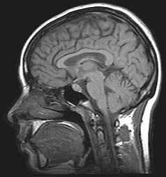
-
Hypokinesia
Wikipedia
The other category of movement disorder is hyperkinesia that features an exaggeration of unwanted movement, such as twitching or writhing in Huntington's disease or Tourette syndrome . [2] Contents 1 Spectrum of disorders 2 Causes 3 Pathophysiology 3.1 Associated neurotransmitters 3.2 Neurobiology 4 Treatments 4.1 Dopaminergic drugs 4.2 Deep brain stimulation 4.3 Methylphenidate 4.4 Stem cells 4.5 NOP receptor antagonists 4.6 Dance therapy 5 Associations 5.1 Cognitive impairment 5.2 Motor motivation 5.3 Demographic differentiation 5.4 Symptoms 6 Connections to other medical conditions 7 See also 8 References Spectrum of disorders [ edit ] Hypokinesia describes a variety of more specific disorders: Hypokinetic disorder Characteristics Akinesia ( α- a- , "without", κίνησις kinēsis , "motion") Inability to initiate voluntary movement. ... In some cases, Parkinson's patients experience dyskinesia as a negative side effect of dopamine medications. [7] Dystonia A movement disorder characterised by sustained muscle contractions, frequently causing twisted and repetitive movements, or abnormal postures. [8] Freezing This is characterized by an inability to move muscles in any desired direction. Neuroleptic malignant syndrome Resulting from heavy exposure to drugs that block dopamine receptors, victims can experience fever, rigidity, mental status change, dysautonomia , tremors, dystonia, and myoclonus . ... Classification D MeSH : D018476 v t e Diseases of the nervous system , primarily CNS Inflammation Brain Encephalitis Viral encephalitis Herpesviral encephalitis Limbic encephalitis Encephalitis lethargica Cavernous sinus thrombosis Brain abscess Amoebic Brain and spinal cord Encephalomyelitis Acute disseminated Meningitis Meningoencephalitis Brain / encephalopathy Degenerative Extrapyramidal and movement disorders Basal ganglia disease Parkinsonism PD Postencephalitic NMS PKAN Tauopathy PSP Striatonigral degeneration Hemiballismus HD OA Dyskinesia Dystonia Status dystonicus Spasmodic torticollis Meige's Blepharospasm Athetosis Chorea Choreoathetosis Myoclonus Myoclonic epilepsy Akathisia Tremor Essential tremor Intention tremor Restless legs Stiff-person Dementia Tauopathy Alzheimer's Early-onset Primary progressive aphasia Frontotemporal dementia / Frontotemporal lobar degeneration Pick's Dementia with Lewy bodies Posterior cortical atrophy Vascular dementia Mitochondrial disease Leigh syndrome Demyelinating Autoimmune Inflammatory Multiple sclerosis For more detailed coverage, see Template:Demyelinating diseases of CNS Episodic/ paroxysmal Seizures and epilepsy Focal Generalised Status epilepticus For more detailed coverage, see Template:Epilepsy Headache Migraine Cluster Tension For more detailed coverage, see Template:Headache Cerebrovascular TIA Stroke For more detailed coverage, see Template:Cerebrovascular diseases Other Sleep disorders For more detailed coverage, see Template:Sleep CSF Intracranial hypertension Hydrocephalus Normal pressure hydrocephalus Choroid plexus papilloma Idiopathic intracranial hypertension Cerebral edema Intracranial hypotension Other Brain herniation Reye syndrome Hepatic encephalopathy Toxic encephalopathy Hashimoto's encephalopathy Both/either Degenerative SA Friedreich's ataxia Ataxia–telangiectasia MND UMN only: Primary lateral sclerosis Pseudobulbar palsy Hereditary spastic paraplegia LMN only: Distal hereditary motor neuronopathies Spinal muscular atrophies SMA SMAX1 SMAX2 DSMA1 Congenital DSMA Spinal muscular atrophy with lower extremity predominance (SMALED) SMALED1 SMALED2A SMALED2B SMA-PCH SMA-PME Progressive muscular atrophy Progressive bulbar palsy Fazio–Londe Infantile progressive bulbar palsy both: Amyotrophic lateral sclerosis v t e Symptoms and signs relating to movement and gait Gait Gait abnormality CNS Scissor gait Cerebellar ataxia Festinating gait Marche à petit pas Propulsive gait Stomping gait Spastic gait Magnetic gait Truncal ataxia Muscular Myopathic gait Trendelenburg gait Pigeon gait Steppage gait Antalgic gait Coordination Ataxia Cerebellar ataxia Dysmetria Dysdiadochokinesia Pronator drift Dyssynergia Sensory ataxia Asterixis Abnormal movement Athetosis Tremor Fasciculation Fibrillation Posturing Abnormal posturing Opisthotonus Spasm Trismus Cramp Tetany Myokymia Joint locking Paralysis Flaccid paralysis Spastic paraplegia Spastic diplegia Spastic paraplegia Syndromes Monoplegia Diplegia / Paraplegia Hemiplegia Triplegia Tetraplegia / Quadruplegia General causes Upper motor neuron lesion Lower motor neuron lesion Weakness Hemiparesis Other Rachitic rosary Hyperreflexia Clasp-knife responseGCH1, OPRL1, APOD, SLC6A3, TH, LRRK2, MAPT, SNCA, ATXN3, ATXN1, FMR1, GBA, PSEN1, TWNK, PLA2G6, KLHL41, ATP6AP2, DNAJC6, KMT2B, SNCAIP, SYNJ1, PDE8B, PRKRA, UCHL1, XPA, FARS2, TPM3, TPM2, TK2, DHDDS, TBP, ACTA1, SPR, SLC20A2, PINK1, WDR45, AFG3L2, DNAJC12, VPS35, SLC30A10, TTC19, VPS13C, C19orf12, CHCHD2, ATL1, RRM2B, HTRA2, COQ2, JPH3, GIGYF2, FBXO7, MYORG, SLC39A14, ATP13A2, DNAJC13, ADGRV1, PARK7, TUBB6, POLG2, TAF1, SCN2A, SCN9A, NR4A2, MYPN, COASY, STX1B, KCNC3, RAB39B, HTT, EARS2, GLUD2, GABRG2, GABRD, FTL, EIF4G1, HCN1, DCTN1, KBTBD13, CSF1R, LYST, ATP1A3, ATXN8, SLC25A4, ADH1C, NEB, PANK2, PRKAR1B, POLG, ATXN2, ATXN8OS, SCN1A, PRNP, PPP2R2B, PTS, SCN1B, PODXL, PDGFRB, PDGFB, PRKN, SF3B1, YWHAE, ASXL1, TET2, PAFAH1B1, REG1A, STXBP3, PSPH, BPIFA2, RIDA, PSPN, HTR2C, MSMB, CCDC62, TMEM240, PPP1R1B, ACO1, TMEM106B, GRIN2A, APOE, CYP1A2, NQO1, DRD2, DRD4, EEF1A2, GRIN2B, MCF2L, IREB2, LRP2, NBN, RGS2, SLC18A2, SOD2, ECT


