For example, if the patient's most anxiety-inducing-component is takeoff, in VRET the patient would be exposed to a plane takeoff repeatedly while in vivo exposure the patient would have to wait for the plane to land and then take off again. [11] Outcomes [ edit ] Studies of interventions like CBT have reported rates of reduction in anxiety of around 80%; however, there is little evidence that any treatment can completely eliminate fear of flying. [3] Epidemiology [ edit ] Estimates for prevalence have ranged between 2.5% and 40%; estimates on the lower end are probably generated through studies where the condition is diagnosed by a professional, and the higher end probably includes people who have diagnosed themselves. [4] History [ edit ] Fear of flying was first discussed in the biomedical literature by a doctor in the UK at the end of World War I , who called it "aero-neurosis" and was describing pilots and crew who were or became anxious about flying.
Initially, people generally are unaware they have cataracts. Blurry vision is one of the first symptoms to appear which gradually worsens as the cataracts develop further.
Manual lifting of the eyelid often resolves the problem and the lid is able to stay open. ALO was first clearly described as a distinct entity in 1965 as “a nonparalytic motor abnormality characterized by the patient’s difficulty in initiating the act of lid elevation present only momentarily at the start of lid opening.” [1] A review of reported cases has shown a 2:1 female to male occurrence, and onset usually in the sixth decade of life. [2] Contents 1 Signs and symptoms 2 Causes 3 Diagnosis 4 Treatment 5 Prognosis 6 See also 7 References Signs and symptoms [ edit ] A person with ALO may complain of occasionally being unable to open the eye at will, typically after prolonged closure.
Defazio et al. (1998) described 10 new patients with 'so-called apraxia of eyelid opening.' They concluded that the term 'apraxia' may not be the correct descriptive term even when the eyelid disturbance occurs without any other central nervous system disease. Familial clustering of the isolated finding in 1 patient was consistent with a genetic contribution: 2 brothers, their father, and 2 paternal aunts were thought to be affected. Combining their 10 patients with 11 previously reported cases of isolated so-called apraxia of eyelid opening, Defazio et al. (1998) found that the peak age at onset was the sixth decade and that there was a female preponderance of 2 to 1. The characteristic inability to initiate lid elevation was frequently associated with failure to sustain lid elevation, thus suggesting that eyelid motor control may be abnormal.
The condition occurs more often in women than men and tends to present first in adulthood, although it has also been reported in children as young as 5 years old. [18] References [ edit ] ^ Mark Thompson, "Hemicrania Continua" ^ Goadsby P, Silberstein S, Dodick D (205).
Hemicrania continua is a rare, indomethacin-responsive, trigeminal autonomic cephalgia characterized by a typically side-locked, continuous, unilateral headache of moderate intensity with recurrent episodes of exacerbations of severe intensity, lasting for more than 3 months, in the absence of structural brain or vascular lesions. Ipsilateral cranial autonomic symptoms (lacrimation, conjunctival injection, nasal congestion or rhinorrhea, ptosis/miosis), restlessness and/or migrainous features are observed during the exacerbations.
Hemicrania continua (HC) is a type of primary headache disorder, which means the headache is not caused by another medical condition. Symptoms of HC include constant mild to moderate pain on one side of the head (unilateral) with periods of more intense, severe, migraine -like pain (exacerbations). These severe pain periods can last from 20 minutes to days. The frequency of exacerbations also varies greatly. The headache stays on the same side of the head and usually without pain free periods. HC is more common in women and most often starts in adulthood, but may begin anywhere from 5 to 67 years of age.
Nelson's syndrome Specialty Endocrinology Nelson's syndrome is a rare disorder that sometimes occurs in patients who have had both adrenal glands removed to treat Cushing's syndrome . [1] During the disorder the patient develops macroadenomas that secrete adrenocorticotropic hormone ( ACTH ). [2] The severity of the disease is dependent upon the effect of ACTH release on the skin, pituitary hormone loss, and the effect the tumor has on the surrounding structures within the body. [2] The first case of Nelson's syndrome was reported in 1958 by Nelson et al. [2] Dr.
A rare, acquired, endocrine disease characterized by the triad of diffuse skin and mucosa hyperpigmentation, markedly elevated serum adrenocorticotropin (ACTH) levels and an enlarging corticotroph adenoma, which manifest following total bilateral adrenalectomy performed for the treatment of Cushing's disease. Additionally, patients may present with headaches, visual field defects, cranial nerve palsy, pituitary apoplexy, diabetes inspidus, panhypopituitarism, and, occasionally, paraovarian or paratesticular tumors.
The causal agent is the Gram negative bacterium Xanthomonas campestris pv. graminis . The first case of bacterial wilt of turf was reported in a cultivar of creeping bentgrass known as Toronto or C-15, which is found throughout the midwestern United States.
History [ edit ] The condition was first described in 1929 by G. Kenneth Mallory and Soma Weiss in 15 alcoholic patients. [10] See also [ edit ] Boerhaave syndrome – Full thickness esophageal ruptures also often secondary to vomiting/retching.
Gold weights can be inserted into the upper eyelid to treat fasial nerve palsy. [9] Treatment [ edit ] Treatment of the cause of the exposure is to be done first. For example, in proptosis due to thyroid eye disease, regulation of thyroid hormone levels may be advised.
The overall live-birth rate in somebody with lupus has been estimated to be 72%. [5] Pregnancy outcome appears to be worse in those with lupus whose disease flares up during pregnancy. [6] Miscarriages in the first trimester appear either to have no known cause or to be associated with signs of active lupus. [7] Later losses appear to occur primarily due to the antiphospholipid syndrome , in spite of treatment with heparin and aspirin . [7] All women with lupus, even those without previous history of miscarriage, are recommended to be screened for antiphospholipid antibodies , both the lupus anticoagulant (the RVVT and sensitive PTT are the best screening battery) and anticardiolipin antibodies . [7] Neonatal lupus [ edit ] Neonatal lupus is the occurrence of lupus symptoms in an infant born from a mother with lupus, most commonly presenting with a rash resembling discoid lupus erythematosus , and sometimes with systemic abnormalities such as heart block or hepatosplenomegaly . [8] Neonatal lupus is usually benign and self-limited. [8] Still, identification of mothers at highest risk for complications allows for prompt treatment before or after birth.
By contrast, ultrasound may permit the injury to be visualized while the mortise is being stressed. [5] Consequently, a diagnostic modality such as ultrasound or magnetic resonance imaging (MRI) [4] that demonstrates the ligament itself may be helpful, if clinical suspicion remains. [6] Treatment [ edit ] Treatment depends on severity and convalescence may be as short as a few days or as long as six months. [7] Rest, icing, compression, and elevation is often recommended. [ citation needed ] Two important issues should be addressed early. First, a determination of whether the ankle is stable or unstable.
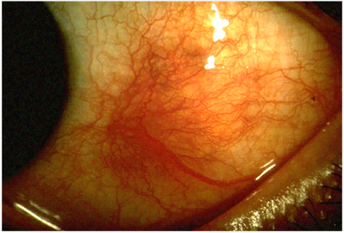
There are three types of scleritis: diffuse scleritis (the most common), nodular scleritis, and necrotizing scleritis (the most severe). Scleritis may be the first symptom of onset of connective tissue disease . [1] Episcleritis is inflammation of the episclera , a less serious condition that seldom develops into scleritis. [2] Contents 1 Signs and symptoms 1.1 Complications 2 Pathophysiology 3 Diagnosis 3.1 Classification 4 Treatment 4.1 Medical 4.2 Surgical Intervention 5 Epidemiology 6 References 7 External links Signs and symptoms [ edit ] Scleral translucency following recurrent scleritis.
With only 0.2 percent of the adult population estimated to be HIV -positive, Nicaragua has one of the lowest HIV prevalence rates in Central America . HIV was first detected in Nicaragua in 1987, after concentrated epidemics had been reported in other Central American nations.
Staging [ edit ] International Federation of Gynecology and Obstetrics (FIGO) staging is done at the time of surgery: Stage 0 :Carcinoma in situ Stage I :Growth limited to fallopian tubes Stage II :Growth involving one or both fallopian tubes with extension to pelvis Stage III:Tumor involving one or both fallopian tubes with spread outside pelvis Stage IV :Growth involving one or more fallopian tubes with distant metastases Treatment [ edit ] The initial approach to tubal cancer is generally surgical, and similar to that of ovarian cancer. As the lesion will spread first to the adjacent uterus and ovary, a total abdominal hysterectomy is an essential part of this approach, removing the ovaries, the tubes, and the uterus with the cervix.
The average intensive care unit (ICU) patient loses up to 660 mL of blood per week to laboratory testing. [3] For each day in the ICU, it is estimated that a person's hemoglobin level falls by 5 g/L (0.5 g/dL), 80% of which is due to phlebotomy. [7] : 20 On the second day of admission to the ICU, more than 70% of adults exhibit anemia, over half of whom will go on to require a blood transfusion . [3] In the neonatal intensive care unit (NICU), the issue is exacerbated by the patients' low body weight: it is estimated that during their first six weeks of life, infants in NICUs may lose 15−30% of their blood volume to blood draws. [3] [8] Premature babies often suffer from anemia of prematurity , which is caused by low production of erythropoietin (a hormone that stimulates red blood cell production) and the short lifespan of neonates' red blood cells, and is worsened by blood loss through phlebotomy. [9] People who are receiving dialysis lose blood not only through sampling for laboratory tests, but from the dialysis process itself and from bleeding caused by accessing veins to attach the dialysis equipment.
Outdoor activities such as hiking, camping, or hunting where plague-infected animals may be found, increase the risk of contracting septicemic plague, and so do certain occupations such as veterinary or other animal-related work. [1] Diagnosis [ edit ] A doctor or veterinarian will perform a physical exam which includes asking about the medical history and possible sources of exposure. [4] The following possible test could include: Blood samples (detecting antibodies) Culture samples of body fluids (check for the bacteria Yersinia pestis ) Kidney and liver testing Checking lymphatic system for signs of infection Examining body fluids for abnormal signs Checking for swelling Checking for signs of dehydration Checking for fever Checking for lung infection [5] Prevention [ edit ] The following steps and precautions should be used to avoid infection of the septicemic plague: [1] [5] Caregivers of infected patients should wear masks, gloves, goggles and gowns Take antibiotics if close contact with infected patient has occurred Use insecticides throughout house Avoid contact with dead rodents or sick cats Set traps if mice or rats are present around the house Do not allow family pets to roam in areas where plague is common Flea control and treatment for animals (especially rodents) Treatment [ edit ] Starting antibiotics early is a first step in treating septicemic plague in humans.
The disorder is sometimes referred to as metaphyseal osteopathy , and typically first presents between the ages of 2 and 7 months. [1] HOD is characterized by decreased blood flow to the metaphysis (the part of the bone adjacent to the joint) leading to a failure of ossification (bone formation) and necrosis and inflammation of cancellous bone . [2] The disease is usually bilateral in the limb bones, especially the distal radius , ulna , and tibia . [3] The Weimaraner, Irish Setter, Boxer, German Shepherd, and Great Dane breeds are heavily represented in case reports of HOD in the veterinary literature, but the severity of symptoms and possible etiology may be different across the breeds.
Genetic disorders , such as hemophilia and Von Willebrand disease , can cause a reduction in clotting factors. [2] Anticoagulants such as warfarin will also prevent clots from forming properly. [2] Coagulopathy may also occur as a result of dysfunction or reduced levels of platelets (small disk-shaped bodies in the bloodstream that aid in the clotting process). [ citation needed ] Acute traumatic coagulopathy [ edit ] In 2003, Karim Brohi, Professor of Trauma Sciences at Queen Mary University of London , first introduced the term Acute Traumatic Coagulopathy (ATC), [3] establishing that coagulopathy induced by trauma results in: more severe bleeding multi-organ failure high mortality Treatment [ edit ] If someone has coagulopathy, their health care provider may help them manage their symptoms with medications or replacement therapy.