Oligodontia-cancer predisposition syndrome is a rare, genetic, odontologic disease characterized by congenital absence of six or more permanent teeth (excluding the third molars) in association with an increased risk for malignancies, ranging from gastrointestinal polyposis to early-onset colorectal cancer and/or breast cancer.
A number sign (#) is used with this entry because of evidence that tooth agenesis-colorectal cancer syndrome is caused by heterozygous mutation in the AXIN2 gene (604025) on chromosome 17q24.
A rare, X-linked, syndromic intellectual disability disease characterized by neonatal hypertonia which evolves to hypotonia and an exaggerated startle response (to sudden visual, auditory or tactile stimuli), followed by the development of early-onset, frequently refractory, tonic or myoclonic seizures.
A number sign (#) is used with this entry because of evidence that early infantile epileptic encephalopathy-8 (EIEE8) is caused by mutation in the ARHGEF9 gene (300429) on chromosome Xq22.1. Description Early infantile epileptic encephalopathy-8 is an X-linked disorder characterized by seizure onset before 2 years of age and severe developmental delay. Some patients have hyperekplexia (summary by Shimojima et al., 2011). For general phenotypic descriptions and discussions of genetic heterogeneity of early infantile epileptic encephalopathy and hyperekplexia, see EIEE1 (308350) and hereditary hyperekplexia (149400), respectively. Clinical Features Harvey et al. (2004) reported a patient with clinical symptoms of both hyperekplexia and early infantile epileptic encephalopathy.
Craniosynostosis-fibular aplasia syndrome is an extremely rare genetic disease, reported in only 2 brothers to date, characterized by the combination of craniosynostosis (involving both coronal sutures), congenital absence of the fibula, cryptorchidism, and bilateral simian creases.
Lowry (1972) described brothers with this combination. The parents were related. Lowry (1993) provided a follow-up of one of the brothers at the age of 25 years. He was of average intelligence and had completed 2 years of college. He showed markedly hypoplastic calves. INHERITANCE - Autosomal recessive HEAD & NECK Head - Craniosynostosis SKELETAL Limbs - Fibular aplasia ▲ Close
Hyperplastic polyposis syndrome is a rare, genetic intestinal disease characterized by the presence of multiple (usually large) hyperplastic/serrated colorectal polyps, usually with a pancolonic distribution.
A number sign (#) is used with this entry because of evidence that sessile serrated polyposis cancer syndrome (SSPCS) is caused by heterozygous mutation in the RNF43 gene (612482) on chromosome 17q22. Description Sessile serrated polyposis cancer syndrome (SSPCS) is a rare disorder characterized by the presence of multiple serrated polyps in the colon and an increased personal and familial risk of colorectal cancer. ... Molecular Genetics Gala et al. (2014) studied 20 probands with serrated polyposis, 16 of whom met the WHO criteria for SSPCS syndrome. A personal history of colon cancer was present in 3, whereas 11 had a family history of colon cancer, and 8 had 1 or more extracolonic neoplasms. ... Four first-degree relatives satisfied the criteria for hyperplastic polyposis syndrome. Based on the estimated HPS prevalence of 1 in 3,000 in the general population, the projected relative risk of HPS in first-degree relatives was 39 (95% CI, 13-121). ... Seven first-degree relatives (9%) had multiple polyps (5 or more); 11 (14%) first-degree relatives fulfilled serrated polyposis syndrome WHO criterion 2 (any number of serrated polyps proximal to the sigmoid colon in a first-degree relative of a patient with serrated polyposis syndrome), of whom 1 sib also met serrated polyposis syndrome WHO criterion 3 (greater than 20 serrated polyps spread throughout the colon).
A rare, syndromic intellectual disability characterized by macrocephaly, short stature, intellectual disability, variable degree of spastic paraplegia, central nervous system malformations (hydrocephalus, Dandy-Walker malformation), and dysmorphic features, such as high and broad forehead, midface hypoplasia, and small and broad hands and feet.
Clinical Features Wolach et al. (1990) described this combination in a 5-year-old daughter of first-cousin, Sephardic Jewish parents. Diffuse skin pigmentation had been present from birth and biopsy confirmed the diagnosis of diffuse cutaneous mastocytosis (see 154800). There was no mental retardation associated with the microcephaly. Wolach et al. (1990) favored autosomal recessive inheritance. Hennekam and Beemer (1992) studied a girl, born of nonconsanguineous parents in the Netherlands, who at birth was noted to have diffuse hyperpigmented, slightly elevated macules on her whole body, especially on the trunk and extremities, with exaggerated dermatographism. She had a large anterior fontanel, slight proptosis, upward slanting palpebral fissures, highly arched palate, receding chin, and clinodactyly of the fifth fingers.
A rare, adrenogenital syndrome characterized by generalized, partial tissue insensitivity to glucocorticoids leading to variable phenotype, including asymptomatic individuals with only biochemical alterations or patients with ambiguous genitalia at birth in females, hypertension, acne, hirsutism, precocious puberty, male-pattern hair loss, anxiety and depression in both sexes, menstrual irregularities in women, and oligospermia in men.
Description Generalized glucocorticoid resistance is an autosomal dominant disease characterized by increased plasma cortisol concentration and high urinary free cortisol, resistance to adrenal suppression by dexamethasone, and the absence of clinical stigmata of Cushing syndrome. The clinical expression of the disease is variable. ... High levels of cortisol (without stigmata of Cushing syndrome), resistance of the hypothalamic-pituitary-adrenal axis to dexamethasone, and an affinity defect of the glucocorticoid receptor characterized the disorder. ... Plasma ACTH concentrations were normal but she was resistant to adrenal suppression by dexamethasone. No stigmata of Cushing syndrome were present. The patient had symptoms of pronounced fatigue. ... The woman and her son instead showed an increased thermolability of the cortisol receptor, a phenomenon also observed with the androgen receptor in patients with the testicular feminization syndrome (300068). Lamberts et al. (1986) described cortisol resistance in a 26-year-old woman with hirsutism, mild virilization, and menstrual difficulties. ... She was initially thought to have Cushing disease, based on high plasma ACTH and serum cortisol levels, increased urinary cortisol secretion, resistance to adrenal suppression with dexamethasone, and bilateral adrenal hyperplasia by computed tomography and scintigraphy; however, she had no clinical signs or symptoms of Cushing syndrome. Laboratory studies indicated that the patient's glucocorticoid resistance was due to a decrease in the affinity of the receptor for glucocorticoids and a decrease in the binding of the GCCR complex to DNA.
Spondylocostal dysostosis-anal and genitourinary malformations syndrome is characterised by the association of spondylocostal dysostosis with anal and genitourinary malformations (anal atresia and agenesis of external and internal genitalia).
Clinical Features In an inbred Mennonite sibship, Casamassima et al. (1981) described 2 brothers with severe vertebral and costal dysplasia of the type variously called spondylothoracic dysplasia, costovertebral dysplasia, or the Jarcho-Levin syndrome (277300). By x-ray, the thorax has a crab-like configuration.
This syndrome is characterized by the association of severe nasal hypoplasia, hypoplasia of the eyes, hyposmia, hypogeusia and hypogonadotropic hypogonadism.
Bosma arhinia microphthalmia syndrome (BAMS) is a rare condition characterized by abnormalities of the nose and eyes and problems with puberty. ... Learn more about the gene associated with Bosma arhinia microphthalmia syndrome SMCHD1 Inheritance Pattern This condition is inherited in an autosomal dominant pattern , which means one copy of the altered SMCHD1 gene in each cell is sufficient to cause the disorder.
A number sign (#) is used with this entry because of evidence that Bosma arhinia microphthalmia syndrome (BAMS) is caused by heterozygous mutation in the SMCHD1 gene (614982) on chromosome 18p11. Description Bosma arhinia microphthalmia syndrome (BAMS) is characterized by severe hypoplasia of the nose and eyes, palatal abnormalities, deficient taste and smell, inguinal hernias, hypogonadotropic hypogonadism with cryptorchidism, and normal intelligence (summary by Graham and Lee, 2006). ... Tryggestad et al. (2013) did not consider this patient to have Bosma syndrome because he did not have microphthalmia. ... The proband's father had been diagnosed with limb-girdle muscular dystrophy, but had no vision abnormalities, anosmia, or other features of Bosma syndrome. The paternal grandmother had coloboma and cataract, and a paternal great-aunt reportedly had coloboma and a paternal great-uncle was reportedly born blind. ... In a 13-year-old boy with arhinia, anosmia, and hypogonadotropic hypogonadism, Tryggestad et al. (2013) analyzed 7 Kallmann syndrome (see 308700)-associated genes, but no mutations were detected.
A malformation disorder characterized by complete or incomplete absence of nose (arrhinia), choanal atresia, microphthalmia, anophthalmia and cleft or high palate.
Clinical description In the infantile form (the most common), the first clinical signs appear between 3 and 6 months of age, with a polyuria-polydipsia syndrome and marked growth delay secondary to a generalized proximal tubular dysfunction with severe fluid-electrolyte balance alterations (renal Fanconi syndrome). ... The first symptoms of juvenile cystinosis (< 5% of patients), typically appear around 6-8 years of age with a milder form of proximal tubulopathy and/or proteinuria in nephrotic syndrome. Progression to renal failure occurs later than in the infantile form. ... Diagnostic methods The diagnosis is based on blood and urine analysis showing features of renal Fanconi syndrome (metabolic acidosis, hypokalemia, hypophosphatemia, hyperaminoaciduria, glycosuria, low molecular weight proteinuria), detection of cystine crystals in the cornea and determination of elevated cystine levels in leucocytes. ... Differential diagnosis Differential diagnosis includes other diseases causing renal Fanconi syndrome (Lowe syndrome, Dent disease, galactosemia, fructose intolerance, thyrosinemia, mitochondrial nephropathies, Wilson disease, Fanconi-Bickel syndrome, lysinuric protein intolerance, idiopathic Fanconi syndromes, secondary Fanconi syndrome due to drug toxicity or substance abuse, recovery of acute tubulus necrosis), diseases causing phosphaturia and rickets, and proteinuria of unknown etiology.
A number sign (#) is used with this entry because ocular nonnephropathic cystinosis is caused by mutation in the gene encoding cystinosin (CTNS; 606272), which maps to chromosome 17p13. Description Ocular nonnephropathic cystinosis, a variant of the classic nephropathic type of cystinosis (219800), is an autosomal recessive lysosomal storage disorder characterized by photophobia due to corneal cystine crystals but absence of renal disease (summary by Anikster et al., 2000). Clinical Features Lietman et al. (1966) observed a benign form of cystinosis in 3 affected sibs of cousin parents. The ages of the patients were 53, 50, and 42 years. Crystals of cystine were demonstrated in the cornea, buffy coat of the blood, and bone marrow. No amino aciduria or impairment of renal function was found. Cogan et al. (1958) described an asymptomatic adult with cystine demonstrable in cornea and bone marrow.
Ocular cystinosis is the benign, adult form of cystinosis (see this term), a metabolic disease characterized by an accumulation of cystine crystals in the cornea and conjunctiva responsible for tearing and photophobia and associated with no other additional manifestations.
Nephropathic cystinosis begins in infancy, causing poor growth and a particular type of kidney damage (renal Fanconi syndrome) in which certain molecules that should be reabsorbed into the bloodstream are instead eliminated in the urine.
With the identification of close linkage of Kallmann syndrome (308700) and X-linked ichthyosis (308100), and the description of deletions leading to the coexistence of these 2 disorders, it becomes a distinct possibility that the affected males in the family reported by Lynch et al. (1960) suffered from a 'contiguous gene syndrome.') ... RUD syndrome is a neurocutaneous disorder characterized by epilepsy, mental retardation, infantilism, congenital ichthyosis, and retinitis pigmentosa. ... Munke et al. (1983) found reports of 28 patients with Rud syndrome. The male:female ratio was 2:1, consistent with some of the cases being instances of an X-linked recessive disorder. ... (The possibility that this family suffered from a 'contiguous gene syndrome' should be investigated with the search for cytogenetic or molecular genetic evidence of deletion in Xp.) ... Traupe (1989) suggested that the designation 'Rud syndrome' be abandoned; from a review of reports he concluded that both the neurologic involvement and the ichthyosis remain ill defined.
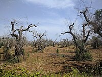
Olive quick decline syndrome An olive grove infested with Xylella fastidiosa in Puglia, Italy in 2019 Common names OQDS Causal agents Xylella fastidiosa Hosts Olive trees Vectors Meadow froghopper Distribution Southern Italy Symptoms Dieback of the leaves, twigs and branches An infected olive grove in Italy in 2019 Disease of olive trees Olive quick decline syndrome ( OQDS ) (in Italian: Complesso da Disseccamento Rapido dell'Olivo , CDRO or CoDiRo ) is a wasting disease of olive trees which causes dieback of the leaves, twigs and branches so that the trees no longer produce crops of olives . ... It was first detected in Italy in 2013, [2] in the Salento Peninsula ; by late 2013, it was estimated that approximately 8,000 hectares were affected. [3] The disease currently threatens olive groves and oil production in Italy, Greece, and Spain, which together account for 95% of European oil production [4] [5] One 2020 model predicts a potential economic impact of the disease for Italy over 50 years between 1.9 billion to 5.6 billion Euros. [4] In addition to Europe, the disease has also been detected in olive crops in California, Argentina and Brazil. [6] Symptoms [ edit ] Symptoms include leaf scorch and desiccation of twigs and branches, beginning at the upper part of the crown and then moving to the rest of the tree, which acquires a burned look. [7] References [ edit ] ^ Olive Quick Decline Syndrome (PDF) , Primary Industries and Regions SA , 2017 ^ Stokstad, Erik (8 May 2015). ... "The current status of the quick decline syndrome of olive in southern Italy". Phytoparasitica . 44 (1): 1–10. doi : 10.1007/s12600-015-0498-6 . ... "Isolation and pathogenicity of Xylella fastidiosa associated to the olive quick decline syndrome in southern Italy" . Scientific Reports . 7 (1): 17723. doi : 10.1038/s41598-017-17957-z . ... P.; Boscia, D.; Porcelli, F.; Saponari, M. (1 February 2016). "The olive quick decline syndrome in south-east Italy: a threatening phytosanitary emergency".
A number sign (#) is used with this entry because of evidence that Tenorio syndrome (TNORS) is caused by heterozygous mutation in the RNF125 gene (610432) on chromosome 18q12. Description Tenorio syndrome is characterized by overgrowth, macrocephaly, and intellectual disability (ID). Some patients may have mild hydrocephaly, hypoglycemia, and inflammatory diseases resembling Sjogren syndrome (270150) (summary by Tenorio et al., 2014). Clinical Features Tenorio et al. (2014) described 6 patients from 4 families with a syndrome of overgrowth, macrocephaly, intellectual disability, mild hydrocephaly, hypoglycemia, and inflammatory diseases resembling Sjogren syndrome. ... Molecular Genetics By SNP-array screening of 270 families from the Spanish Overgrowth Syndrome Registry with no molecular characterization of their disorder, Tenorio et al. (2014) identified 1 patient with a de novo 9-Mb deletion on chromosome 18 encompassing several candidate genes, including RNF125 AND RNF138 (616319).
Clinical Features Gurrieri et al. (1992) described 2 brothers with an orofaciodigital syndrome which did not seem to fit into any of the 7 previously described types (Toriello, 1988; Munke et al., 1990). ... The specific retinal abnormalities in the brothers reported by Gurrieri et al. (1992) were retinochoroidal lacunae of colobomatous type, similar to those seen in Aicardi syndrome (304050). Nevin et al. (1994) reported a female case of OFDS with component manifestations typical of type II (252100), namely median cleft of the upper lip, multiple oral frenula, and lobulated or hamartomatous tongue, associated with retinal abnormalities as distinguishing feature. ... Erickson and Bodensteiner (2007) described 2 pairs of sibs (2 females, and 1 female and 1 male) from the Navajo population with orofaciodigital syndrome. In addition to retinal colobomas and mild digital anomalies, the sibs also had severe microcephaly, mental retardation, and short stature. ... Molecular Genetics For discussion of a possible relationship between variation in the TBC1D32 gene and orofaciodigital syndrome IX, see 615867.0001. For discussion of a possible relationship between variation in the SCLT1 gene and orofaciodigital syndrome IX, see 611399.0001. INHERITANCE - Autosomal recessive GROWTH Height - Short stature HEAD & NECK Head - Microcephaly Eyes - Hypertelorism - Telecanthus - Strabismus - Duane syndrome (rare) - Retinal coloboma Nose - Broad nasal tip - Bifid nasal tip Mouth - Median cleft lip - Tongue lobulation - Tongue hamartoma - Oral frenula - High-arched palate - Cleft palate - Cleft alveolar ridge Teeth - Absent teeth RESPIRATORY Larynx - Hypoplastic epiglottis Lung - Recurrent aspiration pneumonia GENITOURINARY External Genitalia (Female) - Absent clitoris (rare) SKELETAL Limbs - Short tibiae Hands - Polydactyly - Syndactyly - Brachyphalangia - Carpal shortening - Camptodactyly Feet - Bifid halluces - Forked metatarsal - Tarsal shortening - Syndactyly SKIN, NAILS, & HAIR Skin - Milia NEUROLOGIC Central Nervous System - Developmental delay - Brain atrophy - Migrational abnormalities MISCELLANEOUS - Mild manifestations in carrier females (cleft lip, cleft tongue) ▲ Close
Acheiropody is a very rare condition characterized by bilateral, congenital amputations of the hands and feet. Individuals with this condition are born with complete amputation of the distal humeral epiphysis (end of the upper arm bone) and tibial diaphysis (mid-section of the shin bone), and aplasia (lack of development) of the radius , ulna , fibula , and of all the bones of the hands and feet. The condition appears to affect only the extremities, with no other signs and symptoms reported. It is caused by a defect in the LMBR1 gene and is inherited in an autosomal recessive manner. Walking may be possible for individuals with acheiropody with well-fitted prostheses .
An extremely rare developmental disorder characterized by bilateral, congenital and complete amputation of the distal extremities (amputation of distal epiphysis of the humerus, distal portion of the tibial diaphysis, aplasia of the radius, ulna, fibula) and aplasia of hands and feet (aplasia of carpal, metacarpal, tarsal, metatarsal and phalangeal bones). Rarely, an ectopic bone can be found at the distal end of the humerus. No other systemic manifestations have been reported and the disorder follows an autosomal recessive pattern of inheritance.