-
Central Sleep Apnea
Wikipedia
"Association between atrial fibrillation and central sleep apnea" (PDF) . Sleep . 28 (12): 1543–6. doi : 10.1093/sleep/28.12.1543 .TACR1, TAC1, PHOX2B, VAMP1, INPP5E, PSAP, SLC5A7, CCDC47, PRPS1, TECPR2, FGFR3, DKK1, TBR1, NACC1, QRICH1, MYO9A, H3-3A, RPGRIP1L, AHI1, TMEM216, CEP290, COQ2, AGRN, SLC25A1, SYT2, NADK2, COL13A1, WFS1, CHAT, NALCN, TSEN54, SNAP25, RET, SLC35A2, SLC2A1, ASCL1, SLC18A3, CISD2, CSH2, CSH1, REM1, HSPA9, CENPJ, SLC25A38, ERCC8, ALAS2, ALDH2, EPO, ERCC6, BDNF, ABCB1, GZMB, HMOX1, FOXP3, MIR144, GTF2H5, TGFB1, TPO, ERICD, MIRLET7I, VEGFA, TRPV1, XIST, SLC12A9, DLL1, DLK1, PNO1, PPIF, NES, TBC1D9, SUN1, HSPB7, PHF11, AHR, PSG5, SMN2, CYBB, GTF2H4, GTF2H3, GTF2H2, GTF2H1, MS4A2, ERCC5, ERCC3, ERCC2, CYP3A5, CRP, IL2RB, COMT, CDKN1A, CD38, CAT, MYRF, BMP2, APOE, ANXA5, AKR1B1, IL2RA, IL6, SMN1, PKM, SLC5A3, SFRP1, CCL3, CCL2, AKT1, MAPK3, PPID, PON1, PLCB4, PECAM1, IL7R, PCYT1A, OLR1, NOS3, MET, LEP, ISG20, INSRR, IL18, IL10, CERNA3
-
Wheat Allergy
Wikipedia
When 10 foods causing the most reactions were removed migraines fell precipitously, hypertension declined. [27] Some specific instances are attributed to wheat. [28] Autism . Parents of children with autism often ascribe the children's gastrointestinal symptoms to allergies to wheat and other foods.

-
Mitral Valve Stenosis
Wikipedia
. ^ http://cursoenarm.net/UPTODATE/contents/mobipreview.htm?28/3/28733 Archived 2016-11-13 at the Wayback Machine ^ "Mitral Stenosis: Heart Valve Disorders: Merck Manual Home Edition" .PLD1, UBE2A, GBA, FBN1, ADAMTSL2, IFNB1, CHST3, NKX2-5, MCTP2, LZTR1, XYLT1, HAAO, ZNF148, XYLT2, GJA1, ABCC6, NOTCH2, CSF2, LAMC2, MBP, SERPINA1, HLA-DRB1, TNF, NEFL, SPARCL1, TRBV20OR9-2, VDR, IL2RA, IL1RN, IL1B, NOTCH1, IL10, CIITA, TNFRSF11B, MB, IL17A, HLA-DPB1, IL1A, MIR326, LINC01672, IL22, CISH, ACE, MIR155, MIR21, SIRT1, SNCA, SOD1, TAP2, SULT1E1, SELPLG, TGFB1, MIR132, HCAR2, IL27, SLC2A4, CCL2, CCL20, MIR200A, TH, PTX3, PTPN2, PSMB6, CD24, RELN, PRL, MAPK1, PRKAR1A, TGFB3, RBM45, TLR4, RAB3GAP1, DLL1, CXCL13, NR1H4, TNFSF15, FOXP3, ATG5, DESI2, BFAR, PON1, IL23A, IL17D, ADIPOQ, TNFSF11, TUG1, SYN3, MYDGF, IL21, ARHGAP24, YY1, VEGFA, HAVCR2, HELB, TP73, TNFRSF1A, CYP2R1, ADRB2, NFE2L2, PML, ERV3-1, CPB2, CRP, DPP4, DMTN, EPHB2, ERG, GATA3, COL11A2, GFAP, GRM4, GRM8, HCCS, HLA-DQA1, HLA-DRB5, COMT, COL2A1, IFNAR1, BTF3P11, ALB, APOE, B2M, BDKRB1, BDNF, BMP4, C3, CCR5, CAT, CD1D, CD1E, CD14, CD68, CHI3L1, HMOX1, IFNAR2, PLAT, MYT1, MMP3, MPO, MS, MTR, MX1, MYCN, AGER, MIF, NFKB1, NPHS1, NPPB, CLDN11, SERPINE1, PEPD, MMP2, LIF, IFNG, IRS1, IL3, IL4, IL6, IL6ST, IL13, IL18, ISG20, LEP, ITGA4, KLRB1, KNG1, LAG3, LALBA, LBR, LOC102723971

-
Carney Complex
GeneReviews
PMS may occur anywhere in the central and peripheral nervous system; it is most frequently found in the nerves of the gastrointestinal tract (esophagus and stomach) and paraspinal sympathetic chain (28%). The spinal tumors present as pain and radiculopathy in adults (mean age 32 years).PRKAR1A, MEN1, GH1, NR1I3, CXADRP1, CXADR, CNC2, TRIM13, SPG7, CASR, ARR3, SLC8A1, POMC, GNAS, RCVRN, APRT, MFAP1, CDKN1B, AIP, CYP3A4, PDE11A, PRKAR1B, PRL, NR1I2, STK11, SSTR4, HCAR3, SST, UGT1A, SCLC1, ASMTL, SMARCB1, DICER1, USP8, LPAR2, CXCR6, SGSM3, TRAC, GAL, SDF4, UGT1A1, ACKR3, PTBP2, WDR26, ADO, CABLES1, AZIN2, GADL1, SMARCA4, ADRA1A, SDHC, RET, BRS3, CAT, CAV1, CD247, CD19, CETN1, CTNNB1, CYP2B6, CYP19A1, EDNRA, MTOR, GNA11, GPR42, NR3C1, CXCL2, HTC2, IGF1, INHA, ITGA5, ITGAL, ITGAV, ITGB2, KCNJ2, KCNJ5, MAS1, MPO, MSH2, ADRA2B, PTEN, PRKACB
-
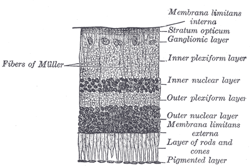
Congenital Stationary Night Blindness
Wikipedia
Specifically, these mutations are the Gly90Asp [26] and the Thr94Ile, which has been the most recent one reported. [27] The third mutation is Ala292Glu, and it is located in the seventh transmembrane helix , in proximity to the site of retinal attachment at Lys-296. [28] Mutations associated with CSNB affect amino acid residues near the protonated Schiff base (PSB) linkage.RHO, GRM6, TRPM1, GNAT1, LRIT3, SLC24A1, PDE6B, GPR179, SAG, CACNA1F, NYX, CABP4, GNB3, CACNA2D4, GRK1, RDH5, RBP4, TRAPPC9, USH2A, ABCA4, RPGR, RBP3, FGFR2, ERG, GRK7, RPE65, USP11, SOCS2, BBS1, GNAZ, GUCY2D, GPR34
-
Delirium
Wikipedia
A 2018 systematic review showed that, broadly, delirium may be associated with neurotransmitter imbalance (namely serotonin and dopamine signaling), reversible fall in somatostatin, and increased cortisol. [27] The leading "neuroinflammatory hypothesis" (where neurodegenerative disease and aging leads the brain to respond to peripheral inflammation with an exaggerated CNS inflammatory response) has been described, [28] but current evidence is still conflicting and fails to concretely support this hypothesis. [27] Neuroimaging [ edit ] Neuroimaging provides an important avenue to explore the mechanisms that are responsible for delirium. [29] [30] Despite progress in the development of magnetic resonance imaging (MRI) , the large variety in imaging-based findings has limited our understanding of the changes in the brain that may be linked to delirium. ... "At the extreme end of the psychoneuroimmunological spectrum: delirium as a maladaptive sickness behaviour response" . Brain, Behavior, and Immunity . 28 : 1–13. doi : 10.1016/j.bbi.2012.07.012 .IL6, APOE, IGF1, SLC6A3, CXCL8, DRD3, IL1RN, DRD2, TH, LIF, SLC25A13, SRPX, DNMT1, NAGS, TRNE, PNN, CALM1, FIG4, CAMKMT, PRDX1, MMACHC, KRIT1, CRP, CALM2, CALM3, ALB, S100B, TNF, LAMC2, BTBD8, BDNF, BCHE, ENO2, CSF2, IL1B, COMT, NEFL, CIT, MTNR1B, CCL2, IL10, ACHE, HCRT, AMBP, IL2, IL17A, PLA2G15, CD2AP, MCF2L, CNOT1, WDHD1, CYP3A4, RRAGA, FGL2, ACSS2, CYP3A5, THOC1, EEF1A2, CCL4L2, ADAMTS2, CD19, PELI1, CCL28, ADIPOQ, CORO7, BCS1L, SARNP, ACCS, PRDM6, NLRP3, GRIN3A, AZGP1, SASS6, NLRP6, CCL4L1, PSS, ABCG2, PER2, IL18, TNFSF14, CXCL10, IL6R, LBP, LEP, LGALS3, LRP2, HTR2C, HTR1A, NPPB, AGT, ABCB1, PIK3CA, PIK3CB, PIK3CD, PIK3CG, NR3C1, S100A12, GRIN2B, GCLC, CCL4, GFAP, SOAT1, TCF21, GABBR1, FCGR3B, TXN, UCHL1, UGCG, FCGR3A, RN7SL263P

-
Pmm2-Cdg (Cdg-Ia)
GeneReviews
Methods used may include: quantitative PCR, long-range PCR, multiplex ligation-dependent probe amplification (MLPA), and a gene-targeted microarray designed to detect single-exon deletions or duplications. 7. A 28-kb deletion that includes exon 8 as well as other novel exon or whole-deletions has been reported [Schollen et al 2007].
-
Mandibulofacial Dysostosis With Microcephaly
GeneReviews
Ear malformations & hearing loss Microtia / Dysplastic pinna(e) 97% Auditory canal atresia or stenosis 68% Preauricular tag 50% Hearing loss 83% Other findings Cardiac anomalies 35% Typically atrial &/or ventricular septal defect Thumb anomalies 34% Typically proximally placed; uncommonly, preaxial polydactyly or hypoplasia Esophageal atresia / Tracheoesophageal fistula 33% Short stature 30% Spine anomalies 28% Incl scoliosis, kyphosis, hemivertebrae, & cervical segmentation anomalies Epilepsy 26% Mandibulofacial dysostosis is characterized by malar and maxillary hypoplasia.
-
Myh9-Related Disorders
GeneReviews
Most affected individuals have no spontaneous bleeding or only easy bruising. About 28% of persons with MYH9RD have spontaneous mucocutaneous bleeding, including epistaxis, gum bleeding, or menorrhagia [Pecci et al 2014a].

-
Incontinentia Pigmenti
GeneReviews
The nail changes may be transient, but a single, chronic, longitudinal ridge in the nail was present in 28% of persons in one study [Phan et al 2005].IKBKG, NSDHL, COX8A, STAT6, IKBKGP1, TNF, TRAF6, EDA, IL4, TRIM13, TNFRSF1A, HAND2, NR1I3, AGT, GJB6, SPG7, SPIN2A, SHARPIN, CYCSP25, EDARADD, PROKR2, SIRT1, PRKAR1A, CCL11, AR, ARR3, CASP3, CASP9, CASR, CD38, CXADR, CYP1B1, G6PD, IFNA1, IFNA13, MPO, MUC1, NF2, NFKB1, PREP, CXADRP1

-
Phelan-Mcdermid Syndrome
GeneReviews
The ADI-R was used to characterize the regression and reported loss of: Motor skills in 50% at mean age 4 years Self-help skill in 50% at mean age 4 years Language in 33% at mean age 3 years Social engagement/responsiveness in 33% at mean age 5 years Purposeful hand movement in 28% at mean age 7 years Constructive/imaginative play in 22% at mean age 7 years The regression in Phelan-McDermid syndrome is distinct from the regression seen in autism and Rett syndrome in that it occurs later in life and has a stronger impact on motor skills and self-help skills [Reierson et al 2017].SHANK3, INS, ARSA, HTC2, CACNA1C, CLK2, IGF1, NOTCH1, PSD, SHANK2, MAPK8IP2, SHANK1, MBD5, PNPLA3, KANSL1

-
Cowden Syndrome 1
OMIM
They concluded that affected persons may have an increased risk of intracranial tumors: a woman in their family had meningothelial meningioma removed at age 28. DiLiberti et al. (1983) described a 7.5-year-old boy with macrocephaly, hamartomatous intestinal polyps, and cafe-au-lait spots on the penis, and referred to the disorder as 'Ruvalcaba-Myhre-Smith syndrome.'
-
Flnb Disorders
GeneReviews
Three individuals with Peipkorn dysplasia have had pathogenic variants in exons 28 and 29. Mosaicism Clinical evidence suggests that somatic mosaicism can complicate the presentation of these conditions [Petrella et al 1993, Bicknell et al 2007, Bernkopf et al 2017].
-
Ezh2-Related Overgrowth
GeneReviews
Note: Five of the individuals presenting with peripheral hypertonia were also reported to have central hypotonia. Poor feeding was reported in 10/28 neonates including one who required nasogastric tube feeding for two weeks.
-
Dystrophinopathies
GeneReviews
Some investigators classify such individuals as having subclinical or benign BMD, whereas others may classify such individuals as having DCM with increased serum CK concentration [Towbin 1998]. In one study of 28 individuals with subclinical and benign BMD between ages six and 48 years, 19 (68%) had myocardial involvement, although only two were symptomatic [Melacini et al 1996]. ... The optimal time to start treatment in DMD is unknown, but most cardiologists will initiate treatment when the left ventricle ejection fraction drops below 55% and fractional shortening is less than 28% [Jefferies et al 2005, Viollet et al 2012].
-
Dystonia/parkinsonism, Hypermanganesemia, Polycythemia, And Chronic Liver Disease
GeneReviews
Prevalence This inborn error of manganese metabolism has only recently been identified. A total of 28 affected individuals from ten families are known worldwide [Quadri et al 2012, Tuschl et al 2012, Avelino et al 2014, Quadri et al 2015, Mukhtiar et al 2016].
- Diabetes Mellitus, 6q24-Related Transient Neonatal GeneReviews
-
Gaucher Disease, Type I
OMIM
-
Search Gaucher's disease case abstracts
The frequency of GD carriers among 308 TSD heterozygotes was 1:28, which is about half that expected (p = 0.03).GBA, SNCA, CHIT1, ACE, SCARB2, FBXO11, MSH6, GBAP1, CYP2D6, TNF, VDR, UGCG, H4C13, H4C5, H4C14, H4C2, SF3B1, BDNF, ABCG8, TET2, GBA3, HAMP, ABCG5, H4C3, PINK1, H4-16, ASXL1, PLF, H4C8, H4C9, H4C11, H4C12, CD34, CTNNB1, DVL1, DVL2, GJB2, HFE, TNFRSF11B, PAFAH1B1, PSAP, SLC6A3, YWHAE, BTF3P11, H4C1, H4C4, H4C6, H4C15
-
Hypodontia
Wikipedia
In hereditary cases, evidence of dental germ developing after surrounding tissues have closed the space required for development may be a huge contributing factor as well such as genetic disorders like down syndrome, [1] ectodermal dysplasia, [28] cleidocranial dysplasia, [29] orofacial clefting, especially cleft lip and palate. [30] Most of the craniofacial characteristics are influenced by the both genetic and environmental factors through complex interactions.MSX1, IRX5, LTBP3, PAX9, EDA, WNT10A, EDAR, DCAF17, NOL11, IFT122, ARHGAP15, WNT5A, CACNA1S, RPS6KA3, NECTIN1, FOXP1, FZD2, FLNA, DVL3, DVL1, EDARADD, FGFR1, FGF8, CHDH, KISS1R, OTOR, UBXN11, CHD7, BMP4, TNF, MBL3P, COTL1, FGF17, CSRP3, TGFA, MBL2, IRF6, IHH, MAPK14, CALML3, BOP

- Arrhythmia Wikipedia


