-
Neurodegeneration With Brain Iron Accumulation
Orphanet
Neurodegeneration with brain iron accumulation (NBIA, formerly Hallervorden-Spatz syndrome) encompasses a group of rare neurodegenerative disorders characterized by progressive extrapyramidal dysfunction (dystonia, rigidity, choreoathetosis), iron accumulation in the brain and the presence of axonal spheroids, usually limited to the central nervous system. ... Brain MRI is standard in the diagnostic evaluation of all forms of NBIA. Individuals with PKAN and HARP syndrome, which is considered part of the PKAN disease spectrum, show a characteristic "eye of the tiger''sign on MRI, a central region of hyperintensity surrounded by a rim of hypointensity on coronal or transverse T2-weighted images of the globus pallidus.
-
Umbilical Cord Prolapse
Wikipedia
External links [ edit ] Classification D ICD - 10 : O69.0 , P02.4 ICD - 9-CM : 663.0 , 762.4 DiseasesDB : 13522 External resources eMedicine : med/3276 v t e Pathology of pregnancy , childbirth and the puerperium Pregnancy Pregnancy with abortive outcome Abortion Ectopic pregnancy Abdominal Cervical Interstitial Ovarian Heterotopic Embryo loss Fetal resorption Molar pregnancy Miscarriage Stillbirth Oedema , proteinuria and hypertensive disorders Gestational hypertension Pre-eclampsia HELLP syndrome Eclampsia Other, predominantly related to pregnancy Digestive system Acute fatty liver of pregnancy Gestational diabetes Hepatitis E Hyperemesis gravidarum Intrahepatic cholestasis of pregnancy Integumentary system / dermatoses of pregnancy Gestational pemphigoid Impetigo herpetiformis Intrahepatic cholestasis of pregnancy Linea nigra Prurigo gestationis Pruritic folliculitis of pregnancy Pruritic urticarial papules and plaques of pregnancy (PUPPP) Striae gravidarum Nervous system Chorea gravidarum Blood Gestational thrombocytopenia Pregnancy-induced hypercoagulability Maternal care related to the fetus and amniotic cavity amniotic fluid Oligohydramnios Polyhydramnios Braxton Hicks contractions chorion / amnion Amniotic band syndrome Chorioamnionitis Chorionic hematoma Monoamniotic twins Premature rupture of membranes Obstetrical bleeding Antepartum placenta Circumvallate placenta Monochorionic twins Placenta accreta Placenta praevia Placental abruption Twin-to-twin transfusion syndrome Labor Amniotic fluid embolism Cephalopelvic disproportion Dystocia Shoulder dystocia Fetal distress Locked twins Nuchal cord Obstetrical bleeding Postpartum Pain management during childbirth placenta Placenta accreta Preterm birth Postmature birth Umbilical cord prolapse Uterine inversion Uterine rupture Vasa praevia Puerperal Breastfeeding difficulties Low milk supply Cracked nipples Breast engorgement Childbirth-related posttraumatic stress disorder Diastasis symphysis pubis Postpartum bleeding Peripartum cardiomyopathy Postpartum depression Postpartum psychosis Postpartum thyroiditis Puerperal fever Puerperal mastitis Other Concomitant conditions Diabetes mellitus Systemic lupus erythematosus Thyroid disorders Maternal death Sexual activity during pregnancy Category v t e Conditions originating in the perinatal period / fetal disease Maternal factors complicating pregnancy, labour or delivery placenta Placenta praevia Placental insufficiency Twin-to-twin transfusion syndrome chorion / amnion Chorioamnionitis umbilical cord Umbilical cord prolapse Nuchal cord Single umbilical artery presentation Breech birth Asynclitism Shoulder presentation Growth Small for gestational age / Large for gestational age Preterm birth / Postterm pregnancy Intrauterine growth restriction Birth trauma scalp Cephalohematoma Chignon Caput succedaneum Subgaleal hemorrhage Brachial plexus injury Erb's palsy Klumpke paralysis Affected systems Respiratory Intrauterine hypoxia Infant respiratory distress syndrome Transient tachypnea of the newborn Meconium aspiration syndrome Pleural disease Pneumothorax Pneumomediastinum Wilson–Mikity syndrome Bronchopulmonary dysplasia Cardiovascular Pneumopericardium Persistent fetal circulation Bleeding and hematologic disease Vitamin K deficiency bleeding HDN ABO Anti-Kell Rh c Rh D Rh E Hydrops fetalis Hyperbilirubinemia Kernicterus Neonatal jaundice Velamentous cord insertion Intraventricular hemorrhage Germinal matrix hemorrhage Anemia of prematurity Gastrointestinal Ileus Necrotizing enterocolitis Meconium peritonitis Integument and thermoregulation Erythema toxicum Sclerema neonatorum Nervous system Perinatal asphyxia Periventricular leukomalacia Musculoskeletal Gray baby syndrome muscle tone Congenital hypertonia Congenital hypotonia Infections Vertically transmitted infection Neonatal infection rubella herpes simplex mycoplasma hominis ureaplasma urealyticum Omphalitis Neonatal sepsis Group B streptococcal infection Neonatal conjunctivitis Other Miscarriage Perinatal mortality Stillbirth Infant mortality Neonatal withdrawal

-
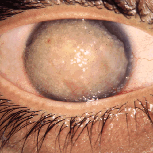
Stye
Wikipedia
Wikimedia Commons has media related to Stye . v t e Diseases of the human eye Adnexa Eyelid Inflammation Stye Chalazion Blepharitis Entropion Ectropion Lagophthalmos Blepharochalasis Ptosis Blepharophimosis Xanthelasma Ankyloblepharon Eyelash Trichiasis Madarosis Lacrimal apparatus Dacryoadenitis Epiphora Dacryocystitis Xerophthalmia Orbit Exophthalmos Enophthalmos Orbital cellulitis Orbital lymphoma Periorbital cellulitis Conjunctiva Conjunctivitis allergic Pterygium Pseudopterygium Pinguecula Subconjunctival hemorrhage Globe Fibrous tunic Sclera Scleritis Episcleritis Cornea Keratitis herpetic acanthamoebic fungal Exposure Photokeratitis Corneal ulcer Thygeson's superficial punctate keratopathy Corneal dystrophy Fuchs' Meesmann Corneal ectasia Keratoconus Pellucid marginal degeneration Keratoglobus Terrien's marginal degeneration Post-LASIK ectasia Keratoconjunctivitis sicca Corneal opacity Corneal neovascularization Kayser–Fleischer ring Haab's striae Arcus senilis Band keratopathy Vascular tunic Iris Ciliary body Uveitis Intermediate uveitis Hyphema Rubeosis iridis Persistent pupillary membrane Iridodialysis Synechia Choroid Choroideremia Choroiditis Chorioretinitis Lens Cataract Congenital cataract Childhood cataract Aphakia Ectopia lentis Retina Retinitis Chorioretinitis Cytomegalovirus retinitis Retinal detachment Retinoschisis Ocular ischemic syndrome / Central retinal vein occlusion Central retinal artery occlusion Branch retinal artery occlusion Retinopathy diabetic hypertensive Purtscher's of prematurity Bietti's crystalline dystrophy Coats' disease Sickle cell Macular degeneration Retinitis pigmentosa Retinal haemorrhage Central serous retinopathy Macular edema Epiretinal membrane (Macular pucker) Vitelliform macular dystrophy Leber's congenital amaurosis Birdshot chorioretinopathy Other Glaucoma / Ocular hypertension / Primary juvenile glaucoma Floater Leber's hereditary optic neuropathy Red eye Globe rupture Keratomycosis Phthisis bulbi Persistent fetal vasculature / Persistent hyperplastic primary vitreous Persistent tunica vasculosa lentis Familial exudative vitreoretinopathy Pathways Optic nerve Optic disc Optic neuritis optic papillitis Papilledema Foster Kennedy syndrome Optic atrophy Optic disc drusen Optic neuropathy Ischemic anterior (AION) posterior (PION) Kjer's Leber's hereditary Toxic and nutritional Strabismus Extraocular muscles Binocular vision Accommodation Paralytic strabismus Ophthalmoparesis Chronic progressive external ophthalmoplegia Kearns–Sayre syndrome palsies Oculomotor (III) Fourth-nerve (IV) Sixth-nerve (VI) Other strabismus Esotropia / Exotropia Hypertropia Heterophoria Esophoria Exophoria Cyclotropia Brown's syndrome Duane syndrome Other binocular Conjugate gaze palsy Convergence insufficiency Internuclear ophthalmoplegia One and a half syndrome Refraction Refractive error Hyperopia Myopia Astigmatism Anisometropia / Aniseikonia Presbyopia Vision disorders Blindness Amblyopia Leber's congenital amaurosis Diplopia Scotoma Color blindness Achromatopsia Dichromacy Monochromacy Nyctalopia Oguchi disease Blindness / Vision loss / Visual impairment Anopsia Hemianopsia binasal bitemporal homonymous Quadrantanopia subjective Asthenopia Hemeralopia Photophobia Scintillating scotoma Pupil Anisocoria Argyll Robertson pupil Marcus Gunn pupil Adie syndrome Miosis Mydriasis Cycloplegia Parinaud's syndrome Other Nystagmus Childhood blindness Infections Trachoma Onchocerciasis

-
Morphea
Wikipedia
Case reports and observational studies suggest there is a higher frequency of family history of autoimmune diseases in patients with morphea. [4] Tests for autoantibodies associated with morphea have shown results in higher frequencies of anti-histone and anti-topoisomerase IIa antibodies. [5] Case reports of morphea co-existing with other systemic autoimmune diseases such as primary biliary cirrhosis, vitiligo, and systemic lupus erythematosus lend support to morphea as an autoimmune disease. [6] [7] [8] Borrelia burgdorferi infection may be relevant for the induction of a distinct autoimmune type of scleroderma; it may be called " Borrelia -associated early onset morphea" and is characterized by the combination of disease onset at younger age, infection with B. burgdorferi , and evident autoimmune phenomena as reflected by high-titer antinuclear antibodies. [9] Diagnosis [ edit ] Classification [ edit ] Morphea–lichen sclerosus et atrophicus overlap is characterized by both lesions of morphea and lichen sclerosus et atrophicus, most commonly seen in women. [2] : 171 Generalized morphea is characterized by widespread indurated plaques and pigmentary changes, sometimes associated with muscle atrophy, but without visceral involvement. [2] : 171 Morphea profunda involves deep subcutaneous tissue , including fascia , and there is a clinical overlap with eosinophilic fasciitis , eosinophilia-myalgia syndrome , and the Spanish toxic oil syndrome . [2] : 171 Morphea profunda shows little response to corticosteroids and tends to run a more chronic debilitating course. [2] : 171 Pansclerotic morphea is manifested by sclerosis of the dermis, panniculus, fascia, muscle, and at times, the bone, all causing disabling limitation of motion of joints. [2] : 171 Linear scleroderma is a type of localised scleroderma [10] which is an autoimmune disease characterized by a line of thickened skin which can affect the bones and muscles underneath it. ... September 2013 External links [ edit ] Classification D ICD - 10 : L94.0 ICD - 9-CM : 701.0 MeSH : D012594 DiseasesDB : 8351 External resources eMedicine : derm/272 Patient UK : Morphea v t e Diseases of the skin and appendages by morphology Growths Epidermal Wart Callus Seborrheic keratosis Acrochordon Molluscum contagiosum Actinic keratosis Squamous-cell carcinoma Basal-cell carcinoma Merkel-cell carcinoma Nevus sebaceous Trichoepithelioma Pigmented Freckles Lentigo Melasma Nevus Melanoma Dermal and subcutaneous Epidermal inclusion cyst Hemangioma Dermatofibroma (benign fibrous histiocytoma) Keloid Lipoma Neurofibroma Xanthoma Kaposi's sarcoma Infantile digital fibromatosis Granular cell tumor Leiomyoma Lymphangioma circumscriptum Myxoid cyst Rashes With epidermal involvement Eczematous Contact dermatitis Atopic dermatitis Seborrheic dermatitis Stasis dermatitis Lichen simplex chronicus Darier's disease Glucagonoma syndrome Langerhans cell histiocytosis Lichen sclerosus Pemphigus foliaceus Wiskott–Aldrich syndrome Zinc deficiency Scaling Psoriasis Tinea ( Corporis Cruris Pedis Manuum Faciei ) Pityriasis rosea Secondary syphilis Mycosis fungoides Systemic lupus erythematosus Pityriasis rubra pilaris Parapsoriasis Ichthyosis Blistering Herpes simplex Herpes zoster Varicella Bullous impetigo Acute contact dermatitis Pemphigus vulgaris Bullous pemphigoid Dermatitis herpetiformis Porphyria cutanea tarda Epidermolysis bullosa simplex Papular Scabies Insect bite reactions Lichen planus Miliaria Keratosis pilaris Lichen spinulosus Transient acantholytic dermatosis Lichen nitidus Pityriasis lichenoides et varioliformis acuta Pustular Acne vulgaris Acne rosacea Folliculitis Impetigo Candidiasis Gonococcemia Dermatophyte Coccidioidomycosis Subcorneal pustular dermatosis Hypopigmented Tinea versicolor Vitiligo Pityriasis alba Postinflammatory hyperpigmentation Tuberous sclerosis Idiopathic guttate hypomelanosis Leprosy Hypopigmented mycosis fungoides Without epidermal involvement Red Blanchable Erythema Generalized Drug eruptions Viral exanthems Toxic erythema Systemic lupus erythematosus Localized Cellulitis Abscess Boil Erythema nodosum Carcinoid syndrome Fixed drug eruption Specialized Urticaria Erythema ( Multiforme Migrans Gyratum repens Annulare centrifugum Ab igne ) Nonblanchable Purpura Macular Thrombocytopenic purpura Actinic/solar purpura Papular Disseminated intravascular coagulation Vasculitis Indurated Scleroderma / morphea Granuloma annulare Lichen sclerosis et atrophicus Necrobiosis lipoidica Miscellaneous disorders Ulcers Hair Telogen effluvium Androgenic alopecia Alopecia areata Systemic lupus erythematosus Tinea capitis Loose anagen syndrome Lichen planopilaris Folliculitis decalvans Acne keloidalis nuchae Nail Onychomycosis Psoriasis Paronychia Ingrown nail Mucous membrane Aphthous stomatitis Oral candidiasis Lichen planus Leukoplakia Pemphigus vulgaris Mucous membrane pemphigoid Cicatricial pemphigoid Herpesvirus Coxsackievirus Syphilis Systemic histoplasmosis Squamous-cell carcinoma v t e Cutaneous keratosis, ulcer, atrophy, and necrobiosis Epidermal thickening keratoderma : Keratoderma climactericum Paraneoplastic keratoderma Acrokeratosis paraneoplastica of Bazex Aquagenic keratoderma Drug-induced keratoderma psoriasis Keratoderma blennorrhagicum keratosis : Seborrheic keratosis Clonal seborrheic keratosis Common seborrheic keratosis Irritated seborrheic keratosis Seborrheic keratosis with squamous atypia Reticulated seborrheic keratosis Dermatosis papulosa nigra Keratosis punctata of the palmar creases other hyperkeratosis : Acanthosis nigricans Confluent and reticulated papillomatosis Callus Ichthyosis acquisita Arsenical keratosis Chronic scar keratosis Hyperkeratosis lenticularis perstans Hydrocarbon keratosis Hyperkeratosis of the nipple and areola Inverted follicular keratosis Lichenoid keratosis Multiple minute digitate hyperkeratosis PUVA keratosis Reactional keratosis Stucco keratosis Thermal keratosis Viral keratosis Warty dyskeratoma Waxy keratosis of childhood other hypertrophy: Keloid Hypertrophic scar Cutis verticis gyrata Necrobiosis / granuloma Necrobiotic/palisading Granuloma annulare Perforating Generalized Subcutaneous Granuloma annulare in HIV disease Localized granuloma annulare Patch-type granuloma annulare Necrobiosis lipoidica Annular elastolytic giant-cell granuloma Granuloma multiforme Necrobiotic xanthogranuloma Palisaded neutrophilic and granulomatous dermatitis Rheumatoid nodulosis Interstitial granulomatous dermatitis / Interstitial granulomatous drug reaction Foreign body granuloma Beryllium granuloma Mercury granuloma Silica granuloma Silicone granuloma Zirconium granuloma Soot tattoo Tattoo Carbon stain Other/ungrouped eosinophilic dermatosis Granuloma faciale Dermis / localized CTD Cutaneous lupus erythematosus chronic: Discoid Panniculitis subacute : Neonatal ungrouped: Chilblain Lupus erythematosus–lichen planus overlap syndrome Tumid Verrucous Rowell's syndrome Scleroderma / Morphea Localized scleroderma Localized morphea Morphea–lichen sclerosus et atrophicus overlap Generalized morphea Atrophoderma of Pasini and Pierini Pansclerotic morphea Morphea profunda Linear scleroderma Atrophic / atrophoderma Lichen sclerosus Anetoderma Schweninger–Buzzi anetoderma Jadassohn–Pellizzari anetoderma Atrophoderma of Pasini and Pierini Acrodermatitis chronica atrophicans Semicircular lipoatrophy Follicular atrophoderma Linear atrophoderma of Moulin Perforating Kyrle disease Reactive perforating collagenosis Elastosis perforans serpiginosa Perforating folliculitis Acquired perforating dermatosis Skin ulcer Pyoderma gangrenosum Other Calcinosis cutis Sclerodactyly Poikiloderma vasculare atrophicans Ainhum / Pseudo-ainhumTNF, LMNA, CSF3, CD34, IL2, FLI1, DCN, IL1A, IFNG, TGFB1, COL1A2, SELE, STAT3, SPARC, TP53, VIM, LTBP4, ABL1, LILRB1, POSTN, CCL18, BTG3, PART1, BNC2, DEFB103B, SKOR1, MIR155, MIR196A1, DEFB103A, CCL19, SMAD7, CCL11, CCL5, BCL2, BMP6, CCR7, COMP, CCN2, GATA3, GRN, IFNA1, IFNA13, IFNB1, IL6, CXCL8, ITGAV, ITGB5, ACTB, CXCL9, MMP1, MIR483

-
Corneal Dystrophy
Wikipedia
PMID 25564336 . v t e Types of corneal dystrophy Epithelial and subepithelial Epithelial basement membrane dystrophy Gelatinous drop-like corneal dystrophy Lisch epithelial corneal dystrophy Meesmann corneal dystrophy Subepithelial mucinous corneal dystrophy Bowman's membrane Reis–Bucklers corneal dystrophy Thiel-Behnke dystrophy Stroma Congenital stromal corneal dystrophy Fleck corneal dystrophy Granular corneal dystrophy Lattice corneal dystrophy Macular corneal dystrophy Posterior amorphous corneal dystrophy Schnyder crystalline corneal dystrophy Descemet's membrane and endothelial Congenital hereditary endothelial dystrophy Fuchs' dystrophy Posterior polymorphous corneal dystrophy X-linked endothelial corneal dystrophy v t e Diseases of the human eye Adnexa Eyelid Inflammation Stye Chalazion Blepharitis Entropion Ectropion Lagophthalmos Blepharochalasis Ptosis Blepharophimosis Xanthelasma Ankyloblepharon Eyelash Trichiasis Madarosis Lacrimal apparatus Dacryoadenitis Epiphora Dacryocystitis Xerophthalmia Orbit Exophthalmos Enophthalmos Orbital cellulitis Orbital lymphoma Periorbital cellulitis Conjunctiva Conjunctivitis allergic Pterygium Pseudopterygium Pinguecula Subconjunctival hemorrhage Globe Fibrous tunic Sclera Scleritis Episcleritis Cornea Keratitis herpetic acanthamoebic fungal Exposure Photokeratitis Corneal ulcer Thygeson's superficial punctate keratopathy Corneal dystrophy Fuchs' Meesmann Corneal ectasia Keratoconus Pellucid marginal degeneration Keratoglobus Terrien's marginal degeneration Post-LASIK ectasia Keratoconjunctivitis sicca Corneal opacity Corneal neovascularization Kayser–Fleischer ring Haab's striae Arcus senilis Band keratopathy Vascular tunic Iris Ciliary body Uveitis Intermediate uveitis Hyphema Rubeosis iridis Persistent pupillary membrane Iridodialysis Synechia Choroid Choroideremia Choroiditis Chorioretinitis Lens Cataract Congenital cataract Childhood cataract Aphakia Ectopia lentis Retina Retinitis Chorioretinitis Cytomegalovirus retinitis Retinal detachment Retinoschisis Ocular ischemic syndrome / Central retinal vein occlusion Central retinal artery occlusion Branch retinal artery occlusion Retinopathy diabetic hypertensive Purtscher's of prematurity Bietti's crystalline dystrophy Coats' disease Sickle cell Macular degeneration Retinitis pigmentosa Retinal haemorrhage Central serous retinopathy Macular edema Epiretinal membrane (Macular pucker) Vitelliform macular dystrophy Leber's congenital amaurosis Birdshot chorioretinopathy Other Glaucoma / Ocular hypertension / Primary juvenile glaucoma Floater Leber's hereditary optic neuropathy Red eye Globe rupture Keratomycosis Phthisis bulbi Persistent fetal vasculature / Persistent hyperplastic primary vitreous Persistent tunica vasculosa lentis Familial exudative vitreoretinopathy Pathways Optic nerve Optic disc Optic neuritis optic papillitis Papilledema Foster Kennedy syndrome Optic atrophy Optic disc drusen Optic neuropathy Ischemic anterior (AION) posterior (PION) Kjer's Leber's hereditary Toxic and nutritional Strabismus Extraocular muscles Binocular vision Accommodation Paralytic strabismus Ophthalmoparesis Chronic progressive external ophthalmoplegia Kearns–Sayre syndrome palsies Oculomotor (III) Fourth-nerve (IV) Sixth-nerve (VI) Other strabismus Esotropia / Exotropia Hypertropia Heterophoria Esophoria Exophoria Cyclotropia Brown's syndrome Duane syndrome Other binocular Conjugate gaze palsy Convergence insufficiency Internuclear ophthalmoplegia One and a half syndrome Refraction Refractive error Hyperopia Myopia Astigmatism Anisometropia / Aniseikonia Presbyopia Vision disorders Blindness Amblyopia Leber's congenital amaurosis Diplopia Scotoma Color blindness Achromatopsia Dichromacy Monochromacy Nyctalopia Oguchi disease Blindness / Vision loss / Visual impairment Anopsia Hemianopsia binasal bitemporal homonymous Quadrantanopia subjective Asthenopia Hemeralopia Photophobia Scintillating scotoma Pupil Anisocoria Argyll Robertson pupil Marcus Gunn pupil Adie syndrome Miosis Mydriasis Cycloplegia Parinaud's syndrome Other Nystagmus Childhood blindness Infections Trachoma OnchocerciasisTGFBI, SLC4A11, COL8A2, ZEB1, CHST6, MBTPS2, UBIAD1, TACSTD2, GLA, KRT12, LRRC6, CCDC40, CFAP298, AGK, DNAAF2, DNAH11, ARMC4, CCNO, PRDM5, DNAAF5, DNAH1, HYDIN, ZMYND10, DNAI2, NME8, EXOSC2, STK36, DNAI1, DNAAF3, GNPTAB, RSPH4A, GRHL2, MCIDAS, CCDC39, DNAJB13, GAS2L2, RSPH9, DNAAF4, PIH1D3, DNAAF1, AGBL1, CCDC151, LRRC56, CCDC114, DRC1, RSPH1, CCDC65, CFAP300, ZNF469, CCDC103, RSPH3, DNAL1, OFD1, TTC25, SLC25A4, KRT6B, OPN1LW, GNAQ, GJA8, KRT3, KRT6A, OPN1MW, KRT16, KRT17, GAS8, MAF, DNAH5, PLOD1, CRYGD, CRYGC, CRYBB2, COL5A2, COL5A1, CRYBB1, CRYBA4, CRYAA, SPAG1, COL1A1, RPGR, TGFB1, TCF4, OVOL2, VSX1, FAS, COL17A1, CDB2, GSN, GCDH, COL8A1, IL2, STS, TGFB2, IL4, SELPLG, WNT7A, ZNF143, MATN4, TBCD, POSTN, SPARC, SOX2, SHH, SELP, CXCL8, PTPRG, HTRA1, CTSA, PAX6, LOX, VPS35, ACD, IL13, GUCY2EP
-
Cerebral Creatine Deficiency Syndrome 1
Omim
A number sign (#) is used with this entry because cerebral creatine deficiency syndrome-1 (CCDS1) is caused by mutation in the creatine transporter gene (SLC6A8; 300036) on chromosome Xq28. Description Cerebral creatine deficiency syndrome-1 is an X-linked disorder of creatine (Cr) transport characterized by mental retardation, severe speech delay, behavioral abnormalities, and seizures. ... Genetic Heterogeneity of Cerebral Creatine Deficiency Syndrome See also CCDS2 (612736), caused by mutation in the GAMT gene (601240) on chromosome 19p13, and CCDS3 (612718), caused by mutation in the AGAT gene (GATM; 602360) on chromosome 15q21. ... Battini et al. (2011) reported a 6.5-year-old boy with X-linked creatine deficiency syndrome confirmed by genetic analysis. ... Together with a fifth case of creatine deficiency due to mutation in the GAMT gene (612736), Lion-Francois et al. (2006) found that the prevalence of cerebral creatine deficiency syndromes was 2.7% in their study population of 188 mentally retarded children.
-
Erotomania
Wikipedia
Primary erotomania is also commonly referred to as de Clerambault's syndrome and Old Maid's Insanity [4] and it exists alone without comorbidities , has a sudden onset and a chronic outcome. [3] The secondary form is found along with mental disorders like paranoid schizophrenia, often includes persecutory delusions , hallucinations , and grandiose ideas, and has a more gradual onset. [3] Patients with a "fixed" condition are more seriously ill with constant delusions and are less responsive to treatment. ... To date, the mainline pharmacological treatments have been pimozide (a typical antipsychotic which was also approved for treating Tourette's Syndrome), [3] [4] and atypical antipsychotics like risperidone and clozapine. [3] [4] Non-pharmacologic treatments that have shown some degree of efficacy are electroconvulsive therapy (ECT), supportive psychotherapy, family and environment therapy, [3] rehousing, risk management and treating underlying disorders in cases of secondary erotomania. [4] ECT may provide temporary remission of delusional beliefs; antipsychotics help attenuate delusions and reduce agitation or associated dangerous behaviors, and SSRIs may be used to treat secondary depression. [3] In delusional disorder there is some evidence that pimozide has superior efficacy compared with other antipsychotics. ... Parisian physician, Bartholomy Pardoux (1545-1611) covered the topics of nymphomania and erotomania. [3] In 1623, erotomania was referred to in a treatise by Jacques Ferrand [3] (Maladie d'amour ou Mélancolie érotique) and has been called "erotic paranoia" and "erotic self-referent delusion" until the common usage of the terms erotomania and de Clérambault's syndrome. In 1971 and 1977, M.V. Seeman referred to the disorder as "phantom lover syndrome" and "psychotic erotic transference reaction and delusional loving". [3] Emil Kraepelin and Bernard also wrote of erotomania and more recently, Winokur, Kendler, and Munro have contributed to knowledge on the disorder. [4] G. ... "Erotomania-A review of De Clerambault's Syndrome". The Journal of the European Psychiatric Association . 33 : 664. ^ a b c Segal, J.H. (1989). ... "Erotomania-A review of De Clerambault's Syndrome". The Journal of the European Psychiatric Association . 33 : S664.

-
Escherichia Coli O104:h4
Wikipedia
Another effect from this bacterial infection is hemolytic uremic syndrome (HUS), which is a condition characterized by destruction of red blood cells, that over a long period of time can cause kidney failure. [13] Some common symptoms of HUS are vomiting, bloody diarrhea, and blood in the urine. [12] Infection [ edit ] A common mode of E. coli O104:H4 infection involves ingestion of fecally contaminated food; the disease can thus be considered a foodborne illness . ... Retrieved 2011-06-15 . ^ "The EU integrated approach to food safety" . ^ "Case Definition for diarrhoea and haemolytic uremic syndrome caused by O104:H4" (PDF) . European Commission . 2011-06-03 . ... (June 30, 2006). "A case of haemolytic uremic syndrome caused by Escherichia coli O104:H4" . ... Web. 08 Nov. 2011. v t e Escherichia coli Outbreaks 1993 Jack in the Box 1996 Odwalla 2000 Walkerton 2005 South Wales (O157) 2006 North American (spinach; O157:H7) 2006 North American (multiple; O157:H7) 2009 United Kingdom 2011 Germany (O104:H4) 2015 United States Genes CPS operon DnaG Fis FNR regulon OmpT RecBCD RpoE RpoF RpoN RpoS Strains Enterohemorrhagic Enteroinvasive Enterotoxigenic O104:H21 O104:H4 O121 O157:H7 Verotoxin-producing Related Aerobactin Coliform index Long-term evolution experiment EcoCyc Enteroaggregative Molecular biology Hok/sok system LacUV5 Min System Pathogenic EnvZ/OmpR Rho factor T4 rII system Theodor Escherich v t e Proteobacteria -associated Gram-negative bacterial infections α Rickettsiales Rickettsiaceae / ( Rickettsioses ) Typhus Rickettsia typhi Murine typhus Rickettsia prowazekii Epidemic typhus , Brill–Zinsser disease , Flying squirrel typhus Spotted fever Tick-borne Rickettsia rickettsii Rocky Mountain spotted fever Rickettsia conorii Boutonneuse fever Rickettsia japonica Japanese spotted fever Rickettsia sibirica North Asian tick typhus Rickettsia australis Queensland tick typhus Rickettsia honei Flinders Island spotted fever Rickettsia africae African tick bite fever Rickettsia parkeri American tick bite fever Rickettsia aeschlimannii Rickettsia aeschlimannii infection Mite-borne Rickettsia akari Rickettsialpox Orientia tsutsugamushi Scrub typhus Flea-borne Rickettsia felis Flea-borne spotted fever Anaplasmataceae Ehrlichiosis : Anaplasma phagocytophilum Human granulocytic anaplasmosis , Anaplasmosis Ehrlichia chaffeensis Human monocytotropic ehrlichiosis Ehrlichia ewingii Ehrlichiosis ewingii infection Rhizobiales Brucellaceae Brucella abortus Brucellosis Bartonellaceae Bartonellosis : Bartonella henselae Cat-scratch disease Bartonella quintana Trench fever Either B. henselae or B. quintana Bacillary angiomatosis Bartonella bacilliformis Carrion's disease , Verruga peruana β Neisseriales M+ Neisseria meningitidis/meningococcus Meningococcal disease , Waterhouse–Friderichsen syndrome , Meningococcal septicaemia M− Neisseria gonorrhoeae/gonococcus Gonorrhea ungrouped: Eikenella corrodens / Kingella kingae HACEK Chromobacterium violaceum Chromobacteriosis infection Burkholderiales Burkholderia pseudomallei Melioidosis Burkholderia mallei Glanders Burkholderia cepacia complex Bordetella pertussis / Bordetella parapertussis Pertussis γ Enterobacteriales ( OX− ) Lac+ Klebsiella pneumoniae Rhinoscleroma , Pneumonia Klebsiella granulomatis Granuloma inguinale Klebsiella oxytoca Escherichia coli : Enterotoxigenic Enteroinvasive Enterohemorrhagic O157:H7 O104:H4 Hemolytic-uremic syndrome Enterobacter aerogenes / Enterobacter cloacae Slow/weak Serratia marcescens Serratia infection Citrobacter koseri / Citrobacter freundii Lac− H2S+ Salmonella enterica Typhoid fever , Paratyphoid fever , Salmonellosis H2S− Shigella dysenteriae / sonnei / flexneri / boydii Shigellosis , Bacillary dysentery Proteus mirabilis / Proteus vulgaris Yersinia pestis Plague / Bubonic plague Yersinia enterocolitica Yersiniosis Yersinia pseudotuberculosis Far East scarlet-like fever Pasteurellales Haemophilus : H. influenzae Haemophilus meningitis Brazilian purpuric fever H. ducreyi Chancroid H. parainfluenzae HACEK Pasteurella multocida Pasteurellosis Actinobacillus Actinobacillosis Aggregatibacter actinomycetemcomitans HACEK Legionellales Legionella pneumophila / Legionella longbeachae Legionnaires' disease Coxiella burnetii Q fever Thiotrichales Francisella tularensis Tularemia Vibrionaceae Vibrio cholerae Cholera Vibrio vulnificus Vibrio parahaemolyticus Vibrio alginolyticus Plesiomonas shigelloides Pseudomonadales Pseudomonas aeruginosa Pseudomonas infection Moraxella catarrhalis Acinetobacter baumannii Xanthomonadaceae Stenotrophomonas maltophilia Cardiobacteriaceae Cardiobacterium hominis HACEK Aeromonadales Aeromonas hydrophila / Aeromonas veronii Aeromonas infection ε Campylobacterales Campylobacter jejuni Campylobacteriosis , Guillain–Barré syndrome Helicobacter pylori Peptic ulcer , MALT lymphoma , Gastric cancer Helicobacter cinaedi Helicobacter cellulitis v t e Consumer food safety Adulterants , food contaminants 3-MCPD Aldicarb Antibiotic use in livestock Cyanide Formaldehyde HGH controversies Lead poisoning Melamine Mercury in fish Sudan I Flavorings Monosodium glutamate (MSG) Salt Sugar High-fructose corn syrup Intestinal parasites and parasitic disease Amoebiasis Anisakiasis Cryptosporidiosis Cyclosporiasis Diphyllobothriasis Enterobiasis Fasciolopsiasis Fasciolosis Giardiasis Gnathostomiasis Paragonimiasis Toxoplasmosis Trichinosis Trichuriasis Microorganisms Botulism Campylobacter jejuni Clostridium perfringens Cronobacter Enterovirus Escherichia coli O104:H4 Escherichia coli O157:H7 Hepatitis A Hepatitis E Listeria Norovirus Rotavirus Salmonella Vibrio cholerae Pesticides Chlorpyrifos DDT Lindane Malathion Methamidophos Preservatives Benzoic acid Ethylenediaminetetraacetic acid (EDTA) Sodium benzoate Sugar substitutes Acesulfame potassium Aspartame Saccharin Sodium cyclamate Sorbitol Sucralose Toxins , poisons , environment pollution Aflatoxin Arsenic contamination of groundwater Benzene in soft drinks Bisphenol A Dieldrin Diethylstilbestrol Dioxin Mycotoxins Nonylphenol Shellfish poisoning Food contamination incidents Devon colic Swill milk scandal Esing Bakery incident 1858 Bradford sweets poisoning 1900 English beer poisoning Morinaga Milk arsenic poisoning incident Minamata disease 1971 Iraq poison grain disaster Toxic oil syndrome 1985 diethylene glycol wine scandal UK mad cow disease outbreak 1993 Jack in the Box E. coli outbreak 1996 Odwalla E. coli outbreak 2006 North American E. coli outbreaks ICA meat repackaging controversy 2008 Canada listeriosis outbreak 2008 Chinese milk scandal 2008 Irish pork crisis 2008 United States salmonellosis outbreak 2011 Germany E. coli outbreak 2011 United States listeriosis outbreak 2013 Bihar school meal poisoning 2013 horse meat scandal 2015 Mozambique beer poisoning 2017 Brazil weak meat scandal 2017–18 South African listeriosis outbreak 2018 Australian rockmelon listeriosis outbreak 2018 Australian strawberry contamination Food safety incidents in China Food safety incidents in Taiwan Food safety in Australia Foodborne illness outbreaks death toll United States Regulation , standards , watchdogs Acceptable daily intake E number Food labeling regulations Food libel laws International Food Safety Network ISO 22000 Nutrition facts label Organic certification The Non-GMO Project Quality Assurance International Food Standards Agency Institutions Institute for Food Safety and Health European Food Safety Authority International Food Safety Network Spanish Agency for Food Safety and Nutrition Food Information and Control Agency (Spain) Centre for Food Safety (Hong Kong) Ministry of Food and Drug Safety (South Korea)
-
Noma (Disease)
Wikipedia
External links [ edit ] Classification D ICD - 9-CM : 528.1 MeSH : D009625 External resources MedlinePlus : 001342 v t e Bacterial diseases due to gram negative non- proteobacteria ( BV4 ) Spirochaete Spirochaetaceae Treponema Treponema pallidum Syphilis / bejel Yaws Treponema carateum ( Pinta ) Treponema denticola Borrelia Borrelia burgdorferi / Borrelia afzelii Lyme disease Erythema migrans Neuroborreliosis Borrelia recurrentis ( Louse borne relapsing fever ) Borrelia hermsii / Borrelia duttoni / Borrelia parkeri ( Tick borne relapsing fever ) Leptospiraceae Leptospira Leptospira interrogans ( Leptospirosis ) Chlamydiaceae Chlamydia Chlamydia psittaci ( Psittacosis ) Chlamydia pneumoniae Chlamydia trachomatis Chlamydia Lymphogranuloma venereum Trachoma Bacteroidetes Bacteroides fragilis Tannerella forsythia Capnocytophaga canimorsus Porphyromonas gingivalis Prevotella intermedia Fusobacteria Fusobacterium necrophorum ( Lemierre's syndrome ) Fusobacterium nucleatum Fusobacterium polymorphum Streptobacillus moniliformis ( Rat-bite fever / Haverhill fever ) v t e Oral and maxillofacial pathology Lips Cheilitis Actinic Angular Plasma cell Cleft lip Congenital lip pit Eclabium Herpes labialis Macrocheilia Microcheilia Nasolabial cyst Sun poisoning Trumpeter's wart Tongue Ankyloglossia Black hairy tongue Caviar tongue Crenated tongue Cunnilingus tongue Fissured tongue Foliate papillitis Glossitis Geographic tongue Median rhomboid glossitis Transient lingual papillitis Glossoptosis Hypoglossia Lingual thyroid Macroglossia Microglossia Rhabdomyoma Palate Bednar's aphthae Cleft palate High-arched palate Palatal cysts of the newborn Inflammatory papillary hyperplasia Stomatitis nicotina Torus palatinus Oral mucosa – Lining of mouth Amalgam tattoo Angina bullosa haemorrhagica Behçet's disease Bohn's nodules Burning mouth syndrome Candidiasis Condyloma acuminatum Darier's disease Epulis fissuratum Erythema multiforme Erythroplakia Fibroma Giant-cell Focal epithelial hyperplasia Fordyce spots Hairy leukoplakia Hand, foot and mouth disease Hereditary benign intraepithelial dyskeratosis Herpangina Herpes zoster Intraoral dental sinus Leukoedema Leukoplakia Lichen planus Linea alba Lupus erythematosus Melanocytic nevus Melanocytic oral lesion Molluscum contagiosum Morsicatio buccarum Oral cancer Benign: Squamous cell papilloma Keratoacanthoma Malignant: Adenosquamous carcinoma Basaloid squamous carcinoma Mucosal melanoma Spindle cell carcinoma Squamous cell carcinoma Verrucous carcinoma Oral florid papillomatosis Oral melanosis Smoker's melanosis Pemphigoid Benign mucous membrane Pemphigus Plasmoacanthoma Stomatitis Aphthous Denture-related Herpetic Smokeless tobacco keratosis Submucous fibrosis Ulceration Riga–Fede disease Verruca vulgaris Verruciform xanthoma White sponge nevus Teeth ( pulp , dentin , enamel ) Amelogenesis imperfecta Ankylosis Anodontia Caries Early childhood caries Concrescence Failure of eruption of teeth Dens evaginatus Talon cusp Dentin dysplasia Dentin hypersensitivity Dentinogenesis imperfecta Dilaceration Discoloration Ectopic enamel Enamel hypocalcification Enamel hypoplasia Turner's hypoplasia Enamel pearl Fluorosis Fusion Gemination Hyperdontia Hypodontia Maxillary lateral incisor agenesis Impaction Wisdom tooth impaction Macrodontia Meth mouth Microdontia Odontogenic tumors Keratocystic odontogenic tumour Odontoma Dens in dente Open contact Premature eruption Neonatal teeth Pulp calcification Pulp stone Pulp canal obliteration Pulp necrosis Pulp polyp Pulpitis Regional odontodysplasia Resorption Shovel-shaped incisors Supernumerary root Taurodontism Trauma Avulsion Cracked tooth syndrome Vertical root fracture Occlusal Tooth loss Edentulism Tooth wear Abrasion Abfraction Acid erosion Attrition Periodontium ( gingiva , periodontal ligament , cementum , alveolus ) – Gums and tooth-supporting structures Cementicle Cementoblastoma Gigantiform Cementoma Eruption cyst Epulis Pyogenic granuloma Congenital epulis Gingival enlargement Gingival cyst of the adult Gingival cyst of the newborn Gingivitis Desquamative Granulomatous Plasma cell Hereditary gingival fibromatosis Hypercementosis Hypocementosis Linear gingival erythema Necrotizing periodontal diseases Acute necrotizing ulcerative gingivitis Pericoronitis Peri-implantitis Periodontal abscess Periodontal trauma Periodontitis Aggressive As a manifestation of systemic disease Chronic Perio-endo lesion Teething Periapical, mandibular and maxillary hard tissues – Bones of jaws Agnathia Alveolar osteitis Buccal exostosis Cherubism Idiopathic osteosclerosis Mandibular fracture Microgenia Micrognathia Intraosseous cysts Odontogenic : periapical Dentigerous Buccal bifurcation Lateral periodontal Globulomaxillary Calcifying odontogenic Glandular odontogenic Non-odontogenic: Nasopalatine duct Median mandibular Median palatal Traumatic bone Osteoma Osteomyelitis Osteonecrosis Bisphosphonate-associated Neuralgia-inducing cavitational osteonecrosis Osteoradionecrosis Osteoporotic bone marrow defect Paget's disease of bone Periapical abscess Phoenix abscess Periapical periodontitis Stafne defect Torus mandibularis Temporomandibular joints , muscles of mastication and malocclusions – Jaw joints, chewing muscles and bite abnormalities Bruxism Condylar resorption Mandibular dislocation Malocclusion Crossbite Open bite Overbite Overeruption Overjet Prognathia Retrognathia Scissor bite Maxillary hypoplasia Temporomandibular joint dysfunction Salivary glands Benign lymphoepithelial lesion Ectopic salivary gland tissue Frey's syndrome HIV salivary gland disease Necrotizing sialometaplasia Mucocele Ranula Pneumoparotitis Salivary duct stricture Salivary gland aplasia Salivary gland atresia Salivary gland diverticulum Salivary gland fistula Salivary gland hyperplasia Salivary gland hypoplasia Salivary gland neoplasms Benign: Basal cell adenoma Canalicular adenoma Ductal papilloma Monomorphic adenoma Myoepithelioma Oncocytoma Papillary cystadenoma lymphomatosum Pleomorphic adenoma Sebaceous adenoma Malignant: Acinic cell carcinoma Adenocarcinoma Adenoid cystic carcinoma Carcinoma ex pleomorphic adenoma Lymphoma Mucoepidermoid carcinoma Sclerosing polycystic adenosis Sialadenitis Parotitis Chronic sclerosing sialadenitis Sialectasis Sialocele Sialodochitis Sialosis Sialolithiasis Sjögren's syndrome Orofacial soft tissues – Soft tissues around the mouth Actinomycosis Angioedema Basal cell carcinoma Cutaneous sinus of dental origin Cystic hygroma Gnathophyma Ludwig's angina Macrostomia Melkersson–Rosenthal syndrome Microstomia Noma Oral Crohn's disease Orofacial granulomatosis Perioral dermatitis Pyostomatitis vegetans Other Eagle syndrome Hemifacial hypertrophy Facial hemiatrophy Oral manifestations of systemic disease

-
Hairy Leukoplakia
Wikipedia
It is caused by Epstein-Barr virus (EBV) and occurs usually in persons who are immunocompromised , especially those with human immunodeficiency virus infection/ acquired immunodeficiency syndrome (HIV/AIDS). The white lesion , which cannot be scraped off, is benign and does not require any treatment, although its appearance may have diagnostic and prognostic implications for the underlying condition. ... External links [ edit ] Classification D ICD - 10 : K13.3 ICD - 9-CM : 528.6 MeSH : D017733 DiseasesDB : 5594 External resources eMedicine : med/938 v t e Oral and maxillofacial pathology Lips Cheilitis Actinic Angular Plasma cell Cleft lip Congenital lip pit Eclabium Herpes labialis Macrocheilia Microcheilia Nasolabial cyst Sun poisoning Trumpeter's wart Tongue Ankyloglossia Black hairy tongue Caviar tongue Crenated tongue Cunnilingus tongue Fissured tongue Foliate papillitis Glossitis Geographic tongue Median rhomboid glossitis Transient lingual papillitis Glossoptosis Hypoglossia Lingual thyroid Macroglossia Microglossia Rhabdomyoma Palate Bednar's aphthae Cleft palate High-arched palate Palatal cysts of the newborn Inflammatory papillary hyperplasia Stomatitis nicotina Torus palatinus Oral mucosa – Lining of mouth Amalgam tattoo Angina bullosa haemorrhagica Behçet's disease Bohn's nodules Burning mouth syndrome Candidiasis Condyloma acuminatum Darier's disease Epulis fissuratum Erythema multiforme Erythroplakia Fibroma Giant-cell Focal epithelial hyperplasia Fordyce spots Hairy leukoplakia Hand, foot and mouth disease Hereditary benign intraepithelial dyskeratosis Herpangina Herpes zoster Intraoral dental sinus Leukoedema Leukoplakia Lichen planus Linea alba Lupus erythematosus Melanocytic nevus Melanocytic oral lesion Molluscum contagiosum Morsicatio buccarum Oral cancer Benign: Squamous cell papilloma Keratoacanthoma Malignant: Adenosquamous carcinoma Basaloid squamous carcinoma Mucosal melanoma Spindle cell carcinoma Squamous cell carcinoma Verrucous carcinoma Oral florid papillomatosis Oral melanosis Smoker's melanosis Pemphigoid Benign mucous membrane Pemphigus Plasmoacanthoma Stomatitis Aphthous Denture-related Herpetic Smokeless tobacco keratosis Submucous fibrosis Ulceration Riga–Fede disease Verruca vulgaris Verruciform xanthoma White sponge nevus Teeth ( pulp , dentin , enamel ) Amelogenesis imperfecta Ankylosis Anodontia Caries Early childhood caries Concrescence Failure of eruption of teeth Dens evaginatus Talon cusp Dentin dysplasia Dentin hypersensitivity Dentinogenesis imperfecta Dilaceration Discoloration Ectopic enamel Enamel hypocalcification Enamel hypoplasia Turner's hypoplasia Enamel pearl Fluorosis Fusion Gemination Hyperdontia Hypodontia Maxillary lateral incisor agenesis Impaction Wisdom tooth impaction Macrodontia Meth mouth Microdontia Odontogenic tumors Keratocystic odontogenic tumour Odontoma Dens in dente Open contact Premature eruption Neonatal teeth Pulp calcification Pulp stone Pulp canal obliteration Pulp necrosis Pulp polyp Pulpitis Regional odontodysplasia Resorption Shovel-shaped incisors Supernumerary root Taurodontism Trauma Avulsion Cracked tooth syndrome Vertical root fracture Occlusal Tooth loss Edentulism Tooth wear Abrasion Abfraction Acid erosion Attrition Periodontium ( gingiva , periodontal ligament , cementum , alveolus ) – Gums and tooth-supporting structures Cementicle Cementoblastoma Gigantiform Cementoma Eruption cyst Epulis Pyogenic granuloma Congenital epulis Gingival enlargement Gingival cyst of the adult Gingival cyst of the newborn Gingivitis Desquamative Granulomatous Plasma cell Hereditary gingival fibromatosis Hypercementosis Hypocementosis Linear gingival erythema Necrotizing periodontal diseases Acute necrotizing ulcerative gingivitis Pericoronitis Peri-implantitis Periodontal abscess Periodontal trauma Periodontitis Aggressive As a manifestation of systemic disease Chronic Perio-endo lesion Teething Periapical, mandibular and maxillary hard tissues – Bones of jaws Agnathia Alveolar osteitis Buccal exostosis Cherubism Idiopathic osteosclerosis Mandibular fracture Microgenia Micrognathia Intraosseous cysts Odontogenic : periapical Dentigerous Buccal bifurcation Lateral periodontal Globulomaxillary Calcifying odontogenic Glandular odontogenic Non-odontogenic: Nasopalatine duct Median mandibular Median palatal Traumatic bone Osteoma Osteomyelitis Osteonecrosis Bisphosphonate-associated Neuralgia-inducing cavitational osteonecrosis Osteoradionecrosis Osteoporotic bone marrow defect Paget's disease of bone Periapical abscess Phoenix abscess Periapical periodontitis Stafne defect Torus mandibularis Temporomandibular joints , muscles of mastication and malocclusions – Jaw joints, chewing muscles and bite abnormalities Bruxism Condylar resorption Mandibular dislocation Malocclusion Crossbite Open bite Overbite Overeruption Overjet Prognathia Retrognathia Scissor bite Maxillary hypoplasia Temporomandibular joint dysfunction Salivary glands Benign lymphoepithelial lesion Ectopic salivary gland tissue Frey's syndrome HIV salivary gland disease Necrotizing sialometaplasia Mucocele Ranula Pneumoparotitis Salivary duct stricture Salivary gland aplasia Salivary gland atresia Salivary gland diverticulum Salivary gland fistula Salivary gland hyperplasia Salivary gland hypoplasia Salivary gland neoplasms Benign: Basal cell adenoma Canalicular adenoma Ductal papilloma Monomorphic adenoma Myoepithelioma Oncocytoma Papillary cystadenoma lymphomatosum Pleomorphic adenoma Sebaceous adenoma Malignant: Acinic cell carcinoma Adenocarcinoma Adenoid cystic carcinoma Carcinoma ex pleomorphic adenoma Lymphoma Mucoepidermoid carcinoma Sclerosing polycystic adenosis Sialadenitis Parotitis Chronic sclerosing sialadenitis Sialectasis Sialocele Sialodochitis Sialosis Sialolithiasis Sjögren's syndrome Orofacial soft tissues – Soft tissues around the mouth Actinomycosis Angioedema Basal cell carcinoma Cutaneous sinus of dental origin Cystic hygroma Gnathophyma Ludwig's angina Macrostomia Melkersson–Rosenthal syndrome Microstomia Noma Oral Crohn's disease Orofacial granulomatosis Perioral dermatitis Pyostomatitis vegetans Other Eagle syndrome Hemifacial hypertrophy Facial hemiatrophy Oral manifestations of systemic disease
-
Coronavirus Diseases
Wikipedia
Human coronavirus 229E ( HCoV-229E ) 1930s ( isolated in 1965) [16] Likely originated from bats . [17] Murine encephalitis JHM (named after John Howard Mueller ), a murine coronavirus [18] 1949 [19] Acute infectious diarrhea Porcine epidemic diarrhea virus (PEDV) 1971 [20] Caused outbreaks in 1972 [21] and 1978, [22] 2010, 2013, 2014, and 2015. [23] Infects pigs and sows. Severe acute respiratory syndrome (SARS) Severe acute respiratory syndrome coronavirus (SARS-CoV), a strain of SARSr-CoV 2002 Caused the 2002–2004 SARS outbreak . Likely originated from horseshoe bats . [24] Common cold Human coronavirus HKU1 ( HCoV-HKU1 ) 2004 Originated from Hong Kong . [25] Respiratory infection Human coronavirus NL63 ( HCoV-NL63 ) 2004 Originated from Amsterdam , Netherlands . [26] Likely originated from tricolored bats . [27] Middle East respiratory syndrome (MERS) Middle East respiratory syndrome–related coronavirus ( MERS-CoV ) 2012 Has caused outbreaks in 2012 , 2015 , and 2018 . Likely originated in the Middle East , particularly Jeddah . [28] Porcine diarrhea HKU15 2014 Discovered in Hong Kong. [29] Coronavirus disease 2019 (COVID-19) severe acute respiratory syndrome coronavirus 2 (SARS-CoV-2), a strain of SARSr-CoV 2019 Cause of ongoing COVID-19 pandemic . ... N. (19 March 1977). "An apparently new syndrome of porcine epidemic diarrhoea" . ... "Possible Bat Origin of Severe Acute Respiratory Syndrome Coronavirus 2" . Emerging Infectious Diseases . 26 (7): 1542–1547. doi : 10.3201/eid2607.200092 .CXCL1, CXCL2, SFTPD, SARS1, SARS2, TMPRSS2, MBL2, TBPL1, TYRP1, VTN, XPC, CDSN, MASP2, MBL3P, AICDA, ACE2, IFNL3, SEC23IP, MAPK3, CCL2, CISH, MX1, MCL1, IL18, IFNG, IFN1@, HLA-B, ERN1, EGFR, DPP4, ACE, CRP, MIR30A

-
Dopamine Beta Hydroxylase Deficiency
Wikipedia
A subset of DβH deficiency patients present with hypermobility . [1] Postural orthostatic tachycardia syndrome , another form of dysautonomia, also sees this comorbidity with hypermobility in the form of a rare connective tissue disorder called Ehlers Danlos syndrome . ... External links [ edit ] Classification D OMIM : 223360 MeSH : C535600 C535600, C535600 DiseasesDB : 33227 SNOMED CT : 237923004 External resources Orphanet : 230 v t e Inborn error of amino acid metabolism K → acetyl-CoA Lysine /straight chain Glutaric acidemia type 1 type 2 Hyperlysinemia Pipecolic acidemia Saccharopinuria Leucine 3-hydroxy-3-methylglutaryl-CoA lyase deficiency 3-Methylcrotonyl-CoA carboxylase deficiency 3-Methylglutaconic aciduria 1 Isovaleric acidemia Maple syrup urine disease Tryptophan Hypertryptophanemia G G→ pyruvate → citrate Glycine D-Glyceric acidemia Glutathione synthetase deficiency Sarcosinemia Glycine → Creatine : GAMT deficiency Glycine encephalopathy G→ glutamate → α-ketoglutarate Histidine Carnosinemia Histidinemia Urocanic aciduria Proline Hyperprolinemia Prolidase deficiency Glutamate / glutamine SSADHD G→ propionyl-CoA → succinyl-CoA Valine Hypervalinemia Isobutyryl-CoA dehydrogenase deficiency Maple syrup urine disease Isoleucine 2-Methylbutyryl-CoA dehydrogenase deficiency Beta-ketothiolase deficiency Maple syrup urine disease Methionine Cystathioninuria Homocystinuria Hypermethioninemia General BC / OA Methylmalonic acidemia Methylmalonyl-CoA mutase deficiency Propionic acidemia G→ fumarate Phenylalanine / tyrosine Phenylketonuria 6-Pyruvoyltetrahydropterin synthase deficiency Tetrahydrobiopterin deficiency Tyrosinemia Alkaptonuria / Ochronosis Tyrosinemia type I Tyrosinemia type II Tyrosinemia type III / Hawkinsinuria Tyrosine → Melanin Albinism : Ocular albinism ( 1 ) Oculocutaneous albinism ( Hermansky–Pudlak syndrome ) Waardenburg syndrome Tyrosine → Norepinephrine Dopamine beta hydroxylase deficiency reverse: Brunner syndrome G→ oxaloacetate Urea cycle / Hyperammonemia ( arginine aspartate ) Argininemia Argininosuccinic aciduria Carbamoyl phosphate synthetase I deficiency Citrullinemia N-Acetylglutamate synthase deficiency Ornithine transcarbamylase deficiency / translocase deficiency Transport / IE of RTT Solute carrier family : Cystinuria Hartnup disease Iminoglycinuria Lysinuric protein intolerance Fanconi syndrome : Oculocerebrorenal syndrome Cystinosis Other 2-Hydroxyglutaric aciduria Aminoacylase 1 deficiency Ethylmalonic encephalopathy Fumarase deficiency Trimethylaminuria

-
Focal Seizure
Wikipedia
Epilepsy syndrome characterised by seizures preceded by isolated disturbances of a cerebral function This article needs additional citations for verification . ... External links [ edit ] Classification D ICD - 10 : G40.0 - G40.2 ICD - 9-CM : 345.4 - 345.5 MeSH : D004828 External resources MedlinePlus : 000697 v t e Diseases of the nervous system , primarily CNS Inflammation Brain Encephalitis Viral encephalitis Herpesviral encephalitis Limbic encephalitis Encephalitis lethargica Cavernous sinus thrombosis Brain abscess Amoebic Brain and spinal cord Encephalomyelitis Acute disseminated Meningitis Meningoencephalitis Brain / encephalopathy Degenerative Extrapyramidal and movement disorders Basal ganglia disease Parkinsonism PD Postencephalitic NMS PKAN Tauopathy PSP Striatonigral degeneration Hemiballismus HD OA Dyskinesia Dystonia Status dystonicus Spasmodic torticollis Meige's Blepharospasm Athetosis Chorea Choreoathetosis Myoclonus Myoclonic epilepsy Akathisia Tremor Essential tremor Intention tremor Restless legs Stiff-person Dementia Tauopathy Alzheimer's Early-onset Primary progressive aphasia Frontotemporal dementia / Frontotemporal lobar degeneration Pick's Dementia with Lewy bodies Posterior cortical atrophy Vascular dementia Mitochondrial disease Leigh syndrome Demyelinating Autoimmune Inflammatory Multiple sclerosis For more detailed coverage, see Template:Demyelinating diseases of CNS Episodic/ paroxysmal Seizures and epilepsy Focal Generalised Status epilepticus For more detailed coverage, see Template:Epilepsy Headache Migraine Cluster Tension For more detailed coverage, see Template:Headache Cerebrovascular TIA Stroke For more detailed coverage, see Template:Cerebrovascular diseases Other Sleep disorders For more detailed coverage, see Template:Sleep CSF Intracranial hypertension Hydrocephalus Normal pressure hydrocephalus Choroid plexus papilloma Idiopathic intracranial hypertension Cerebral edema Intracranial hypotension Other Brain herniation Reye syndrome Hepatic encephalopathy Toxic encephalopathy Hashimoto's encephalopathy Both/either Degenerative SA Friedreich's ataxia Ataxia–telangiectasia MND UMN only: Primary lateral sclerosis Pseudobulbar palsy Hereditary spastic paraplegia LMN only: Distal hereditary motor neuronopathies Spinal muscular atrophies SMA SMAX1 SMAX2 DSMA1 Congenital DSMA Spinal muscular atrophy with lower extremity predominance (SMALED) SMALED1 SMALED2A SMALED2B SMA-PCH SMA-PME Progressive muscular atrophy Progressive bulbar palsy Fazio–Londe Infantile progressive bulbar palsy both: Amyotrophic lateral sclerosis v t e Seizures and epilepsy Basics Seizure types Aura (warning sign) Postictal state Epileptogenesis Neonatal seizure Epilepsy in children Management Anticonvulsants Investigations Electroencephalography Epileptologist Personal issues Epilepsy and driving Epilepsy and employment Seizure types Focal Seizures Simple partial Complex partial Gelastic seizure Epilepsy Temporal lobe epilepsy Frontal lobe epilepsy Rolandic epilepsy Nocturnal epilepsy Panayiotopoulos syndrome Vertiginous epilepsy Generalised Tonic–clonic Absence seizure Atonic seizure Automatism Benign familial neonatal seizures Lennox–Gastaut syndrome Myoclonic astatic epilepsy Epileptic spasms Status epilepticus Epilepsia partialis continua Complex partial status epilepticus Myoclonic epilepsy Progressive myoclonus epilepsy Dentatorubral–pallidoluysian atrophy Unverricht–Lundborg disease MERRF syndrome Lafora disease Juvenile myoclonic epilepsy Non-epileptic seizure Febrile seizure Psychogenic non-epileptic seizure Related disorders Sudden unexpected death in epilepsy Todd's paresis Landau–Kleffner syndrome Epilepsy in animals Organizations Citizens United for Research in Epilepsy (US) Epilepsy Action (UK) Epilepsy Action Australia Epilepsy Foundation (US) Epilepsy Outlook (UK) Epilepsy Research UK Epilepsy Society (UK)KCNT1, SCN3A, FGFR3, CNR1, MMP9, SCN1A, SCN2A, LGI1, SLC33A1, CDKL5, MECP2, SPTAN1, SCN8A, DEPDC5, SNAP25-AS1, NID1, ADCY9, SNAP25, SCN1A-AS1, NPRL3, MTOR, NPRL2, GRIN2A, TBC1D24, CHRNA4, FAM20C, CPA6, TSC1, CNTNAP2, ELP4, PRRT2, TSC2, SLC2A1, SYN1, SMUG1, LGI2, BDNF, MVP, RBFOX1, EJM2, TESC, BRAF, TRPV4, WDR59, SCGB1D4, FECD3, ARHGEF9, TBC1D9, MSE, GRIN2B, RGS6, FOXG1, LMNA, NR3C2, GDNF, GAD1, ABCB1, PTEN, PTPN4, RASA1, SCN1B, DAG1, BABAM2, CREB1, SLC12A3, AKT1, COL4A1, ABCC2, TIMP4, KCNAB1, NR3C1, CHRNB2, CLCN6
-
Buccal Exostosis
Wikipedia
Dental panoramic tomography and cone beam tomography can be used to confirm diagnosis. [13] An additional biopsy for diagnosis of buccal exostosis is not usually recommended, however it is important to rule out the possibility of early osseosarcomas and chondrosarcomas. [6] In addition, it is recommended that patients who present with multiple growths showing similar characteristics but not in the classic exostoses locations should be evaluated for Gardner syndrome. [7] Management [ edit ] Currently, buccal exostoses do not commonly require treatment. ... External links [ edit ] Classification D ICD - 10 : K10.8 v t e Oral and maxillofacial pathology Lips Cheilitis Actinic Angular Plasma cell Cleft lip Congenital lip pit Eclabium Herpes labialis Macrocheilia Microcheilia Nasolabial cyst Sun poisoning Trumpeter's wart Tongue Ankyloglossia Black hairy tongue Caviar tongue Crenated tongue Cunnilingus tongue Fissured tongue Foliate papillitis Glossitis Geographic tongue Median rhomboid glossitis Transient lingual papillitis Glossoptosis Hypoglossia Lingual thyroid Macroglossia Microglossia Rhabdomyoma Palate Bednar's aphthae Cleft palate High-arched palate Palatal cysts of the newborn Inflammatory papillary hyperplasia Stomatitis nicotina Torus palatinus Oral mucosa – Lining of mouth Amalgam tattoo Angina bullosa haemorrhagica Behçet's disease Bohn's nodules Burning mouth syndrome Candidiasis Condyloma acuminatum Darier's disease Epulis fissuratum Erythema multiforme Erythroplakia Fibroma Giant-cell Focal epithelial hyperplasia Fordyce spots Hairy leukoplakia Hand, foot and mouth disease Hereditary benign intraepithelial dyskeratosis Herpangina Herpes zoster Intraoral dental sinus Leukoedema Leukoplakia Lichen planus Linea alba Lupus erythematosus Melanocytic nevus Melanocytic oral lesion Molluscum contagiosum Morsicatio buccarum Oral cancer Benign: Squamous cell papilloma Keratoacanthoma Malignant: Adenosquamous carcinoma Basaloid squamous carcinoma Mucosal melanoma Spindle cell carcinoma Squamous cell carcinoma Verrucous carcinoma Oral florid papillomatosis Oral melanosis Smoker's melanosis Pemphigoid Benign mucous membrane Pemphigus Plasmoacanthoma Stomatitis Aphthous Denture-related Herpetic Smokeless tobacco keratosis Submucous fibrosis Ulceration Riga–Fede disease Verruca vulgaris Verruciform xanthoma White sponge nevus Teeth ( pulp , dentin , enamel ) Amelogenesis imperfecta Ankylosis Anodontia Caries Early childhood caries Concrescence Failure of eruption of teeth Dens evaginatus Talon cusp Dentin dysplasia Dentin hypersensitivity Dentinogenesis imperfecta Dilaceration Discoloration Ectopic enamel Enamel hypocalcification Enamel hypoplasia Turner's hypoplasia Enamel pearl Fluorosis Fusion Gemination Hyperdontia Hypodontia Maxillary lateral incisor agenesis Impaction Wisdom tooth impaction Macrodontia Meth mouth Microdontia Odontogenic tumors Keratocystic odontogenic tumour Odontoma Dens in dente Open contact Premature eruption Neonatal teeth Pulp calcification Pulp stone Pulp canal obliteration Pulp necrosis Pulp polyp Pulpitis Regional odontodysplasia Resorption Shovel-shaped incisors Supernumerary root Taurodontism Trauma Avulsion Cracked tooth syndrome Vertical root fracture Occlusal Tooth loss Edentulism Tooth wear Abrasion Abfraction Acid erosion Attrition Periodontium ( gingiva , periodontal ligament , cementum , alveolus ) – Gums and tooth-supporting structures Cementicle Cementoblastoma Gigantiform Cementoma Eruption cyst Epulis Pyogenic granuloma Congenital epulis Gingival enlargement Gingival cyst of the adult Gingival cyst of the newborn Gingivitis Desquamative Granulomatous Plasma cell Hereditary gingival fibromatosis Hypercementosis Hypocementosis Linear gingival erythema Necrotizing periodontal diseases Acute necrotizing ulcerative gingivitis Pericoronitis Peri-implantitis Periodontal abscess Periodontal trauma Periodontitis Aggressive As a manifestation of systemic disease Chronic Perio-endo lesion Teething Periapical, mandibular and maxillary hard tissues – Bones of jaws Agnathia Alveolar osteitis Buccal exostosis Cherubism Idiopathic osteosclerosis Mandibular fracture Microgenia Micrognathia Intraosseous cysts Odontogenic : periapical Dentigerous Buccal bifurcation Lateral periodontal Globulomaxillary Calcifying odontogenic Glandular odontogenic Non-odontogenic: Nasopalatine duct Median mandibular Median palatal Traumatic bone Osteoma Osteomyelitis Osteonecrosis Bisphosphonate-associated Neuralgia-inducing cavitational osteonecrosis Osteoradionecrosis Osteoporotic bone marrow defect Paget's disease of bone Periapical abscess Phoenix abscess Periapical periodontitis Stafne defect Torus mandibularis Temporomandibular joints , muscles of mastication and malocclusions – Jaw joints, chewing muscles and bite abnormalities Bruxism Condylar resorption Mandibular dislocation Malocclusion Crossbite Open bite Overbite Overeruption Overjet Prognathia Retrognathia Scissor bite Maxillary hypoplasia Temporomandibular joint dysfunction Salivary glands Benign lymphoepithelial lesion Ectopic salivary gland tissue Frey's syndrome HIV salivary gland disease Necrotizing sialometaplasia Mucocele Ranula Pneumoparotitis Salivary duct stricture Salivary gland aplasia Salivary gland atresia Salivary gland diverticulum Salivary gland fistula Salivary gland hyperplasia Salivary gland hypoplasia Salivary gland neoplasms Benign: Basal cell adenoma Canalicular adenoma Ductal papilloma Monomorphic adenoma Myoepithelioma Oncocytoma Papillary cystadenoma lymphomatosum Pleomorphic adenoma Sebaceous adenoma Malignant: Acinic cell carcinoma Adenocarcinoma Adenoid cystic carcinoma Carcinoma ex pleomorphic adenoma Lymphoma Mucoepidermoid carcinoma Sclerosing polycystic adenosis Sialadenitis Parotitis Chronic sclerosing sialadenitis Sialectasis Sialocele Sialodochitis Sialosis Sialolithiasis Sjögren's syndrome Orofacial soft tissues – Soft tissues around the mouth Actinomycosis Angioedema Basal cell carcinoma Cutaneous sinus of dental origin Cystic hygroma Gnathophyma Ludwig's angina Macrostomia Melkersson–Rosenthal syndrome Microstomia Noma Oral Crohn's disease Orofacial granulomatosis Perioral dermatitis Pyostomatitis vegetans Other Eagle syndrome Hemifacial hypertrophy Facial hemiatrophy Oral manifestations of systemic disease

-
Breastfeeding And Medications
Wikipedia
External links [ edit ] La Leche League International US Clinical trials related to breastfeeding v t e Breastfeeding Anatomy, physiology and immunolgy Anti inflammatory agents in breast milk Areola Areolar gland (gland of Montgomery) Breast Breast anatomy Breast crawl Breast milk Breastfeeding and medications Dysphoric milk ejection reflex Frenulum of tongue Galactose Human milk oligosaccharide Hypothalamic–pituitary–prolactin_axis Latching on Lactation suppression Lactiferous duct Lactose Milk Overactive let-down Oxytocin Passive immunity Prolactin Prolactin modulator Colostrum Lactation Nipple Mammary gland Mammary alveoli Mammary lobule Disorders and difficulties Blocked milk duct Breastfeeding difficulties Breast engorgement Low milk supply Cracked nipple Mastitis Milk blister (milk bleb) Nipple vasospasm Neonatal jaundice Breastfeeding infertility Breastfeeding and HIV Prolactinoma Breastfeeding and mental health Culture and support Breastfeeding advocacy Breastfeeding in art Breastfeeding in public Breastfeeding in the United States Breastfeeding organizations Breastfeeding promotion Child's Right to Nurse Act Diana West (lactation consultant) History and culture of breastfeeding Human-animal breastfeeding Human milk bank Human milk banking in North America International Breastfeeding Symbol Jack Newman (doctor) International Breast Milk Project La Leche League Lactation room Lactivism Mothers' rights Pat Shelly Amy Spangler Mary Rose Tully World Breastfeeding Week Roles Doula Midwifery Lactation consultant Lactation counselor List of breastfeeding activists Wet nurse Equipment used with breastfeeding Breast pump Nipple shield Nursing bra Nursing chair Supplemental nursing system Other topics Baby-led weaning Weaning Extended breastfeeding v t e Infants and their care Health ( Pediatrics ) Baby food Birth weight Breast pump Breastfeeding Breastfeeding and medications Bottle feeding Colic Immunizations Cradle cap Cross eyed Failure to thrive Immunization Infant and toddler safety Infant bathing Infant food safety Infant formula Infant massage Infant food safety Infant nutrition Infant respiratory distress syndrome Infant sleep training Neo-natal intensive care unit Newborn care and safety Oral rehydration therapy Pedialyte Preterm birth Shaken baby syndrome Soy formula Sudden infant death syndrome Breastfeeding and mental health Development Attachment parenting Baby-led weaning Baby talk Babbling Childbirth Congenital disorder Crawling Infant visual development Diaper rash Gestational age Infant cognitive development Kangaroo care Mother Nursery Rhyme Object permanence Parent Parenting Peekaboo Play Prenatal development Prenatal development table Teething Types of crying Walking Weaning Socialization and Culture Attachment Babysitting Child abuse Child custody Child's rights UN Child rights Circumcision Daycare Foster care Grandparent visitation Infant swimming Milk bank Nanny Wet nurse Infant care and equipment Baby bouncer Baby gate Baby monitor / Hidden camera Baby powder Baby shampoo Baby toy Baby walker Bib Baby swing Baby transport Bassinet Car seat safety Cloth diaper Cradle board Diaper Diaper bag Baby wipes Haberman Feeder High chair Infant bed (American 'crib' and 'cradle', British 'cot') Infant carrier Infant clothing Pacifier Playpen Stroller Supplemental nursing system Swaddling Swim diaper Teether Travel cot Other topics Baby shower Babywearing Child neglect Closed adoption Cry room Infant ear piercing Open adoption Prenatal cocaine exposure Neonatal withdrawal syndrome Parental child abduction Parental responsibility Parenting plan Paternity Paternity fraud v t e Breast disease Inflammation Mastitis Nonpuerperal mastitis Subareolar abscess Granulomatous mastitis Physiological changes and conditions Benign mammary dysplasia Duct ectasia of breast Chronic cystic mastitis Mammoplasia Gynecomastia Adipomastia (lipomastia, pseudogynecomastia) Breast hypertrophy Breast atrophy Micromastia Amastia Anisomastia Breast engorgement Nipple Nipple discharge Galactorrhea Inverted nipple Cracked nipples Nipple pigmentation Masses Galactocele Breast cyst Breast hematoma Breast lump Pseudoangiomatous stromal hyperplasia Other Pain Tension Ptosis Fat necrosis Amazia v t e Pathology of pregnancy , childbirth and the puerperium Pregnancy Pregnancy with abortive outcome Abortion Ectopic pregnancy Abdominal Cervical Interstitial Ovarian Heterotopic Embryo loss Fetal resorption Molar pregnancy Miscarriage Stillbirth Oedema , proteinuria and hypertensive disorders Gestational hypertension Pre-eclampsia HELLP syndrome Eclampsia Other, predominantly related to pregnancy Digestive system Acute fatty liver of pregnancy Gestational diabetes Hepatitis E Hyperemesis gravidarum Intrahepatic cholestasis of pregnancy Integumentary system / dermatoses of pregnancy Gestational pemphigoid Impetigo herpetiformis Intrahepatic cholestasis of pregnancy Linea nigra Prurigo gestationis Pruritic folliculitis of pregnancy Pruritic urticarial papules and plaques of pregnancy (PUPPP) Striae gravidarum Nervous system Chorea gravidarum Blood Gestational thrombocytopenia Pregnancy-induced hypercoagulability Maternal care related to the fetus and amniotic cavity amniotic fluid Oligohydramnios Polyhydramnios Braxton Hicks contractions chorion / amnion Amniotic band syndrome Chorioamnionitis Chorionic hematoma Monoamniotic twins Premature rupture of membranes Obstetrical bleeding Antepartum placenta Circumvallate placenta Monochorionic twins Placenta accreta Placenta praevia Placental abruption Twin-to-twin transfusion syndrome Labor Amniotic fluid embolism Cephalopelvic disproportion Dystocia Shoulder dystocia Fetal distress Locked twins Nuchal cord Obstetrical bleeding Postpartum Pain management during childbirth placenta Placenta accreta Preterm birth Postmature birth Umbilical cord prolapse Uterine inversion Uterine rupture Vasa praevia Puerperal Breastfeeding difficulties Low milk supply Cracked nipples Breast engorgement Childbirth-related posttraumatic stress disorder Diastasis symphysis pubis Postpartum bleeding Peripartum cardiomyopathy Postpartum depression Postpartum psychosis Postpartum thyroiditis Puerperal fever Puerperal mastitis Other Concomitant conditions Diabetes mellitus Systemic lupus erythematosus Thyroid disorders Maternal death Sexual activity during pregnancy Category
-
Hypocalcaemia
Wikipedia
Hypocalcemia Other names Hypocalcemia Calcium within the periodic table Specialty Endocrinology Symptoms Numbness , muscle spasms , seizures , confusion [1] [2] Complications Cardiac arrest . [1] [2] Causes Hypoparathyroidism , vitamin D deficiency , kidney failure , pancreatitis , calcium channel blocker overdose , rhabdomyolysis , tumor lysis syndrome , bisphosphonates [1] [2] Diagnostic method Blood serum < 2.1 mmol/L ( corrected calcium or ionized calcium ) [1] [2] [3] Treatment Calcium supplements , vitamin D , magnesium sulfate . [1] [2] Frequency ~18% of people in hospital [4] Hypocalcaemia is low calcium levels in the blood serum . [5] The normal range is 2.1–2.6 mmol/L (8.8–10.7 mg/dl , 4.3–5.2 mEq/L ) with levels less than 2.1 mmol/l defined as hypocalcemia. [1] [3] [6] Mildly low levels that develop slowly often have no symptoms. [2] [4] Otherwise symptoms may include numbness , muscle spasms , seizures , confusion, or cardiac arrest . [1] [2] Common causes include hypoparathyroidism and vitamin D deficiency . [2] Others causes include kidney failure , pancreatitis , calcium channel blocker overdose , rhabdomyolysis , tumor lysis syndrome , and medications such as bisphosphonates . [1] Diagnosis should generally be confirmed with a corrected calcium or ionized calcium level. [2] Specific changes may be seen on an electrocardiogram (ECG). [1] Initial treatment for severe disease is with intravenous calcium chloride and possibly magnesium sulfate . [1] Other treatments may include vitamin D , magnesium , and calcium supplements . [2] If due to hypoparathyroidism , hydrochlorothiazide , phosphate binders , and a low salt diet may also be recommended. [2] About 18% of people who are being treated in hospital have hypocalcemia. [4] Contents 1 Signs and symptoms 2 Causes 3 Mechanism 4 Diagnosis 5 Management 6 See also 7 References 8 External links Signs and symptoms [ edit ] Purpura The neuromuscular symptoms of hypocalcemia are caused by a positive bathmotropic effect (i.e. increased responsiveness) due to the decreased interaction of calcium with sodium channels . ... Hypoparathyroidism is commonly due to surgical destruction of the parathyroid glands. [9] Hypoparathyroidism may also be due to autoimmune problem. [11] [12] Some causes of hypocalcaemia are as follows: [ citation needed ] Hyperphosphatemia [13] Vitamin D deficiency Chronic liver disease Edetate disodium [13] Magnesium deficiency [14] Prolonged use of medications/laxatives (magnesium) [15] Osteomalacia [14] Chronic kidney failure [15] Ineffective active vitamin D [15] Hypoparathyroidism /genetic [15] After surgery hypoparathyroidism [15] Hungry bone syndrome Tumour lysis syndrome [16] Acute kidney injury [15] Rhabdomyolysis (initial stage) [14] As a complication of pancreatitis [14] Alkalosis [13] Massive red blood cell transfusion due to excess citrate in the blood As blood plasma hydrogen ion concentration decreases, caused by respiratory or metabolic alkalosis, the concentration of freely ionized calcium, the biologically active component of blood calcium, decreases. ... External links [ edit ] Classification D ICD - 10 : E83.5 MeSH : D006996 SNOMED CT : 5291005 External resources eMedicine : article/241893 Patient UK : Hypocalcaemia Scholia has a topic profile for Hypocalcaemia . v t e Electrolyte imbalances Sodium High Salt poisoning Low Hypotonic Isotonic Cerebral salt-wasting syndrome Potassium High Low Chloride High Low Calcium High Low Symptoms and signs Chvostek sign Trousseau sign Milk-alkali syndrome Disorders of calcium metabolism Calcinosis ( Calciphylaxis , Calcinosis cutis ) Calcification ( Metastatic calcification , Dystrophic calcification ) Familial hypocalciuric hypercalcemia Phosphate High Low Magnesium High Low v t e Metal deficiency and toxicity disorders Iron excess: Iron overload Hemochromatosis Hemochromatosis/HFE1 Juvenile/HFE2 HFE3 African iron overload/HFE4 Aceruloplasminemia Atransferrinemia Hemosiderosis deficiency: Iron deficiency Copper excess: Copper toxicity Wilson's disease deficiency: Copper deficiency Menkes disease / Occipital horn syndrome Zinc excess: Zinc toxicity deficiency: Acrodermatitis enteropathica Other Inborn errors of metabolismPTH, CASR, POMC, VDR, TRPM6, GNAS, TBX1, GNA11, SLC12A3, STX16, GCM2, RMRP, ORAI1, ALG12, FAM111A, RREB1, SLC3A1, JMJD1C, SLC4A1, UBR1, TBCE, HIRA, UFD1, GNAS-AS1, TNFSF11, CCBE1, ADAMTS3, PREPL, SEC24C, SEMA3E, PLVAP, CAMKMT, FAT4, TCIRG1, OSTM1, SNX10, FOXP3, CHD7, IFT122, FARSB, ACADVL, PIK3C2A, HLA-DQB1, HADHB, BTK, HADHA, PDE4D, GP1BB, CAV1, HLA-DQA1, CLCN7, CLCNKB, ARVCF, PPM1B, COMT, PRKAR1A, HPT, ALB, CYP27B1, DGCR, FGF23, KL, TCF21, CORO7, STIM1, CHDH, ATP4A, COASY, PTRH1, CALCA, ATP2B1, CD36, ATP12A, ADH1B, TRPV6, PTHLH, DDIT4, CRP, PKIB, PKD1, ADH1A, SRY, PER1, TSC2, PDCD1, LRP2, GPT, NUAK1, RET, MICU1, CLDN16, CYP2B6, CTLA4, NPS
-
Intestinal Atresia
Wikipedia
It has a strong association with Down syndrome . [14] The second-most common type is ileal atresia. 95% of congenital jejunoileal obstructions are atresias; only 5% are stenoses. [2] Prevalence of jejunoileal atresia is 1 to 3 in 10,000 live births. ... "Multiple intestinal atresia with apple peel syndrome: successful treatment by five end-to-end anastomoses, jejunostomy, and transanastomotic silicone stent". ... S2CID 45391970 . ^ Blyth, H; Dickson, J A (September 1969). "Apple peel syndrome (congenital intestinal atresia): a family study of seven index patients" . ... "Multiple intestinal atresia with apple peel syndrome: successful treatment by five end-to-end anastomoses, jejunostomy, and transanastomotic silicone stent" . ... External links [ edit ] Classification D ICD - 10 : Q41 , Q42 ICD - 9-CM : 751.1 751.2 OMIM : 223400 243600 MeSH : D007409 DiseasesDB : 31514 v t e Congenital malformations and deformations of digestive system Upper GI tract Tongue , mouth and pharynx Cleft lip and palate Van der Woude syndrome tongue Ankyloglossia Macroglossia Hypoglossia Esophagus EA/TEF Esophageal atresia: types A, B, C, and D Tracheoesophageal fistula: types B, C, D and E esophageal rings Esophageal web (upper) Schatzki ring (lower) Stomach Pyloric stenosis Hiatus hernia Lower GI tract Intestines Intestinal atresia Duodenal atresia Meckel's diverticulum Hirschsprung's disease Intestinal malrotation Dolichocolon Enteric duplication cyst Rectum / anal canal Imperforate anus Rectovestibular fistula Persistent cloaca Rectal atresia Accessory Pancreas Annular pancreas Accessory pancreas Johanson–Blizzard syndrome Pancreas divisum Bile duct Choledochal cysts Caroli disease Biliary atresia Liver Alagille syndrome Polycystic liver diseaseFGF10, FGFR2, CHRM3, ITGA6, ITGB4, STPG4, ERGIC1, TTC7A, SIN3A, MIR211, SLC2A2, SLC5A1, SLC2A1, MEG3, PLAGL1, NOTCH1, SLC2A7, RFX6, MYCN, SLC2A5
-
Desquamative Gingivitis
Wikipedia
External links [ edit ] Classification D ICD - 10 : K05.1 ( ILDS K05.130) ICD - 9-CM : 523.10 v t e Oral and maxillofacial pathology Lips Cheilitis Actinic Angular Plasma cell Cleft lip Congenital lip pit Eclabium Herpes labialis Macrocheilia Microcheilia Nasolabial cyst Sun poisoning Trumpeter's wart Tongue Ankyloglossia Black hairy tongue Caviar tongue Crenated tongue Cunnilingus tongue Fissured tongue Foliate papillitis Glossitis Geographic tongue Median rhomboid glossitis Transient lingual papillitis Glossoptosis Hypoglossia Lingual thyroid Macroglossia Microglossia Rhabdomyoma Palate Bednar's aphthae Cleft palate High-arched palate Palatal cysts of the newborn Inflammatory papillary hyperplasia Stomatitis nicotina Torus palatinus Oral mucosa – Lining of mouth Amalgam tattoo Angina bullosa haemorrhagica Behçet's disease Bohn's nodules Burning mouth syndrome Candidiasis Condyloma acuminatum Darier's disease Epulis fissuratum Erythema multiforme Erythroplakia Fibroma Giant-cell Focal epithelial hyperplasia Fordyce spots Hairy leukoplakia Hand, foot and mouth disease Hereditary benign intraepithelial dyskeratosis Herpangina Herpes zoster Intraoral dental sinus Leukoedema Leukoplakia Lichen planus Linea alba Lupus erythematosus Melanocytic nevus Melanocytic oral lesion Molluscum contagiosum Morsicatio buccarum Oral cancer Benign: Squamous cell papilloma Keratoacanthoma Malignant: Adenosquamous carcinoma Basaloid squamous carcinoma Mucosal melanoma Spindle cell carcinoma Squamous cell carcinoma Verrucous carcinoma Oral florid papillomatosis Oral melanosis Smoker's melanosis Pemphigoid Benign mucous membrane Pemphigus Plasmoacanthoma Stomatitis Aphthous Denture-related Herpetic Smokeless tobacco keratosis Submucous fibrosis Ulceration Riga–Fede disease Verruca vulgaris Verruciform xanthoma White sponge nevus Teeth ( pulp , dentin , enamel ) Amelogenesis imperfecta Ankylosis Anodontia Caries Early childhood caries Concrescence Failure of eruption of teeth Dens evaginatus Talon cusp Dentin dysplasia Dentin hypersensitivity Dentinogenesis imperfecta Dilaceration Discoloration Ectopic enamel Enamel hypocalcification Enamel hypoplasia Turner's hypoplasia Enamel pearl Fluorosis Fusion Gemination Hyperdontia Hypodontia Maxillary lateral incisor agenesis Impaction Wisdom tooth impaction Macrodontia Meth mouth Microdontia Odontogenic tumors Keratocystic odontogenic tumour Odontoma Dens in dente Open contact Premature eruption Neonatal teeth Pulp calcification Pulp stone Pulp canal obliteration Pulp necrosis Pulp polyp Pulpitis Regional odontodysplasia Resorption Shovel-shaped incisors Supernumerary root Taurodontism Trauma Avulsion Cracked tooth syndrome Vertical root fracture Occlusal Tooth loss Edentulism Tooth wear Abrasion Abfraction Acid erosion Attrition Periodontium ( gingiva , periodontal ligament , cementum , alveolus ) – Gums and tooth-supporting structures Cementicle Cementoblastoma Gigantiform Cementoma Eruption cyst Epulis Pyogenic granuloma Congenital epulis Gingival enlargement Gingival cyst of the adult Gingival cyst of the newborn Gingivitis Desquamative Granulomatous Plasma cell Hereditary gingival fibromatosis Hypercementosis Hypocementosis Linear gingival erythema Necrotizing periodontal diseases Acute necrotizing ulcerative gingivitis Pericoronitis Peri-implantitis Periodontal abscess Periodontal trauma Periodontitis Aggressive As a manifestation of systemic disease Chronic Perio-endo lesion Teething Periapical, mandibular and maxillary hard tissues – Bones of jaws Agnathia Alveolar osteitis Buccal exostosis Cherubism Idiopathic osteosclerosis Mandibular fracture Microgenia Micrognathia Intraosseous cysts Odontogenic : periapical Dentigerous Buccal bifurcation Lateral periodontal Globulomaxillary Calcifying odontogenic Glandular odontogenic Non-odontogenic: Nasopalatine duct Median mandibular Median palatal Traumatic bone Osteoma Osteomyelitis Osteonecrosis Bisphosphonate-associated Neuralgia-inducing cavitational osteonecrosis Osteoradionecrosis Osteoporotic bone marrow defect Paget's disease of bone Periapical abscess Phoenix abscess Periapical periodontitis Stafne defect Torus mandibularis Temporomandibular joints , muscles of mastication and malocclusions – Jaw joints, chewing muscles and bite abnormalities Bruxism Condylar resorption Mandibular dislocation Malocclusion Crossbite Open bite Overbite Overeruption Overjet Prognathia Retrognathia Scissor bite Maxillary hypoplasia Temporomandibular joint dysfunction Salivary glands Benign lymphoepithelial lesion Ectopic salivary gland tissue Frey's syndrome HIV salivary gland disease Necrotizing sialometaplasia Mucocele Ranula Pneumoparotitis Salivary duct stricture Salivary gland aplasia Salivary gland atresia Salivary gland diverticulum Salivary gland fistula Salivary gland hyperplasia Salivary gland hypoplasia Salivary gland neoplasms Benign: Basal cell adenoma Canalicular adenoma Ductal papilloma Monomorphic adenoma Myoepithelioma Oncocytoma Papillary cystadenoma lymphomatosum Pleomorphic adenoma Sebaceous adenoma Malignant: Acinic cell carcinoma Adenocarcinoma Adenoid cystic carcinoma Carcinoma ex pleomorphic adenoma Lymphoma Mucoepidermoid carcinoma Sclerosing polycystic adenosis Sialadenitis Parotitis Chronic sclerosing sialadenitis Sialectasis Sialocele Sialodochitis Sialosis Sialolithiasis Sjögren's syndrome Orofacial soft tissues – Soft tissues around the mouth Actinomycosis Angioedema Basal cell carcinoma Cutaneous sinus of dental origin Cystic hygroma Gnathophyma Ludwig's angina Macrostomia Melkersson–Rosenthal syndrome Microstomia Noma Oral Crohn's disease Orofacial granulomatosis Perioral dermatitis Pyostomatitis vegetans Other Eagle syndrome Hemifacial hypertrophy Facial hemiatrophy Oral manifestations of systemic disease v t e Dentistry involving supporting structures of teeth ( Periodontology ) Anatomy Periodontium Alveolar bone Biologic width Bundle bone Cementum Free gingival margin Gingiva Gingival fibers Gingival sulcus Junctional epithelium Mucogingival junction Periodontal ligament Sulcular epithelium Stippling Disease Diagnoses Chronic periodontitis Localized aggressive periodontitis Generalized aggressive periodontitis Periodontitis as a manifestation of systemic disease Periodontosis Necrotizing periodontal diseases Abscesses of the periodontium Combined periodontic-endodontic lesions Infection A. actinomycetemcomitans Capnocytophaga sp.
-
Idiopathic Orbital Inflammatory Disease
Wikipedia
It is the most common painful orbital mass in the adult population, and is associated with proptosis , cranial nerve palsy ( Tolosa–Hunt syndrome ), uveitis , and retinal detachment . Idiopathic orbital inflammatory syndrome, also known as orbital pseudotumor, was first described by Gleason [1] in 1903 and by Busse and Hochhmein. [2] It was then characterized as a distinct entity in 1905 by Birch-Hirschfeld. [3] [4] It is a benign, nongranulomatous orbital inflammatory process characterized by extraocular orbital and adnexal inflammation with no known local or systemic cause. [5] Its diagnosis is of exclusion once neoplasm, primary infection and systemic disorders have been ruled-out. ... Overall, radiographic features for idiopathic orbital inflammatory syndrome vary widely. They include inflammation of the extraocular muscles (myositis) with tendinous involvement, orbital fat stranding, lacrimal gland inflammation and enlargement (dacryoadenitis), involvement of the optic sheath complex, uvea, and sclera, a focal intraorbital mass or even diffuse orbital involvement. [5] Bone destruction and intracranial extension is rare, but has been reported. [9] Depending on the area of involvement, IOI may be categorized as: Myositic Lacrimal Anterior – Involvement of the globe, retrobulbar orbit Diffuse – Multifocal intraconal involvement with or without an extraconal component Apical – Involving the orbital apex and with intracranial involvement Tolosa–Hunt syndrome is a variant of orbital pseudotumor in which there is extension into the cavernous sinus through the superior orbital fissure. ... Findings on STIR (Short T1 Inversion Recovery) are similar to those on T2WI FS. In Tolosa–Hunt syndrome, findings include enhancement and fullness of the anterior cavernous sinus and superior orbital fissure in T1WI with contrast, while MRA may show narrowing of cavernous sinus internal carotid artery (ICA). ... Receptor expression in orbital inflammatory syndromes and impli- cations for targeted therapy.

-
Ranula
Wikipedia
DermAtlas 259163774 v t e Oral and maxillofacial pathology Lips Cheilitis Actinic Angular Plasma cell Cleft lip Congenital lip pit Eclabium Herpes labialis Macrocheilia Microcheilia Nasolabial cyst Sun poisoning Trumpeter's wart Tongue Ankyloglossia Black hairy tongue Caviar tongue Crenated tongue Cunnilingus tongue Fissured tongue Foliate papillitis Glossitis Geographic tongue Median rhomboid glossitis Transient lingual papillitis Glossoptosis Hypoglossia Lingual thyroid Macroglossia Microglossia Rhabdomyoma Palate Bednar's aphthae Cleft palate High-arched palate Palatal cysts of the newborn Inflammatory papillary hyperplasia Stomatitis nicotina Torus palatinus Oral mucosa – Lining of mouth Amalgam tattoo Angina bullosa haemorrhagica Behçet's disease Bohn's nodules Burning mouth syndrome Candidiasis Condyloma acuminatum Darier's disease Epulis fissuratum Erythema multiforme Erythroplakia Fibroma Giant-cell Focal epithelial hyperplasia Fordyce spots Hairy leukoplakia Hand, foot and mouth disease Hereditary benign intraepithelial dyskeratosis Herpangina Herpes zoster Intraoral dental sinus Leukoedema Leukoplakia Lichen planus Linea alba Lupus erythematosus Melanocytic nevus Melanocytic oral lesion Molluscum contagiosum Morsicatio buccarum Oral cancer Benign: Squamous cell papilloma Keratoacanthoma Malignant: Adenosquamous carcinoma Basaloid squamous carcinoma Mucosal melanoma Spindle cell carcinoma Squamous cell carcinoma Verrucous carcinoma Oral florid papillomatosis Oral melanosis Smoker's melanosis Pemphigoid Benign mucous membrane Pemphigus Plasmoacanthoma Stomatitis Aphthous Denture-related Herpetic Smokeless tobacco keratosis Submucous fibrosis Ulceration Riga–Fede disease Verruca vulgaris Verruciform xanthoma White sponge nevus Teeth ( pulp , dentin , enamel ) Amelogenesis imperfecta Ankylosis Anodontia Caries Early childhood caries Concrescence Failure of eruption of teeth Dens evaginatus Talon cusp Dentin dysplasia Dentin hypersensitivity Dentinogenesis imperfecta Dilaceration Discoloration Ectopic enamel Enamel hypocalcification Enamel hypoplasia Turner's hypoplasia Enamel pearl Fluorosis Fusion Gemination Hyperdontia Hypodontia Maxillary lateral incisor agenesis Impaction Wisdom tooth impaction Macrodontia Meth mouth Microdontia Odontogenic tumors Keratocystic odontogenic tumour Odontoma Dens in dente Open contact Premature eruption Neonatal teeth Pulp calcification Pulp stone Pulp canal obliteration Pulp necrosis Pulp polyp Pulpitis Regional odontodysplasia Resorption Shovel-shaped incisors Supernumerary root Taurodontism Trauma Avulsion Cracked tooth syndrome Vertical root fracture Occlusal Tooth loss Edentulism Tooth wear Abrasion Abfraction Acid erosion Attrition Periodontium ( gingiva , periodontal ligament , cementum , alveolus ) – Gums and tooth-supporting structures Cementicle Cementoblastoma Gigantiform Cementoma Eruption cyst Epulis Pyogenic granuloma Congenital epulis Gingival enlargement Gingival cyst of the adult Gingival cyst of the newborn Gingivitis Desquamative Granulomatous Plasma cell Hereditary gingival fibromatosis Hypercementosis Hypocementosis Linear gingival erythema Necrotizing periodontal diseases Acute necrotizing ulcerative gingivitis Pericoronitis Peri-implantitis Periodontal abscess Periodontal trauma Periodontitis Aggressive As a manifestation of systemic disease Chronic Perio-endo lesion Teething Periapical, mandibular and maxillary hard tissues – Bones of jaws Agnathia Alveolar osteitis Buccal exostosis Cherubism Idiopathic osteosclerosis Mandibular fracture Microgenia Micrognathia Intraosseous cysts Odontogenic : periapical Dentigerous Buccal bifurcation Lateral periodontal Globulomaxillary Calcifying odontogenic Glandular odontogenic Non-odontogenic: Nasopalatine duct Median mandibular Median palatal Traumatic bone Osteoma Osteomyelitis Osteonecrosis Bisphosphonate-associated Neuralgia-inducing cavitational osteonecrosis Osteoradionecrosis Osteoporotic bone marrow defect Paget's disease of bone Periapical abscess Phoenix abscess Periapical periodontitis Stafne defect Torus mandibularis Temporomandibular joints , muscles of mastication and malocclusions – Jaw joints, chewing muscles and bite abnormalities Bruxism Condylar resorption Mandibular dislocation Malocclusion Crossbite Open bite Overbite Overeruption Overjet Prognathia Retrognathia Scissor bite Maxillary hypoplasia Temporomandibular joint dysfunction Salivary glands Benign lymphoepithelial lesion Ectopic salivary gland tissue Frey's syndrome HIV salivary gland disease Necrotizing sialometaplasia Mucocele Ranula Pneumoparotitis Salivary duct stricture Salivary gland aplasia Salivary gland atresia Salivary gland diverticulum Salivary gland fistula Salivary gland hyperplasia Salivary gland hypoplasia Salivary gland neoplasms Benign: Basal cell adenoma Canalicular adenoma Ductal papilloma Monomorphic adenoma Myoepithelioma Oncocytoma Papillary cystadenoma lymphomatosum Pleomorphic adenoma Sebaceous adenoma Malignant: Acinic cell carcinoma Adenocarcinoma Adenoid cystic carcinoma Carcinoma ex pleomorphic adenoma Lymphoma Mucoepidermoid carcinoma Sclerosing polycystic adenosis Sialadenitis Parotitis Chronic sclerosing sialadenitis Sialectasis Sialocele Sialodochitis Sialosis Sialolithiasis Sjögren's syndrome Orofacial soft tissues – Soft tissues around the mouth Actinomycosis Angioedema Basal cell carcinoma Cutaneous sinus of dental origin Cystic hygroma Gnathophyma Ludwig's angina Macrostomia Melkersson–Rosenthal syndrome Microstomia Noma Oral Crohn's disease Orofacial granulomatosis Perioral dermatitis Pyostomatitis vegetans Other Eagle syndrome Hemifacial hypertrophy Facial hemiatrophy Oral manifestations of systemic disease


