Differential diagnosis Differential diagnoses include other causes of megacystis including posterior urethral valve, anterior urethral valve, urethral stenosis, urethral agenesis, double urethra, cloacal malformation and, in rare and severe cases, different forms of voiding dysfunction or megacystis megaureter syndrome. Atresia of urethra can occur in combination with several other conditions including caudal dysplasia, cloacal extrophy, DiGeorge, prune belly syndrome, Fraser cryptophtalmus, Johnson-Munson, Meckel-Gruber, Sirenomelia, and Townes Brocks.
Other more variable features included panic attacks and hyperventilation syndrome. Brain imaging was normal in 2 patients; 1 patient had mild cerebral and cerebellar atrophy. ... INHERITANCE - Autosomal dominant HEAD & NECK Head - Head tremor (variable) Face - Facial dystonia Neck - Cervical dystonia - Torticollis - Hypertrophy of the sternocleidomastoid muscle CARDIOVASCULAR Heart - Arrhythmias (in some patients) SKELETAL Limbs - Cramping of the limbs Hands - Writer's cramp Feet - Dystonic foot posturing NEUROLOGIC Central Nervous System - Cervical dystonia - Axial dystonia - Limb dystonia - Myoclonus - Action-induced dystonia and myoclonus - Gait difficulties - Lower limb myoclonus upon standing - Cortical atrophy (1 patient) - Cerebellar atrophy (1 patient) Behavioral Psychiatric Manifestations - Panic attacks (in some patients) - Hyperventilation syndrome (in some patients) VOICE - Dysphonia MISCELLANEOUS - Variable age at onset - Progressive disorder - Myoclonus occurs at rest and with action - One family with a CACNA1B mutation has been reported (last curated March 2015) MOLECULAR BASIS - Caused by mutation in the voltage-dependent calcium channel, N type, alpha-1B subunit gene (CACNA1B, 601012.0001 ) ▲ Close
A rare, genetic, isolated dystonia characterized by adult-onset, non-progressive, focal cervical dystonia typically manifesting with torticollis and occasionally accompanied by mild head tremor and essential-type limb tremor.
The first group of disorders includes Fanconi syndrome, Wilson's disease, syndrome of inappropriate secretion of vasopressin (SIADH), neoplasia, some drugs (as probenecid), diabetes mellitus.
Description Renal hypouricemia is characterized by impaired uric acid reabsorption at the apical membrane of proximal renal tubule cells. The syndrome is not lethal and may be asymptomatic.
Hypouricemia occurs with xanthine oxidase deficiency (278300), Wilson disease (277900), and Fanconi renotubular syndrome (134600) and as a primary renal hypouricemia (220150).
Renal hypouricemia is a kidney (renal) disorder that results in a reduced amount of urate in the blood. Urate is a byproduct of certain normal chemical reactions in the body. In the bloodstream it acts as an antioxidant, protecting cells from the damaging effects of unstable molecules called free radicals. However, having too much urate in the body is toxic, so excess urate is removed from the body in urine. People with renal hypouricemia have little to no urate in their blood; they release an excessive amount of it in the urine.
A number sign (#) is used with this entry because of evidence that renal hypouricemia-2 (RHUC2) is caused by homozygous mutation in the SLC2A9 gene (606142) on chromosome 4p16. Some patients have been reported with heterozygous mutations in SLC2A9. Description Renal hypouricemia is a common inherited disorder characterized by impaired renal urate reabsorption and subsequent low serum urate levels. It may be associated with severe complications such as exercise-induced acute renal failure (EIARF) and nephrolithiasis (summary by Matsuo et al., 2008). For additional phenotypic information and a discussion of genetic heterogeneity of renal hypouricemia, see RHUC1 (220150).
Probenecid and pyrazinamide are the drugs most widely used in the evaluation of the renal handling of urate. By application of these drugs, three types of tubular defects responsible for renal hypouricemia have been identified (De Vries and Sperling, 1979). They include presecretory, postsecretory, and combined urate reabsorption in the kidney (see 220150). A fourth type of renal hypouricemia was described by Shichiri et al. (1982), Dumont and Decaux (1983), and Sanz et al. (1983). In this type of hypouricemia, responses of renal urate clearance to probenecid or pyrazinamide are normal, sometimes even exaggerated, and the hypouricemia appears to be due to tubular hypersecretion.
Differential diagnosis Differential diagnosis includes epilepsy with myoclonic absences, Jeavons syndrome, juvenile absence epilepsy, perioral myoclonia with absences, juvenile myoclonic epilepsy, encephalopathy due to GLUT1 deficiency as well as absences associated with chromosomal anomalies (ring chromosome 20, 15q13.3 microdeletion syndrome) (see these terms).
Childhood absence epilepsy is a condition characterized by recurrent seizures (epilepsy). This condition begins in childhood, usually between ages 3 and 8. Affected children have absence seizures (also known as petit mal seizures), which are brief episodes of impaired consciousness that look like staring spells. During seizures, children are not aware of and do not respond to people or activities around them. The seizures usually last several seconds and they occur often, up to 200 times each day. Some affected individuals have febrile seizures before they develop childhood absence epilepsy.
Winawer et al. (2003) concluded that there are distinct genetic effects on absence and myoclonic seizures, and suggested that examining seizure types as opposed to syndromes may be more useful in linkage studies.
This disorder is sometimes mistaken for Reye syndrome, a severe disorder that may develop in children while they appear to be recovering from viral infections such as chicken pox or flu. Most cases of Reye syndrome are associated with the use of aspirin during these viral infections.
Omphalocele is found in 88-100% of patients and gastrointestinal (GI) malrotation/duplication and short bowel syndrome (see this term) are present in 46%, with absorptive dysfunction in some cases. ... The main aims of management are secure abdominal wall closure, prevention of short bowel syndrome, urinary and fecal continence, preserved renal function, and adequate cosmetic and functional genital reconstruction.
Cloacal exstrophy Other names Omphalocele-cloacal exstrophy-imperforate anus-spinal defect syndrome Specialty Medical genetics Complications limb deformities, open neural tube defects [1] Treatment Surgical intervention Cloacal exstrophy ( EC ) is a severe birth defect wherein much of the abdominal organs (the bladder and intestines ) are exposed.
Deficiencies [ edit ] Proteins/fats/carbohydrates [ edit ] Protein malnutrition Kwashiorkor Marasmus Dietary vitamins and minerals [ edit ] Calcium Osteoporosis Rickets Tetany Iodine deficiency Goiter Selenium deficiency Keshan disease Iron deficiency Iron deficiency anemia Zinc Growth retardation Thiamine (Vitamin B 1 ) Beriberi Niacin (Vitamin B 3 ) Pellagra Vitamin C Scurvy Vitamin D Osteoporosis Rickets Vitamin A Night Blindness Vitamin K Haemophilia v t e Malnutrition Protein-energy malnutrition Kwashiorkor Marasmus Catabolysis Vitamin deficiency B vitamins B 1 Beriberi Wernicke–Korsakoff syndrome Wernicke's encephalopathy Korsakoff's syndrome B 2 Riboflavin deficiency B 3 Pellagra B 6 Pyridoxine deficiency B 7 Biotin deficiency B 9 Folate deficiency B 12 Vitamin B 12 deficiency Other A: Vitamin A deficiency Bitot's spots C: Scurvy D: Vitamin D deficiency Rickets Osteomalacia Harrison's groove E: Vitamin E deficiency K: Vitamin K deficiency Mineral deficiency Sodium Potassium Magnesium Calcium Iron Zinc Manganese Copper Iodine Chromium Molybdenum Selenium Keshan disease Growth Delayed milestone Failure to thrive Short stature Idiopathic General Anorexia Weight loss Cachexia Underweight
Dengue fever (DF), caused by dengue virus, is an arboviral disease characterized by an initial non-specific febrile illness that can sometimes progress to more severe forms manifesting capillary leakage and hemorrhage (dengue hemorrhagic fever, or DHF) and shock (dengue shock syndrome, or DSS). Epidemiology DF is found in the tropics worldwide, especially in Southeast Asia, the Pacific region, and the Americas, with 40% of the global population at risk. ... However, in a small minority of patients, a brief period of deffervescence is followed by worsening abdominal symptoms (pain, nausea, vomiting, diarrhea), thrombocytopenia, hemorrhage (DHF: epistaxis, bleeding gums, gastrointestinal bleeding) and a capillary leak syndrome (DSS: hemoconcentration, hypoalbuminemia, pleural effusion, shock).
Associated problems [ edit ] Dengue can occasionally affect several other body systems , [19] either in isolation or along with the classic dengue symptoms. [24] A decreased level of consciousness occurs in 0.5–6% of severe cases, which is attributable either to inflammation of the brain by the virus or indirectly as a result of impairment of vital organs, for example, the liver . [24] [29] [31] Other neurological disorders have been reported in the context of dengue, such as transverse myelitis and Guillain–Barré syndrome . [24] [31] Infection of the heart and acute liver failure are among the rarer complications. [13] [19] A pregnant woman who develops dengue is at higher risk of miscarriage , low birth weight birth, and premature birth . [32] Cause [ edit ] Virology [ edit ] Main article: Dengue virus A TEM micrograph showing dengue virus virions (the cluster of dark dots near the center) Dengue fever virus (DENV) is an RNA virus of the family Flaviviridae ; genus Flavivirus . ... It is not entirely clear why secondary infection with a different strain of dengue virus places people at risk of dengue hemorrhagic fever and dengue shock syndrome. The most widely accepted hypothesis is that of antibody-dependent enhancement (ADE). ... Grade I is the presence only of easy bruising or a positive tourniquet test in someone with fever, grade II is the presence of spontaneous bleeding into the skin and elsewhere, grade III is the clinical evidence of shock, and grade IV is shock so severe that blood pressure and pulse cannot be detected. [53] Grades III and IV are referred to as "dengue shock syndrome". [47] [53] Laboratory tests [ edit ] Graph of when laboratory tests for dengue fever become positive. ... This severe form of the disease was first reported in the Philippines in 1953; by the 1970s, it had become a major cause of child mortality and had emerged in the Pacific and the Americas. [14] Dengue hemorrhagic fever and dengue shock syndrome were first noted in Central and South America in 1981, as DENV-2 was contracted by people who had previously been infected with DENV-1 several years earlier. [29] Etymology [ edit ] The origins of the Spanish word dengue are not certain, but it is possibly derived from dinga in the Swahili phrase Ka-dinga pepo , which describes the disease as being caused by an evil spirit . [78] Slaves in the West Indies having contracted dengue were said to have the posture and gait of a dandy , and the disease was known as "dandy fever". [80] [81] The term break-bone fever was applied by physician and United States Founding Father Benjamin Rush , in a 1789 report of the 1780 epidemic in Philadelphia . ... "Dengue hemorrhagic fever and shock syndromes". Pediatric Critical Care Medicine . 12 (1): 90–100. doi : 10.1097/PCC.0b013e3181e911a7 .
This is called severe dengue, dengue hemorrhagic fever or dengue shock syndrome. Severe dengue happens when your blood vessels become damaged and leaky.
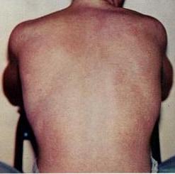
Eosinophilic gastroenteritis occurs when certain white blood cells known as eosinophils get into the digestive tract and cause damage. Symptoms of eosinophilic gastroenteritis usually start in adulthood and may include stomach pain, nausea, vomiting, and the inability to absorb nutrients from food. Sometimes, a blockage in the intestines occurs. In most people, symptoms occur from time to time and may go away completely with treatment. The exact cause of eosinophilic gastroenteritis is unknown, but it may be due to an abnormal response of the immune system to food allergies. Diagnosis is based on the symptoms, a clinical exam, laboratory tests, and by excluding other more common conditions.
Radio isotope scan using technetium ( 99m Tc) exametazime -labeled leukocyte SPECT may be useful in assessing the extent of disease and response to treatment but has little value in diagnosis, as the scan does not help differentiating EG from other causes of inflammation. [24] [25] When eosinophilic gastroenteritis is observed in association with eosinophilic infiltration of other organ systems, the diagnosis of idiopathic hypereosinophilic syndrome should be considered. [26] Management [ edit ] Corticosteroids are the mainstay of therapy with a 90% response rate in some studies.
Differential diagnoses include acquired generalized lipodystrophy (which occurs mainly in the context of auto-immune diseases), monogenic syndromes of insulin resistance, autoinflammatory diseases, partial forms of lipodystrophy and premature ageing syndromes.
Lipoatrophic diabetes Specialty Endocrinology Lipoatrophic diabetes is a type of diabetes mellitus presenting with severe lipodystrophy in addition to the traditional signs of diabetes. See also [ edit ] Familial partial lipodystrophy Congenital generalized lipodystrophy External links [ edit ] Classification D OMIM : 151660 269700 MeSH : D003923 Iglesias P, Alvarez Fidalgo P, Codoceo R, Díez J (2004). "Lipoatrophic diabetes in an elderly woman: clinical course and serum adipocytokine concentrations" . Endocr J . 51 (3): 279–86. doi : 10.1507/endocrj.51.279 . PMID 15256772 . Morse A, Whitaker M (2000). "Successful pregnancy in a woman with lipoatrophic diabetes mellitus.
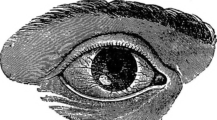
Phlyctenular keratoconjunctivitis Other names Phlyctenulosis Illustration of a corneal phlyctenule Specialty Neurology Phlyctenular keratoconjunctivitis ,is an inflammatory syndrome caused by a delayed (aka type-IV) hypersensitivity reaction to one or more antigens. The triggering antigen is usually a bacterial protein (particularly from Staphylococcus aureus ), but may also be a virus, fungus (particularly Candida albicans ), or nematode . [1] Contents 1 Symptoms 1.1 Presentation 2 Diagnosis 3 Treatment 4 See also 5 References Symptoms [ edit ] Irritation [1] Discomfort or pain [1] [2] Foreign-body sensation [2] Tearing [1] [2] Blepharospasm [2] Photophobia [1] [2] Mucopurulent discharge (rarely) [2] In cases where the cornea is affected, pain and photophobia are more likely, [1] [2] and corneal scarring can occur (potentially impairing vision). [1] Presentation [ edit ] The syndrome is marked by the appearance of characteristic lesions, known as phlyctenules , on the cornea and/or conjunctiva.
Jamaican vomiting sickness Specialty Toxicology Jamaican vomiting sickness (also known as toxic hypoglycemic syndrome (THS) , [1] acute ackee fruit intoxication , [2] or ackee poisoning [1] ) is an acute illness caused by the toxins hypoglycin A and hypoglycin B , which are present in fruit of the ackee tree . ... You can help by adding to it . ( December 2018 ) In popular culture [ edit ] The disease appears in the ER episode " Great Expectations ", where the symptoms are recognised by Dr Mallucci who, it is later revealed, attended medical school in Grenada . [ citation needed ] References [ edit ] ^ a b Gordon, André (2015-01-01), Gordon, André (ed.), "Chapter 4 - Biochemistry of Hypoglycin and Toxic Hypoglycemic Syndrome" , Food Safety and Quality Systems in Developing Countries , San Diego: Academic Press, pp. 47–61, doi : 10.1016/b978-0-12-801227-7.00004-4 , ISBN 978-0-12-801227-7 , retrieved 2020-07-05 ^ "The portal for rare diseases and orphan drugs" .
A rare disease caused by the ingestion of unripe Blighia sapida fruits. It is a serious intoxication that is frequent in certain countries in the Caribbean and Western Africa. In contrast, it is rare in France and other Western countries. Intoxication leads to toxic hypoglycaemia and inhibition of neoglucogenesis. The hypoglycaemia is caused by the effect of hypoglycin A, which is found in the arils. Clinical description The clinical manifestations are severe (coma, convulsions, delirium, toxic hepatitis, acute dehydration and a state of shock) and may lead to death.