Differential diagnosis Differential diagnoses include in utero diagnosis of chondrodysplasia, idiopathic juvenile osteoporosis, osteoporosis-pseudoglioma syndrome, Cole-Carpenter and Bruck syndromes, hyper or hypophosphatasia, panostotic form of polyostotic fibrous dysplasia (see these terms), non-accidental injury (multiple fractures without osteoporosis), and osteoporosis due to medication, nutritional deficiency, metabolic disease, or leukemia.
Osteogenesis imperfecta type I is a mild type of osteogenesis imperfecta (OI; see this term), a genetic disorder characterized by increased bone fragility, low bone mass and susceptibility to bone fractures. Epidemiology The overall prevalence of OI is estimated at between 1/10,000 and 1/20,000 but the prevalence of type I is unknown. Clinical description OI type I is nondeforming with normal height or mild short stature, blue sclera, and no dentinogenesis imperfecta (DI; see this term). Etiology OI type I is caused by mutations in the COL1A1 and COL1A2 genes (17q21.31-q22 and 7q22.1 respectively). Genetic counseling Transmission is autosomal dominant.
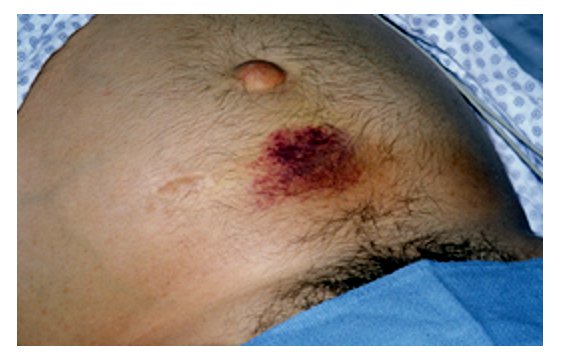
Osteogenesis imperfecta (OI) is a group of genetic disorders that mainly affect the bones. The term "osteogenesis imperfecta" means imperfect bone formation. People with this condition have bones that break (fracture) easily, often from mild trauma or with no apparent cause. Multiple fractures are common, and in severe cases, can occur even before birth. Milder cases may involve only a few fractures over a person's lifetime. There are at least 19 recognized forms of osteogenesis imperfecta, designated type I through type XIX.
In a likely heterogeneous group of 16 patients with OI syndromes, Kaiser-Kupfer et al. (1981) found low ocular rigidity and small corneal diameter and globe length; no correlation was found between rigidity of the eyeball and blueness of the sclera. ... Aortic root dilatation was found by echocardiogram to be present in 8 of 66 persons with OI syndrome; dilatation was mild and unrelated to age of the patient but was strikingly aggregated in families. ... Hortop et al. (1986) stated that aortic root dilatation was seen in each of the different OI syndromes but strikingly segregated within certain families. ... CLASSIFICATION Using clinical, radiographic, and genetic criteria, Sillence et al. (1979) developed the classification currently in use into types I to IV: a dominant form with blue sclerae, type I; a dominant form with normal sclerae, type IV (166220); a perinatally lethal OI syndrome, type II (166210); and a progressively deforming form with normal sclerae, type III. ... Assessing reports of biochemical findings in the OI syndromes is difficult because the phenotype and genetics generally are not specified.
Osteogenesis imperfecta (OI) is a group of genetic disorders that mainly affect the bones. Osteogenesis imperfecta type 1 is the mildest form of OI and is characterized by bone fractures during childhood and adolescence that often result from minor trauma. Fractures occur less frequently in adulthood. People with mild forms of the condition typically have a blue or grey tint to the part of the eye that is usually white (the sclera), and may develop hearing loss in adulthood. Affected individuals are usually of normal or near normal height. Most of the mutations that cause osteogenesis imperfecta type 1 occur in the COL1A1 gene. These genetic changes reduce the amount of type I collagen produced in the body, which causes bones to be brittle and to fracture easily.
Differential diagnosis Differential diagnosis includes retinitis pigmentosa and Goldmann-Favre syndrome. The presence of an autosomal recessive inheritance, severe nyctalopia, pigmentary retinopathy, and reduced alpha- and beta-waves on the electroretinogram (ERG) help to differentiate Goldmann-Favre syndrome from XLRS.
Juvenile retinoschisis is an eye condition characterized by impaired vision that begins in childhood and occurs almost exclusively in males. The condition affects the retina, which is a specialized light-sensitive tissue that lines the back of the eye. This affects the sharpness of vision. Central vision is more commonly affected. Vision often deteriorates early in life, but then usually becomes stable until late adulthood. A second decline in vision typically occurs in a man's fifties or sixties.
Several of the patients had previously been diagnosed with macular degeneration (see 603075), Stargardt disease, or Goldmann-Favre syndrome (268100). All 7 had fine white dots resembling drusen-like deposits, which were sometimes associated with retinal pigment epithelial abnormalities, in the maculas.
External links [ edit ] Classification D ICD - 10 : E61.3 v t e Malnutrition Protein-energy malnutrition Kwashiorkor Marasmus Catabolysis Vitamin deficiency B vitamins B 1 Beriberi Wernicke–Korsakoff syndrome Wernicke's encephalopathy Korsakoff's syndrome B 2 Riboflavin deficiency B 3 Pellagra B 6 Pyridoxine deficiency B 7 Biotin deficiency B 9 Folate deficiency B 12 Vitamin B 12 deficiency Other A: Vitamin A deficiency Bitot's spots C: Scurvy D: Vitamin D deficiency Rickets Osteomalacia Harrison's groove E: Vitamin E deficiency K: Vitamin K deficiency Mineral deficiency Sodium Potassium Magnesium Calcium Iron Zinc Manganese Copper Iodine Chromium Molybdenum Selenium Keshan disease Growth Delayed milestone Failure to thrive Short stature Idiopathic General Anorexia Weight loss Cachexia Underweight
CS1 maint: bot: original URL status unknown ( link ) External links [ edit ] Wikiquote has quotations related to: Overeating Media related to Overeating at Wikimedia Commons Overeating at Curlie v t e Malnutrition Protein-energy malnutrition Kwashiorkor Marasmus Catabolysis Vitamin deficiency B vitamins B 1 Beriberi Wernicke–Korsakoff syndrome Wernicke's encephalopathy Korsakoff's syndrome B 2 Riboflavin deficiency B 3 Pellagra B 6 Pyridoxine deficiency B 7 Biotin deficiency B 9 Folate deficiency B 12 Vitamin B 12 deficiency Other A: Vitamin A deficiency Bitot's spots C: Scurvy D: Vitamin D deficiency Rickets Osteomalacia Harrison's groove E: Vitamin E deficiency K: Vitamin K deficiency Mineral deficiency Sodium Potassium Magnesium Calcium Iron Zinc Manganese Copper Iodine Chromium Molybdenum Selenium Keshan disease Growth Delayed milestone Failure to thrive Short stature Idiopathic General Anorexia Weight loss Cachexia Underweight
Pseudohypertension , also known as pseudohypertension in the elderly , noncompressibility artery syndrome , and Osler's sign of pseudohypertension is a falsely elevated blood pressure reading obtained through sphygmomanometry due to calcification of blood vessels which cannot be compressed. [1] There is normal blood pressure when it is measured from within the artery. [2] This condition however is associated with significant cardiovascular disease risk. [2] Because the stiffened arterial walls of arteriosclerosis do not compress with pressure normally, the blood pressure reading is theoretically higher than the true intra-arterial measurement. ... PMID 7825617 . v t e Symptoms and signs relating to the circulatory system Chest pain Referred pain Angina Levine's sign Auscultation Heart sounds Split S2 S3 S4 Gallop rhythm Heart murmur Systolic Functional murmur Still's murmur Diastolic Pulmonary insufficiency Graham Steell murmur Continuous Carey Coombs murmur Mitral insufficiency Presystolic murmur Pericardial friction rub Heart click Bruit carotid Pulse Tachycardia Bradycardia Pulsus paradoxus doubled Pulsus bisferiens Pulsus bigeminus Pulsus alternans Other Palpitations Apex beat Cœur en sabot Jugular venous pressure Cannon A waves Hyperaemia Shock Cardiogenic Obstructive Hypovolemic Distributive See further Template:Shock Cardiovascular disease Aortic insufficiency Collapsing pulse De Musset's sign Duroziez's sign Müller's sign Austin Flint murmur Mayne's sign Other endocardium endocarditis : Roth's spot Janeway lesion / Osler's node Bracht–Wachter bodies Pericardium Cardiac tamponade / Pericardial effusion : Beck's triad Ewart's sign Other rheumatic fever : Anitschkow cell Aschoff body EKG J wave Gallavardin phenomenon Vascular disease Arterial aortic aneurysm Cardarelli's sign Oliver's sign pulmonary embolism Right heart strain radial artery sufficiency Allen's test pseudohypertension thrombus Lines of Zahn Adson's sign arteriovenous fistula Nicoladoni sign Venous Friedreich's sign Caput medusae Kussmaul's sign Trendelenburg test superior vena cava syndrome Pemberton's sign This medical sign article is a stub .
Goldmuntz et al. (2002) noted that cardiac laterality defects, including dextro-looped TGA (DTGA) and DORV, are a characteristic finding in visceral or thoracic heterotaxy syndromes, although they are most frequently seen in isolation. ... Since CFC1 mutations had been identified in patients with heterotaxy syndrome, all of whom had congenital cardiac malformations, Goldmuntz et al. (2002) analyzed the CFC1 gene in 86 patients with these cardiac disorders and found that 2 had heterozygous pathogenic mutations (605194.0002 and 605194.0004).
A number sign (#) is used with this entry because visceral heterotaxy-5 (HTX5) is caused by heterozygous mutation in the NODAL gene (601265) on chromosome 10q22. Description Heterotaxy ('heter' meaning 'other' and 'taxy' meaning 'arrangement'), or situs ambiguus, is a developmental condition characterized by randomization of the placement of visceral organs, including the heart, lungs, liver, spleen, and stomach. The organs are oriented randomly with respect to the left-right axis and with respect to one another (Srivastava, 1997). Heterotaxy is a clinically and genetically heterogeneous disorder. For a discussion of genetic heterogeneity of visceral heterotaxy, see HTX1 (306955). Clinical Features Zlotogora et al. (1987) described a family in which 4 out of 7 children had situs inversus and/or congenital heart disease; more specifically, 3 had situs inversus with a normal heart in one and 3 had heart defects with normal organ orientation in one.
A number sign (#) is used with this entry because of evidence that autosomal visceral heterotaxy-6 (HTX6) is caused by homozygous mutation in the CCDC11 gene (614759) on chromosome 18q21. For a discussion of the genetic heterogeneity of visceral heterotaxy, see HTX1 (306955). Clinical Features Perles et al. (2012) reported 2 brothers, born of consanguineous Arab-Muslim parents, with variable manifestations of visceral heterotaxy. The younger brother, aged 14 years, presented with congenital heart disease and severe cyanosis. Echocardiography showed a complex cardiovascular defect and abdominal situs abnormalities.
A number sign (#) is used with this entry because X-linked heterotaxy-1 (HTX1) and multiple types of congenital heart defects-1 (CHTD1) are caused by mutation in the ZIC3 gene (300265) on chromosome Xq26. Mutation in the ZIC3 gene can also cause VACTERL with or without hydrocephalus (VACTERLX; 314390), a disorder with overlapping features. Description Heterotaxy Heterotaxy ('heter' meaning 'other' and 'taxy' meaning 'arrangement'), or situs ambiguus, is a developmental condition characterized by randomization of the placement of visceral organs, including the heart, lungs, liver, spleen, and stomach. The organs are oriented randomly with respect to the left-right axis and with respect to one another (Srivastava, 1997). Heterotaxy is a clinically and genetically heterogeneous disorder. Multiple Types of Congenital Heart Defects Congenital heart defects (CHTD) are among the most common congenital defects, occurring with an incidence of 8/1,000 live births.
A number sign (#) is used with this entry because visceral heterotaxy-4 (HTX4) is caused by heterozygous mutation in the ACVR2B gene (602730). Description Heterotaxy ('heter' meaning 'other' and 'taxy' meaning 'arrangement'), or situs ambiguus, is a developmental condition characterized by randomization of the placement of visceral organs, including the heart, lungs, liver, spleen, and stomach. The organs are oriented randomly with respect to the left-right axis and with respect to one another (Srivastava, 1997). Heterotaxy is a clinically and genetically heterogeneous disorder. For a discussion of the genetic heterogeneity of visceral heterotaxy, see HTX1 (306955). Clinical Features Kosaki et al. (1999) reported 3 unrelated patients with left-right axis malformations.
A number sign (#) is used with this entry because of evidence that autosomal visceral heterotaxy-8 (HTX8) is caused by homozygous mutation in the PKD1L1 gene (609721) on chromosome 7p12. Description Autosomal visceral heterotaxy-8 is an autosomal recessive developmental disorder characterized by visceral situs inversus associated with complex congenital heart malformations caused by defects in the normal left-right asymmetric positioning of internal organs (summary by Vetrini et al., 2016). For a discussion of the genetic heterogeneity of visceral heterotaxy, see HTX1 (306955). Clinical Features Vetrini et al. (2016) reported 2 male infants, born of unrelated parents of northern European descent, with heterotaxy and complex congenital heart disease resulting in death shortly after birth and at age 3 weeks. In 1 infant, the abnormalities were apparent on prenatal ultrasound at 21 weeks' gestation.
A number sign (#) is used with this entry because of evidence that autosomal visceral heterotaxy-7 (HTX7) is caused by homozygous or compound heterozygous mutation in the MMP21 gene (608416) on chromosome 10q26. Description Autosomal visceral heterotaxy-7 is an autosomal recessive developmental disorder characterized by complex congenital heart malformations and/or situs inversus and caused by defects in the normal left-right asymmetric positioning of internal organs. The phenotype is variable (summary by Guimier et al., 2015). For a discussion of the genetic heterogeneity of visceral heterotaxy, see HTX1 (306955). Clinical Features Perles et al. (2015) reported 3 sibs, born of consanguineous parents, with a congenital laterality disorder. One patient presented with cyanosis at age 3 months and was found to have dextrocardia with atrial situs inversus, complete atrioventricular canal defect, transposition of the great arteries (TGA) and pulmonary atresia with a duct-like aortopulmonary collateral.
An autosomal form of visceral heterotaxy, designated HTX3, has been mapped to chromosome 6q21. Description Heterotaxy ('heter' meaning 'other' and 'taxy' meaning 'arrangement'), or situs ambiguus, is a developmental condition characterized by randomization of the placement of visceral organs, including the heart, lungs, liver, spleen, and stomach. The organs are oriented randomly with respect to the left-right axis and with respect to one another (Srivastava, 1997). Heterotaxy is a clinically and genetically heterogeneous disorder. For a discussion of the genetic heterogeneity of visceral heterotaxy, see HTX1 (306955). Clinical Features Peeters et al. (2001) reported a patient in whom routine ultrasound examination at gestational age 28 weeks had shown atrioventricular septal defect and abdominal situs inversus.
Two additional family members had isolated cardiac defects without any other L/R abnormality: one had ventricular inversion in combination with transposition of the great arteries, whereas the other had hypoplastic left heart syndrome. Maclean and Dunwoodie (2004) reviewed the evidence that mutations in genes and pathways critical for L-R patterning are involved in common isolated congenital malformations such as congenital heart disease, biliary tract anomalies, renal polycystic disease, and malrotation of the intestine, indicating that disorders of L-R development are far more common than a 1 in 10,000 incidence of heterotaxia might suggest.
In 1955, Björn Ivemark described a series of autopsies of hearts from patients with asplenia. Thus, Ivemark syndrome corresponds to heterotaxia with asplenia.
Some people with corticosteroid-binding globulin deficiency also have a condition called chronic fatigue syndrome. The features of chronic fatigue syndrome are prolonged fatigue that interferes with daily activities, as well as general symptoms, such as sore throat or headaches.
Causes of hyperprolactinemia without pituitary adenoma include: pregnancy, lactation, exercise, stress and polycystic ovary syndrome. Pituitary lesions that do not produce prolactin can also cause hyperprolactinemia by pituitary stalk impingement (stalk effect). Genetic counseling Prolactinomas usually occur sporadically and with the exception of prolactinoma due to MEN1 or FIPA, the occurrence of prolactinomas as part of inherited syndromes is exceptionally rare. In patients with a pathogenic variant of AIP , the inheritance is autosomal dominant and thus offspring of an affected individual have a 50% risk of inheriting the mutation; however, penetrance is variable.
Prolactinoma Specialty Oncology , endocrinology , neurology , neurosurgery A prolactinoma is a benign tumor ( adenoma ) of the pituitary gland that produces a hormone called prolactin . It is the most common type of functioning pituitary tumor . [1] Symptoms of prolactinoma are due to too much prolactin in the blood ( hyperprolactinemia ), or those caused by pressure of the tumor on surrounding tissues. Prolactin stimulates the breast to produce milk, and has many other functions such as regulation of mood. Hence prolactin levels are usually higher during pregnancy and after childbirth. After delivery of a baby, a mother's prolactin levels come down to normal a few weeks after breastfeeding is discontinued.
Prolactinoma is a tumor of the pituitary gland that causes increased levels of the hormone prolactin . This hormone normally stimulates breast development and milk production in women. Prolactinoma can affect men or women. In women, the symptoms may include unusual milk production (galactorrhea) when not pregnant or nursing and having no menstrual cycles ( amenorrhea ). In men, the most common symptoms are decreased sex drive and infertility. Most prolactinomas occur by chance (sporadically) in people with no family history.
Overview Prolactinoma is a noncancerous tumor of the pituitary gland. This tumor causes the pituitary gland to make too much of a hormone called prolactin. The major effect of a prolactinoma is decreased levels of some sex hormones — namely, estrogen and testosterone. Prolactinoma A prolactinoma is a type of tumor that develops in the pituitary gland at the base of your brain. A prolactinoma isn't life-threatening. But it can cause vision difficulties, infertility and other problems.
Contents 1 Classification 2 See also 3 References 4 External links Classification [ edit ] Although a clear understanding of the various skin lesions in IgG4-related disease is a work in progress, skin lesions have been classified into subtypes based on documented cases: [2] Angiolymphoid hyperplasia with eosinophilia (or lesions that mimic it) [3] and cutaneous pseudolymphoma Cutaneous plasmacytosis [Note 1] Eyelid swelling (as part of Mikulicz's disease ) Psoriasis -like eruptions Unspecified maculopapular or erythematous eruptions Hypergammaglobulinemic purpura and urticarial vasculitis Impaired blood supply to fingers or toes, leading to Raynaud's phenomenon or gangrene In addition, Wells syndrome has also been reported in a case of IgG4-related disease. [5] Note : ^ Some do not consider cutaneous plasmacytosis to be a feature of IgG4-related disease for reasons that include: a lack of systemic features, no response to steroid therapy and a different histological pattern. [4] See also [ edit ] IgG4-related disease References [ edit ] ^ John H. ... "IgG4-related disease of the paratestis in a patient with Wells syndrome: a case report" . Diagnostic Pathology . 9 : 225. doi : 10.1186/s13000-014-0225-5 .
Differential diagnosis The diagnosis of CDA should be considered following exclusion of other causes of macrocytosis (B12 deficiency, folic acid deficiency or other megaloblastic anemias such as pernicious anemia or thiamine-responsive megaloblastic anemia syndrome; see this term), acquired dyserythropoiesis (myelodysplastic disorders) and hemolytic anemias. Gilbert syndrome (see this term) and infections should be also excluded.
The diagnosis of CDA should be considered following exclusion of other causes of macrocytosis (mainly B 12 deficiency and folic acid deficiency) and dyserythropoiesis, including thalassemia syndromes and hereditary sideroblastic anemia.
A number sign (#) is used with this entry because congenital dyserythropoietic anemia type Ib (CDAN1B) is caused by homozygous mutation in the C15ORF41 gene (615626) on chromosome 15q14. Description Congenital dyserythropoietic anemia type I is an autosomal recessive hematologic disorder characterized by congenital macrocytic anemia secondary to ineffective erythropoiesis. The bone marrow shows erythroid hyperplasia, with nuclear abnormalities in most erythroblasts. Up to 3% of erythroblasts have interchromatin bridges, and erythroblast nuclei are abnormally electron dense with spongy ('Swiss cheese-like') heterochromatin on electron microscopy. Some reported patients have distal digital abnormalities (summary by Ahmed et al., 2006).
Congenital dyserythropoietic anemia (CDA) type 1 is an inherited blood disorder characterized by moderate to severe anemia . It is usually diagnosed in childhood or adolescence, although in some cases, the condition can be detected before birth. Many affected individuals have yellowing of the skin and eyes (jaundice) and an enlarged liver and spleen (hepatosplenomegaly). This condition also causes the body to absorb too much iron, which builds up and can damage tissues and organs. In particular, iron overload can lead to an abnormal heart rhythm (arrhythmia), congestive heart failure, diabetes, and chronic liver disease (cirrhosis).
Congenital dyserythropoietic anemia (CDA) is an inherited blood disorder that affects the development of red blood cells. This disorder is one of many types of anemia , which is a condition characterized by a shortage of red blood cells. This shortage prevents the blood from carrying an adequate supply of oxygen to the body's tissues. The resulting symptoms can include tiredness (fatigue), weakness, pale skin, and other complications. Researchers have identified three major types of CDA: type I, type II, and type III.
A number sign (#) is used with this entry because congenital dyserythropoietic anemia type Ia (CDAN1A) is caused by homozygous or compound heterozygous mutation in the gene encoding codanin-1 (CDAN1; 607465) on chromosome 15q15. Description CDA type I is a rare inherited red blood cell disorder characterized by macrocytic anemia, ineffective erythropoiesis, and secondary hemochromatosis. It is occasionally associated with bone abnormalities, especially of the hands and feet (acrodysostosis), nail hypoplasia, and scoliosis (Tamary et al., 2005). Striking morphologic abnormalities of erythroblasts, reviewed by Wickramasinghe and Wood (2005), include the 'Swiss-cheese' abnormality of erythroblasts on electron microscopy. Four types of CDA, all of which show show ineffective erythropoiesis and multinuclear erythroblasts, have been characterized by clinical and hematopoietic findings.
Congenital dyserythropoietic anemia type I (CDA I) is a disorder of blood cell production, particularly of the production of erythroblasts, which are the precursors of the red blood cells (RBCs). [1] Congenital dyserythropoietic anemia type I Specialty Hematology Contents 1 Presentation 2 Genetics 3 Diagnosis 4 Treatment 5 See also 6 References 7 Further reading 8 External links Presentation [ edit ] Many affected individuals have yellowing of the skin and eyes (jaundice) and an enlarged liver and spleen (hepatosplenomegaly). This condition also causes the body to absorb too much iron, which builds up and can damage tissues and organs. In particular, iron overload can lead to an abnormal heart rhythm (arrhythmia), congestive heart failure, diabetes, and chronic liver disease (cirrhosis). Rarely, people with CDA type I are born with skeletal abnormalities, most often involving the fingers and/or toes. [2] Genetics [ edit ] CDA type I is transmitted by both parents autosomal recessively and usually results from mutations in the CDAN1 gene. Little is known about the function of this gene, and it is unclear how mutations cause the characteristic features of CDA type I.
They demonstrated a complex (core plus secondary) binding site for TCF8 in the promoter of the COL4A3 gene (120070), which is mutant in Alport syndrome (203780), and presented immunohistochemical evidence of ectopic expression of COL4A3 in corneal endothelium of the proband of the original PPCD3 family. ... This study identified TCF8 as the gene responsible for approximately half of the cases of PPCD, implicated TCF8 mutations in developmental abnormalities outside the eye, and revealed COL4A3, the TCF8 regulatory target, as a key, shared molecular component of 2 different diseases, PPCD and Alport syndrome. Molecular Genetics Krafchak et al. (2005) identified a heterozygous frameshift mutation in the TCF8 (ZEB1) gene (189909.0001) in affected members of the family with PPCD reported by Moroi et al. (2003) and 4 different heterozygous nonsense and frameshift mutations in TCF8 in 4 other PPCD probands.
A rare mild subtype of posterior corneal dystrophy characterized by small aggregates of apparent vesicles bordered by a gray haze at the level of Descemet membrane, generally with no effect on vision. Epidemiology Prevalence of this form of corneal dystrophy is unknown. Clinical description Lesions generally develop in early childhood and are mostly bilateral but may be asymmetrical or unilateral in some cases. Most patients are asymptomatic with no corneal edema, but progressive visual impairment due to stromal clouding may rarely occur. Etiology PPCD is a genetically heterogenous condition with extremely variable expression.
None of the PPCD-affected individuals reported hearing loss or other features of ectodermal dysplasia syndrome (ECTDS; 616029). Mapping Liskova et al. (2018) genotyped 9 affected and 7 unaffected members of a large Czech family (C15) segregating autosomal dominant PPCD, and identified a single locus on chromosome 8 (chr8:100,821,039-119,725,923; GRCh38), spanning 8q22.3-q24.12, with a maximum lod score of 4.38.
In a photo essay, Anderson et al. (2001) reviewed the clinical and histopathologic overlaps between posterior polymorphous membranous dystrophy and iridocorneal endothelial syndrome. PPCD is bilateral, usually asymptomatic, and usually nonprogressive; it occurs at all ages and shows no sex predilection. Sporadic iridocorneal endothelial syndrome is usually unilateral, symptomatic, and progressive; it presents at middle age and is more common in women. Corneal edema, glaucoma, and iris changes are more common in the iridocorneal endothelial syndrome. In PPCD, endothelial cells are more likely to display epithelial-like characteristics. ... They thought that an insult during embryogenesis might result in PPCD, whereas an insult later in corneal development might result in the iridocorneal endothelial syndrome. They also noted that the herpes simplex virus had been implicated as a cause in the iridocorneal endothelial syndrome.
Posterior polymorphous corneal dystrophy Other names Ophthalmology Appearance of the abnormal corneal endothelial cells that have become transformed into stratified squamous epithelium. Periodic acid Schiff (PAS) stain Posterior Polymorphous Corneal Dystrophy ( PPCD ; sometimes also Schlichting dystrophy ) is a type of corneal dystrophy, characterised by changes in Descemet's membrane and endothelial layer . Symptoms mainly consist of decreased vision due to corneal edema. In some cases they are present from birth, other patients are asymptomatic. Histopathological analysis shows that the cells of endothelium have some characteristics of epithelial cells and have become multilayered. The disease was first described in 1916 by Koeppe as keratitis bullosa interna . [1] Contents 1 Genetics 2 Pathophysiology 3 Diagnosis 4 See also 5 References 6 External links Genetics [ edit ] PPCD type 2 is linked to the mutations in COL8A2 , and PPCD type 3 mutations in ZEB1 gene, but the underlying genetic disturbance in PPCD type 1 is unknown.
A number sign (#) is used with this entry because of evidence that posterior polymorphous corneal dystrophy-2 (PPCD2) is caused by heterozygous mutation in the COL8A2 gene (120252) on chromosome 1p34. For a phenotypic description and a discussion of genetic heterogeneity of PPCD, see PPCD1 (122000). Clinical Features Biswas et al. (2001) reported a father and daughter with posterior polymorphous corneal dystrophy. Bilateral penetrating keratoplasty was performed in the proband in her twenties and in her father in his fifties. Molecular Genetics In 2 affected members of a family with posterior polymorphous corneal dystrophy, Biswas et al. (2001) identified a missense mutation in the triple helical domain of the COL8A2 gene (120252.0001), which encodes the alpha-2 chain of type VIII collagen, a short-chain collagen that is a component of endothelial basement membranes.
Eosinophilic pustular folliculitis (EPF) affects the skin causing itchy, red or skin-colored bumps and pustules (bumps containing pus). The papules mostly appear on the face, scalp, neck and trunk and may last for weeks or months. EPF affects males more than females. There are several forms of EPF. Classic eosinophilic pustular folliculitis mainly occurs in Japan. Immunosuppression-associated EPF is mainly associated with HIV infection, but has also been associated with certain cancers and medications.[16046] The infantile form of EPF is seen in infants from birth or within the first year of life. The underlying cause of EFP is unknown. All of these forms have similar skin findings.
The other had an extremely severe syndrome dominated by lactic acidosis and rapidly fatal encephalopathy, with diffuse cystic leukodystrophy and micropolygyria, a developmental abnormality of the brain that occurs well before birth. ... INHERITANCE - Autosomal recessive GROWTH Other - Intrauterine growth retardation HEAD & NECK Head - Microcephaly (patient A) Eyes - Nystagmus (patient A) RESPIRATORY - Respiratory failure MUSCLE, SOFT TISSUES - Hypotonia, neonatal NEUROLOGIC Central Nervous System - Developmental regression - Encephalopathy (patient A) - Hypertonia (patient A) - Spasticity (patient A) - Cystic leukodystrophy (patient A) - Micropolygyria (patient A) - Abnormalities of the basal ganglia on brain imaging - Leigh syndrome METABOLIC FEATURES - Lactic acidosis LABORATORY ABNORMALITIES - Increased serum lactate - Hyperammonemia MISCELLANEOUS - Onset in infancy - Death in infancy may occur MOLECULAR BASIS - Caused by mutation in the mitochondrial Tu translation elongation factor gene (TUFM, 602389.0001 ) ▲ Close
Combined oxidative phosphorylation defect type 4 is a rare mitochondrial disorder due to a defect in mitochondrial protein synthesis characterized by a neonatal onset of severe metabolic acidosis and respiratory distress, persistent lactic acidosis with episodes of metabolic crises, developmental regression, microcephaly, abnormal gaze fixation and pursuit, axial hypotonia with limb spasticity and reduced spontaneous movements. Neuroimaging studies reveal polymicrogyria, white matter abnormalities and multiple cystic brain lesions, including basal ganglia, and cerebral atrophy. Decreased activity of complex I and IV have been determined in muscle biopsy.
A group of rare acute leukemias of ambiguous lineage characterized by the presence of separate populations of blasts of more than one lineage (bilineal), a single population of blasts coexpressing antigens of more than one lineage (biphenotypic), or a combination thereof. The diagnosis relies on immunophenotyping, the T-cell component being characterized by strong expression of cytoplasmic CD3, usually in the absence of surface CD3, the B-cell component expressing CD19, almost always together with CD10, cCD79a, CD22, or PAX5, while the most specific hallmark of the myeloid component is the presence of myeloperoxidase in the blast cytoplasm.
Differential diagnosis Differential diagnosis includes megaloblastic anaemia, myelodysplastic syndromes, acute lymphoblastic leukemia, acute biphenotypic leukemia, chronic myeloid leukemia (myeloid blast phase), and metastases of tumors such rhabdomyosarcoma and neuroblastoma (see these terms).
Core binding factor acute myeloid leukemia (CBF-AML) is one form of a cancer of the blood-forming tissue (bone marrow) called acute myeloid leukemia. In normal bone marrow, early blood cells called hematopoietic stem cells develop into several types of blood cells: white blood cells (leukocytes) that protect the body from infection, red blood cells (erythrocytes) that carry oxygen, and platelets (thrombocytes) that are involved in blood clotting . In acute myeloid leukemia , the bone marrow makes large numbers of abnormal, immature white blood cells called myeloid blasts. Instead of developing into normal white blood cells, the myeloid blasts develop into cancerous leukemia cells. The large number of abnormal cells in the bone marrow interferes with the production of functional white blood cells, red blood cells, and platelets.
A subtype of acute myeloid leukemia with recurrent genetic abnormalities, characterized by clonal proliferation of myeloid blasts harboring somatic mutations of the CEBPA gene in the bone marrow, blood and, rarely, other tissues. It can present with anemia, thrombocytopenia, and other nonspecific symptoms related to ineffective hematopoesis (fatigue, bleeding and bruising, recurrent infections, bone pain) and/or extramedullary site involvement (gingivitis, splenomegaly).
Familial acute myeloid leukemia with mutated CEBPA is one form of a cancer of the blood-forming tissue (bone marrow ) called acute myeloid leukemia. In normal bone marrow, early blood cells called hematopoietic stem cells develop into several types of blood cells : white blood cells (leukocytes) that protect the body from infection; red blood cells (erythrocytes) that carry oxygen; and platelets (thrombocytes), which are involved in blood clotting . In acute myeloid leukemia, the bone marrow makes large numbers of abnormal, immature white blood cells called myeloid blasts. Instead of developing into normal white blood cells, the myeloid blasts develop into cancerous leukemia cells. The large number of abnormal cells in the bone marrow interferes with the production of functional white blood cells, red blood cells, and platelets.
There are many potential causes of AML such as certain blood disorders, inherited syndromes, environmental exposures, and drug exposures; however, most people who develop AML have no identifiable risk factor.
Cytogenetically normal acute myeloid leukemia (CN-AML) is one form of a cancer of the blood-forming tissue (bone marrow) called acute myeloid leukemia. In normal bone marrow, early blood cells called hematopoietic stem cells develop into several types of blood cells: white blood cells (leukocytes) that protect the body from infection, red blood cells (erythrocytes) that carry oxygen, and platelets (thrombocytes) that are involved in blood clotting . In acute myeloid leukemia , the bone marrow makes large numbers of abnormal, immature white blood cells called myeloid blasts. Instead of developing into normal white blood cells, the myeloid blasts develop into cancerous leukemia cells. The large number of abnormal cells in the bone marrow interferes with the production of functional white blood cells, red blood cells, and platelets.
Acatalasemia is a condition characterized by very low levels of an enzyme called catalase. Many people with acatalasemia never have any health problems related to the condition and are diagnosed because they have affected family members. Some of the first reported individuals with acatalasemia developed open sores (ulcers) inside the mouth that led to the death of soft tissue (gangrene). When mouth ulcers and gangrene occur with acatalasemia, the condition is known as Takahara disease. These complications are rarely seen in more recent cases of acatalasemia, probably because of improvements in oral hygiene.
A rare congenital disorder resulting from a deficiency in erythrocyte catalase, an enzyme responsible for the breakdown of hydrogen peroxide. Epidemiology The disorder is very rare in the general population with an estimated prevalence of 1 in 31250. Clinical description The disorder is usually asymptomatic but may be associated with oral ulcerations and gangrene, or diabetes mellitus and atherosclerosis in certain populations. Genetic counseling Transmission is autosomal recessive.
A number sign (#) is used with this entry because acatalasemia is caused by homozygous mutation in the CAT gene (115500) on chromosome 11p13. Description Acatalasemia, also known as acatalasia, is a metabolic disorder characterized by a total or near total loss of catalase activity in erythrocytes. About half of cases originate from ulcerating oral gangrenes, and these cases are referred to as having Takahara disease. Half-normal levels of catalase in heterozygotes is referred to as hypocatalasemia or hypocatalasia (Ogata, 1991). Clinical Features Acatalasemia was first discovered in Japan by Takahara, an otolaryngologist who found that in cases of progressive oral gangrene, hydrogen peroxide applied to the ulcerated areas did not froth in the usual manner (Takahara and Miyamoto, 1948).