-
Functional Neurologic Disorder
Wikipedia
Wessely and White have argued that FND may merely be an unexplained somatic illness (like fibromyalgia, irritable bowel syndrome, or chronic fatigue syndrome) single disorder than separate disorders. [28] References [ edit ] ^ a b Carson, A.; et al.
- Ritscher-Schinzel Syndrome GeneReviews
-
Abrasion (Dental)
Wikipedia
Chemical [ edit ] The current selection of dentifrice should also be critically analysed and changed to include a less abrasive and gentler paste such as sensitive toothpaste as evidence suggests that a very abrasive toothpaste would lead to loss of tooth structure. [27] A toothpaste containing increased fluoride will also help combat the increased sensitivity and risk to dental decay. [28] Fluoride varnish is known to alleviate hypersensitivity in teeth and can be used as a preventive measure for high risk patients of dental erosion with abrasion because fluoride varnish is reported to have an effect on the surface and subsurface of the tooth. [29] Treatment in the dental chair may include a fluoride application or the placement of a restoration in more severe cases.

-
Micropsia
Wikipedia
This study was the first ever to prove that dietary supplements can alter the natural progression and complications of a disease state. [26] Laser treatments also look promising but are still in clinical stages. [26] Epidemiology [ edit ] Episodes of micropsia or macropsia occur in 9% of adolescents. [27] 10-35% of migraine sufferers experience auras, with 88% of these patients experiencing both visual auras (which include micropsia) and neurological auras. [28] Micropsia seems to be slightly more common in boys than in girls among children who experience migraines. [29] Approximately 80% of temporal lobe seizures produce auras that may lead to micropsia or macropsia.

-
Bovine Viral Diarrhea
Wikipedia
Vaccination [ edit ] Modern vaccination programmes aim not only to provide a high level of protection from clinical disease for the dam, but, crucially, to protect against viraemia and prevent the production of PIs. [28] While the immune mechanisms involved are the same, the level of immune protection required for foetal protection is much higher than for prevention of clinical disease. [29] While challenge studies indicate that killed, as well as live, vaccines prevent foetal infection under experimental conditions, the efficacy of vaccines under field conditions has been questioned. [30] The birth of PI calves into vaccinated herds suggests that killed vaccines do not stand up to the challenge presented by the viral load excreted by a PI in the field. [31] See also [ edit ] Animal viruses References [ edit ] ^ a b c d Fray; et al. (2000).
-
Central Nervous System Disease
Wikipedia
., Lorazepam ) in pill and I.V. form. [28] Depression [ edit ] Main article: Major depressive disorder Major depressive disorder, otherwise known as depression, is a disorder that is characterized by a pervasive and persistent low mood that is accompanied by low self-esteem and by a loss of interest or pleasure in normally enjoyable activities.EPO, IDH2, IL17A, SOD2, BDNF, MOG, GFAP, GSK3B, AQP4, GRM5, HMOX1, IL6, TSPO, NFE2L2, NLRP3, APOE, P2RX7, PDE4A, APP, ALK, PARP1, MMP9, GABPA, ABCB1, PDE10A, PLP1, PDXP, CYP2D6, IL33, SLC6A3, PTHLH, CSF2, CCL2, PRDX5, TNF, ERBB2, LAMC2, PRNP, CXCR3, USP18, GCG, HRH3, FGF2, LPAR3, GPR42, SSTR4, GRN, GRM1, MAPK3, KEAP1, LPAR2, ICAM1, IFNG, FZD4, EGFR, CXCL8, KCNK2, TTC4, TRPM2, TRAF6, TLR4, HTR1A, CXCR6, MRGPRX1, ADRA1A, MRGPRX4, CD44, EDNRA, CCR5, GPR151, BRS3, BRAF, OXER1, GPRC6A, LGR6, CSF1R, CSF3, ADRA2B, ACKR3, MRGPRX3, GPR166P, VN1R17P, WWOX, ATN1, RSS, REN, TNFRSF1A, TNFRSF1B, RAG1, NPS, TSHR, CD200R1, RAC2, SGSH, ADGRG3, TXN, NR1H2, VCP, RBM45, VEGFA, BHLHE23, VIM, PTPRZ1, PTPN6, TTR, TLR2, MIR34A, TIMP1, SLC6A4, SLC6A9, CXCL12, SMS, SNCA, SOD1, MIR21, MIR132, GTF2H5, SPP1, SST, SCN2A, MTDH, ROS1, SYN1, TACR1, TCF4, TGFB1, TGIF1, PSAT1, KRT90P, XPA, ZFP36, PTEN, NGB, EBP, CYSLTR1, SLC12A5, RIPK3, TPPP, PTPRT, NDRG2, PDCD10, GJC2, ELP2, FEZF2, MGLL, MPRIP, SLC52A1, CYFIP1, BRD4, PANX1, TREM2, SAMHD1, ADGRA2, ELP4, GPR88, TMEM97, CXCL13, SIGMAR1, BCAP31, XPR1, CXCR4, NR0B2, ITGA10, RIPK1, NR1I2, ORAI1, MEGF10, SOCS3, CH25H, PLVAP, MYOM2, CD276, NYX, PNMA1, ADIPOQ, ABCG2, ROCK2, ADAMTS4, ADAMTS3, NPEPPS, RTN4R, VSIR, SV2A, HDAC6, AAVS1, OAS3, PTAFR, ESR2, CYP2B6, CYP2C9, CYP3A4, ACE, DDIT3, DRD2, TSC22D3, TOR1A, ECHS1, EDNRB, ELF2, ERCC2, ERCC3, ERN1, EWSR1, GRIN2B, F2R, F2RL1, FANCB, FPR2, MTOR, GAD2, GC, GDF2, GIP, GJA1, GLB1, GLP1R, GNA12, GPR3, CYP1A2, CTSS, CST3, CSF1, ACHE, ACTB, ADAM8, ADORA2A, AGT, AGTR1, AGTR2, KLK3, ARSB, ALDH7A1, BCL2, DST, SERPING1, C5, C5AR1, CACNA1D, CAV1, CD19, CD34, CD36, CD38, CDH11, CHAT, CHRNA4, CNR2, CNTF, ATF2, CRH, CRHBP, GPR17, GRM3, KLK6, PDE1C, MME, MMP2, MMP12, CD200, MST1R, MYCL, MYD88, NCAM1, NEFL, NPC1, NTS, OAS2, ASIC1, OPRK1, PDE4B, GRM4, PDE4D, PGR, SERPINA1, SERPINI1, PIK3CD, PLAG1, PMP22, POMC, PPARA, PPARG, MAPK8, MAP2K7, PROS1, LGMN, NR3C2, MGAT1, MEF2C, CD46, GRM7, GRP, GTF2H1, GTF2H2, GTF2H3, GTF2H4, HINT1, HLA-DRB1, HMGB1, HMGCR, NR4A1, HP, HSPA9, HTR2A, HTR2C, IDS, IDUA, IFNA1, IFNA13, IGF1, IGHM, IL1B, IL3, CXCL10, L1CAM, MAOB, MAPT, MBL2, MBP, CCR2

-
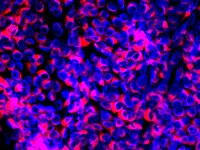
Neuroinflammation
Wikipedia
Current treatments for multiple sclerosis include interferon-B, Glatiramer acetate, and Mitoxantrone, which function by reducing or inhibiting T Cell activation, but have the side effect of systemic immunosuppression [28] In Alzheimer's disease, the use of non-steroidal anti-inflammatory drugs decreases the risk of developing the disease.
-
Cyclic Vomiting Syndrome
Wikipedia
"Cyclic vomiting syndrome: epidemiology, diagnosis, and treatment". Clinical Autonomic Research . 28 (2): 203–209. doi : 10.1007/s10286-018-0506-2 .
-
Duarte Galactosemia
Wikipedia
The first [12] report published in 2008 was a pilot study that looked at biochemical markers and developmental outcomes in a group of 28 toddlers and young children with DG, some of whom had drunk milk through infancy and some of whom had drunk low-galactose formula.

-
Viral Meningitis
Wikipedia
For herpes the treatment of choice is aciclovir . [28] If encephalitis is suspected, empiric treatment with IV aciclovir is often warranted. [16] Surgical management is indicated where there is extremely increased intracranial pressure, infection of an adjacent bony structure (e.g. mastoiditis ), skull fracture, or abscess formation. [12] The majority of people that have viral meningitis get better within 7–10 days. [29] Epidemiology [ edit ] From 1988–1999, about 36,000 cases occurred each year. [30] As recently as 2017, the incidence in the U.S. alone increased to 75,000 cases per year for enteroviral meningitis. [10] With the advent and implementation of vaccinations for organisms such as Streptococcus pneumoniae, Haemophilus influenza type B, and Neisseria meningitis , rates of bacterial meningitis have been in decline, making viral meningitis more common. [16] Countries without high rates of immunization still carry higher rates of bacterial disease. [16] While the disease can occur in both children and adults, it is more common in children. [1] Rates of infection tend to reach a peak in the summer and fall. [31] During an outbreak in Romania and in Spain viral meningitis was more common among adults. [32] While, people aged younger than 15 made up 33.8% of cases. [32] In contrast in Finland in 1966 and in Cyprus in 1996, Gaza 1997, China 1998 and Taiwan 1998, the incidences of viral meningitis were more common among children. [33] [34] [35] [36] Recent research [ edit ] It has been proposed that viral meningitis might lead to inflammatory injury of the vertebral artery wall . [37] The Meningitis Research Foundation is conducting a study to see if new genomic techniques can improve the speed, accuracy and cost of diagnosing meningitis in children in the UK.

-
Auditory Fatigue
Wikipedia
As blood temperature rises, TTS increases when paired with high-frequency noise exposure. [12] It is hypothesized that hair cells for high-frequency transduction require a greater oxygen supply than others, and the two simultaneous metabolic processes can deplete any oxygen reserves of the cochlea. [27] In this case, the auditory system undergoes temporary changes caused by a decrease in the oxygen tension of the cochlear endolymph that leads to vasoconstriction of the local vessels. [28] Further research could be done to see if this is a reason for the increased TTS during physical exercise that is during continued noise-exposure as well.
- Purpura Fulminans Wikipedia
-
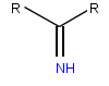
Iminoglycinuria
Wikipedia
Here, cotransporters such as sodium or chloride (part of the system of Na-K-Cl cotransporters ) couple with the amino or imino acids on the molecular level and transport them through specific integral membrane proteins that form ion channels , which are located within the cell membrane . [27] [28] From the cells, the absorbed or reabsorbed amino and imino acids eventually reach the blood.
-
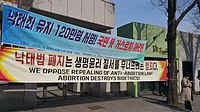
Abortion In South Korea
Wikipedia
. ^ a b c d e f "South Korea: Stop criminalization of abortion" . Amnesty International . 28 October 2016. doi : 10.1163/2210-7975_hrd-9211-2016094 .
-
Limited-Stage Small Cell Lung Carcinoma
Wikipedia
Comparing to continued smokers, patients who quit at or after diagnosis lower the risk of death by 45%. [28] References [ edit ] ^ a b c Stinchcombe TE, Gore EM (2010).

- Hemiballismus Wikipedia
-
Atopy
Wikipedia
In addition, several studies have documented that an IgE-mediated response to S. aureus is present in people with atopic eczema. [27] [28] Changes in prevalence [ edit ] In adults, the prevalence of IgE sensitization to allergens from house dust mite and cat, but not grass, seem to decrease over time as people age. [29] However, the biological reasons for these changes are not fully understood.

-
Soil-Transmitted Helminthiasis
Wikipedia
. ^ "Neglected Tropical Diseases" . cdc.gov . June 6, 2011 . Retrieved 28 November 2014 . ^ London Declaration (2012) (30 January 2012).

-
Genital Wart
Wikipedia
. ^ a b c d e f g h i "CDC - Genital Warts - 2010 STD Treatment Guidelines" . www.cdc.gov . 28 January 2011. Archived from the original on 8 July 2018 .TP53, PDXP, CDKN2A, IFNB1, IL10, PIK3CG, PIK3CA, PIK3CB, IFNG, IFNA13, IFNA1, PIK3CD, ZAP70, BCL2, SULT2A1, JUN, NDRG1, PYCARD, ARTN, NR0B2, UBL4A, WNT1, VEGFA, YAP1, PDZD2, CADM1, AGRP, CD274, IL22, FOXP3, DNAJA4, PNPLA2, MIB1, IL21, BIRC7, IL33, MPEG1, MIR99B, HNP1, MIR551B, DEFA1B, DEFB4B, TTK, PTPN11, TGFB1, IL6, XIAP, STS, DEFA1, DEFB4A, EGFR, FCGRT, G6PD, GLB1, ICAM3, IDUA, IGF1R, IL2, IL18, STAT2, CXCL10, JAK1, MBL2, MKI67, PCNA, SERPINF1, PIN1, PTPN6, BIRC3, RPS27, XCL1, STAT1, H3P10

-
Congenital Heart Block
Wikipedia
See also: Neonatal lupus erythematosus Congenital heart block The conduction system of the heart (shown in yellow) Specialty Medical genetics Symptoms slow heart rate [1] Usual onset in utero. [1] Diagnostic method fetal echocardiogram and Doppler and ELISA for the mother [1] Treatment fluorinated steroids, beta agonists, IVIG, HCQ, pace maker implantation and maternal plasmapheresis. [1] [2] Frequency 1 child in every 15000-20000 [3] The congenital heart block ( CHB ) is the heart block that is diagnosed in fetus ( in utero ) or within the first 28 days after birth [1] [4] (neonatal period), some studies also include the diagnosis during early childhood to the definition of CHB. [5] It refers to the disorder in the electrical conduction system within the heart muscle, [4] which leads to the failure in pumping the blood efficiently into the aorta and the pulmonary trunk .


