Rare isolated myopia is a rare, genetic, refraction anomaly disorder characterized by non-syndromic severe myopia, which may be associated with cataract and vitreoretinal degeneration (retinal detachment) that may lead to blindness.
Borish and Duke-Elder classified myopia by these physical causes: [32] [33] Axial myopia is attributed to an increase in the eye's axial length [34] Refractive myopia is attributed to the condition of the refractive elements of the eye. [34] Borish further subclassified refractive myopia: [32] Curvature myopia is attributed to excessive, or increased, curvature of one or more of the refractive surfaces of the eye, especially the cornea. [34] In those with Cohen syndrome , myopia appears to result from high corneal and lenticular power. [35] Index myopia is attributed to variation in the index of refraction of one or more of the ocular media. [34] As with any optical system experiencing a defocus aberration , the effect can be exaggerated or masked by changing the aperture size . ... Myopia is often induced this way in various animal models to study the pathogenesis and mechanism of myopia development. [50] Degree [ edit ] The degree of myopia is described in terms of the power of the ideal correction , which is measured in diopters : [51] Low myopia usually describes myopia of −3.00 diopters or less (i.e. closer to 0.00). [34] Moderate myopia usually describes myopia between −3.00 and −6.00 diopters . [34] Those with moderate amounts of myopia are more likely to have pigment dispersion syndrome or pigmentary glaucoma . [52] High myopia usually describes myopia of −6.00 or more. [34] [53] People with high myopia are more likely to have retinal detachments [54] and primary open angle glaucoma . [55] They are also more likely to experience floaters , shadow-like shapes which appear in the field of vision . [56] Age at onset [ edit ] Myopia is sometimes classified by the age at onset: [51] Congenital myopia, also known as infantile myopia, is present at birth and persists through infancy. [40] Youth onset myopia occurs in early childhood or teenage, and the ocular power can keep varying until the age of 21, before which any form of corrective surgery is usually not recommended by ophthalmic specialists around the world. [40] School myopia appears during childhood, particularly the school-age years. [57] This form of myopia is attributed to the use of the eyes for close work during the school years. [34] Adult onset myopia Early adult onset myopia occurs between ages 20 and 40. [40] Late adult onset myopia occurs after age 40. [40] Prevention [ edit ] Some suggest that more time spent outdoors during childhood is effective for prevention. [4] Various methods have been employed in an attempt to decrease the progression of myopia, although studies show mixed results. [58] Many myopia treatment studies have a number of design drawbacks: small numbers , lack of adequate control group, and failure to mask examiners from knowledge of treatments used. ... ISBN 978-0-7506-9895-5 . ^ Summanen P, Kivitie-Kallio S, Norio R, Raitta C, Kivelä T (May 2002). "Mechanisms of myopia in Cohen syndrome mapped to chromosome 8q22". Investigative Ophthalmology & Visual Science . 43 (5): 1686–93.
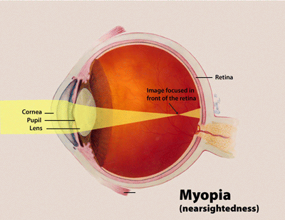
Overview Nearsightedness (myopia) is a common vision condition in which near objects appear clear, but objects farther away look blurry. It occurs when the shape of the eye — or the shape of certain parts of the eye — causes light rays to bend (refract) inaccurately. Light rays that should be focused on nerve tissues at the back of the eye (retina) are focused in front of the retina. Nearsightedness usually develops during childhood and adolescence, and it usually becomes more stable between the ages of 20 and 40. Myopia tends to run in families. A basic eye exam can confirm nearsightedness.
A number sign (#) is used with this entry because of evidence that high myopia with cataract and vitreoretinal degeneration (MCVD) is caused by homozygous mutation in the LEPREL1 gene (P3H2; 610341) on chromosome 3q28. Clinical Features Mordechai et al. (2011) studied a large consanguineous Israeli Bedouin kindred segregating autosomal recessive nonsyndromic severe myopia with variable expressivity of cataract and vitreoretinal degeneration. The 13 affected family members all presented with poor eyesight in childhood, and all had axial myopia, with increased axial lengths ranging between 25.1 mm and 30.5 mm. Eleven patients developed cataracts that were significant enough to warrant surgery in 1 or both eyes, usually in the first or second decade of life. In 3 patients, subluxated lenses were detected, and were associated with cataract in 2 patients and with lens coloboma in 1 patient.
A number sign (#) is used with this entry because of evidence that myopia-23 (MYP23) is caused by homozygous mutation in the LRPAP1 gene (104225) on chromosome 4p16. Description Myopia, or nearsightedness, is a refractive error of the eye. Light rays from a distant object are focused in front of the retina and those from a near object are focused in the retina; therefore distant objects are blurry and near objects are clear (summary by Kaiser et al., 2004). For a discussion of genetic heterogeneity of myopia, see 160700. Molecular Genetics In 3 consanguineous Saudi Arabian families in which multiple sibs, aged 2 to 16 years, had nonsyndromic extreme myopia with spherical equivalents of -17 diopters or greater and subnormal best-corrected visual acuity, Aldahmesh et al. (2013) performed autozygome analysis and identified only 1 interval exclusively shared among all 8 affected individuals. Linkage analysis confirmed the autozygous interval, and exome sequencing revealed 2 homozygous truncating mutations in the LRPAP1 gene: a 1-bp deletion in 1 family (104225.0001) and a 2-bp deletion in the other 2 families (104225.0002).
A number sign (#) is used with this entry because of evidence that myopia-6 (MYP6) is caused by heterozygous mutation in the SCO2 gene (604272) on chromosome 22q13. Description Myopia, or nearsightedness, is a refractive error of the eye. Light rays from a distant object are focused in front of the retina and those from a near object are focused in the retina; therefore distant objects are blurry and near objects are clear (summary by Kaiser et al., 2004). For a discussion of genetic heterogeneity of susceptibility to myopia, see 160700. Mapping Stambolian et al. (2004) sought to identify a myopia susceptibility gene related to mild/moderate myopia, which is a very common disorder particularly in Chinese and Japanese populations (Saw et al., 1996) and Ashkenazi Jews.
A group of rare arthrogryposis syndromes characterized by congenital contractures of two or more areas of the body, primarily involving the hands and feet, while the proximal joints are largely spared, in the absence of primary neurologic and/or muscle disease affecting limb function.
Clinical description Intelligence is usually normal. Etiology The syndrome is caused by a mutation in the TRPV4 gene (12q24.1) and is transmitted in an autosomal dominant manner.
Spondylometaphyseal dysplasia, Kozlowski type is a bone disease characterized by short stature involving the trunk. "Spondylo"refers to the spine (vertebrae), "metaphysis" refers to the wide part of the bone that contains the growth plate (the part of the bone that grows during childhood), and "dysplasia" means abnormal growth. It usually starts in early childhood when poor growth with uneven stature and a waddling gait with bow legs (genu varum) is noticed. Early osteoarthritis of the joints is also common. Other signs and symptoms include small hands and fingers, spine deformities, and X-ray showing short vertebra, mild metaphyseal changes, severe delay in ossification , square, short, flared iliac wings (the broadest part of the pelvic bone) and a flat and irregular hipbone. Spondylometaphyseal dysplasia, Kozlowski type is caused by mutations in the TRPV4 gene.
3-Phosphoglycerate dehydrogenase deficiency (3-PGDH deficiency) is an autosomal recessive form of serine deficiency syndrome (see this term) characterized clinically in the few reported cases by congenital microcephaly, psychomotor retardation and intractable seizures in the infantile form and by absence seizures, moderate developmental delay and behavioral disorders in the juvenile form
A number sign (#) is used with this entry because phosphoglycerate dehydrogenase deficiency (PHGDHD) is caused by homozygous or compound heterozygous mutation in the PHGDH gene (606879) on chromosome 1p12. See also Neu-Laxova syndrome (NLS; 256520), an allelic disorder with a much more severe phenotype that usually results in neonatal death.
It typically affects older patients and clinical presentation includes signs and symptoms of renal dysfunction, sometimes leading to nephrotic syndrome and end stage renal disease. Cardiac, liver and nerves involvement has also been described.
The spectrum of BSCL2 -related neurologic disorders includes Silver syndrome and variants of Charcot-Marie-Tooth neuropathy type 2, distal hereditary motor neuropathy (dHMN) type V, and spastic paraplegia 17. ... Diagnosis The phenotypic spectrum of BSCL2 -related neurologic disorders includes Silver syndrome and variants of Charcot-Marie-Tooth disease type 2, distal hereditary motor neuropathy (dHMN) type V, and spastic paraplegia 17. ... Muscle tone is normal; tendon reflexes may be preserved or slightly brisk. Subtype 4. Silver syndrome phenotype [Silver 1966]. Findings are mild-to-severe symmetric or unilateral amyotrophy of the small muscles of the hand, variable spasticity of the lower limbs, and other signs of pyramidal tract disturbance (very brisk tendon reflexes and/or extensor plantar responses and/or increased muscle tone). ... Depending on the absence or presence of clinical and electrophysiologic sensory abnormalities, affected individuals may show spinal CMT syndrome or hereditary motor and sensory neuropathy (HMSN) type II. ... A detailed genotype-phenotype correlation study in 90 individuals with the p.Asn88Ser pathogenic variant demonstrated that 24.4% of individuals with the variant remained asymptomatic (subtype 1) or were only subclinically affected (subtype 2) [Auer-Grumbach et al 2005]. Nomenclature Silver syndrome was first described in 1966 in two British families [Silver 1966].
Find sources: "Pathological jealousy" – news · newspapers · books · scholar · JSTOR ( December 2011 ) ( Learn how and when to remove this template message ) Pathological jealousy , also known as morbid jealousy , Othello syndrome or delusional jealousy , is a psychological disorder in which a person is preoccupied with the thought that their spouse or sexual partner is being unfaithful without having any real proof, [1] along with socially unacceptable or abnormal behaviour related to these thoughts. [1] The most common cited forms of psychopathology in morbid jealousy are delusions and obsessions. ... Parkinson's). The name "Othello Syndrome" comes from the character in Shakespeare's play Othello , who murders his wife as a result of a false belief that she has been unfaithful. ... ISSN 2056-4678 . ^ http://www.freethoughtlebanon.net/2012/10/pathological-jealousy-its-symptoms-and-definition/ ^ Crichton, P. Did Othello have 'the Othello Syndrome? Journal of Forensic Psychiatry & Psychology . 1996; 7(1) :161-9. ^ Cobb, J (1979). ... Aspects of morbid jealous. http://apt.rcpsych.org/content/10/3/207.full.pdf+html ^ Enoch, M.D (1979). Uncommon Psychiatric Syndromes . Bristol: John Wright. pp. 25–40. ^ Easton, Judith, and Todd Shackelford. ... Sources [ edit ] Enoch, D. & Ball, H. (2001) The Othello Syndrome. In Enoch, D. & Ball, H. Uncommon psychiatric syndromes (fourth edition) pp50–73.
Over time this may lead to an Eisenmenger's syndrome the original VSD operating with a left-to-right shunt, now becomes a right-to-left shunt because of the increased pressures in the pulmonary vascular bed. Cause [ edit ] Congenital VSDs are frequently associated with other congenital conditions, such as Down syndrome . [5] A VSD can also form a few days after a myocardial infarction [6] (heart attack) due to mechanical tearing of the septal wall, before scar tissue forms, when macrophages start remodeling the dead heart tissue. ... "Congenital heart disease in infants with Down's syndrome". Southern Medical Journal . 87 (7): 724–7. doi : 10.1097/00007611-199407000-00010 . ... External links [ edit ] Classification D ICD - 10 : Q21.0 ICD - 9-CM : 745.4 MeSH : D006345 DiseasesDB : 13808 External resources MedlinePlus : 001099 eMedicine : med/3517 v t e Congenital heart defects Heart septal defect Aortopulmonary septal defect Double outlet right ventricle Taussig–Bing syndrome Transposition of the great vessels dextro levo Persistent truncus arteriosus Aortopulmonary window Atrial septal defect Sinus venosus atrial septal defect Lutembacher's syndrome Ventricular septal defect Tetralogy of Fallot Atrioventricular septal defect Ostium primum Consequences Cardiac shunt Cyanotic heart disease Eisenmenger syndrome Valvular heart disease Right pulmonary valves stenosis insufficiency absence tricuspid valves stenosis atresia Ebstein's anomaly Left aortic valves stenosis insufficiency bicuspid mitral valves stenosis regurgitation Other Underdeveloped heart chambers right left Uhl anomaly Dextrocardia Levocardia Cor triatriatum Crisscross heart Brugada syndrome Coronary artery anomaly Anomalous aortic origin of a coronary artery Ventricular inversion
Risk factors Risk factors for ventricular septal defect include: Premature birth Down syndrome and other genetic conditions Family history of heart problems present at birth (congenital heart defects) A baby born with ventricular septal defect may have other heart problems, such as: Atrial septal defect Coarctation of the aorta Double outlet syndrome Patent ductus arteriosus Tetralogy of Fallot If you already have a child with a congenital heart defect, a genetic counselor can discuss the risk of your next child having one. ... Without treatment, heart failure can develop. Eisenmenger syndrome. An unrepaired hole in the heart can lead to this complication after many years. ... Blood pressure rises in the lungs' arteries (pulmonary hypertension). This syndrome permanently damages the blood vessels in the lungs. ... Ask your health care provider which sports and types of exercise are safe for you or your child. People with Eisenmenger syndrome should avoid strenuous physical activity. ... Pregnancy is considered very high risk for those with Eisenmenger syndrome and is not recommended. Coping and support You may find that talking with others who've experienced similar events or situations can be helpful.
Further investigation has shown that only some people drinking this sort of beer get an iron overload syndrome, and that a similar syndrome occurred in people of African descent who have had no contact with this kind of beer (e.g., African Americans ). [6] This led investigators to the discovery of a gene polymorphism in the gene for ferroportin , which predisposes some people of African descent to iron overload. [7] Genetics [ edit ] SLC40A1 gene encodes for ferroportin . ... External links [ edit ] Bantu siderosis at NIH 's Office of Rare Diseases Classification D OMIM : 601195 MeSH : C537904 DiseasesDB : 35029 v t e Metal deficiency and toxicity disorders Iron excess: Iron overload Hemochromatosis Hemochromatosis/HFE1 Juvenile/HFE2 HFE3 African iron overload/HFE4 Aceruloplasminemia Atransferrinemia Hemosiderosis deficiency: Iron deficiency Copper excess: Copper toxicity Wilson's disease deficiency: Copper deficiency Menkes disease / Occipital horn syndrome Zinc excess: Zinc toxicity deficiency: Acrodermatitis enteropathica Other Inborn errors of metabolism v t e Genetic disorder , membrane: Solute carrier disorders 1-10 SLC1A3 Episodic ataxia 6 SLC2A1 De Vivo disease SLC2A5 Fructose malabsorption SLC2A10 Arterial tortuosity syndrome SLC3A1 Cystinuria SLC4A1 Hereditary spherocytosis 4 / Hereditary elliptocytosis 4 SLC4A11 Congenital endothelial dystrophy type 2 Fuchs' dystrophy 4 SLC5A1 Glucose-galactose malabsorption SLC5A2 Renal glycosuria SLC5A5 Thyroid dyshormonogenesis type 1 SLC6A19 Hartnup disease SLC7A7 Lysinuric protein intolerance SLC7A9 Cystinuria 11-20 SLC11A1 Crohn's disease SLC12A3 Gitelman syndrome SLC16A1 HHF7 SLC16A2 Allan–Herndon–Dudley syndrome SLC17A5 Salla disease SLC17A8 DFNA25 21-40 SLC26A2 Multiple epiphyseal dysplasia 4 Achondrogenesis type 1B Recessive multiple epiphyseal dysplasia Atelosteogenesis, type II Diastrophic dysplasia SLC26A4 Pendred syndrome SLC35C1 CDOG 2C SLC39A4 Acrodermatitis enteropathica SLC40A1 African iron overload see also solute carrier family
Description African iron overload is a distinct iron-loading disorder prevalent in Africa. Formerly termed Bantu siderosis, the disorder results from a predisposition to iron loading that is exacerbated by excessive intake of dietary iron. It is particularly a problem among Africans who drink a traditional beer brewed in non-galvanized steel drums. Although the disorder was once attributed to dietary excess alone, serious iron overload does not develop in all beer drinkers, and not all patients with iron overload consume excessive amounts of the beer (summary by Andrews, 1999). Clinical Features The pattern of iron deposition among persons with African iron overload differs from that among those with hereditary hemochromatosis (see 235200) (Gangaidzo et al., 1999).
African iron overload is a condition that involves absorption of too much iron from the diet. The excess iron is stored in the body's tissues and organs, particularly the liver, bone marrow , and spleen. Humans cannot increase the excretion of iron, although some iron is lost through bleeding or when cells of the intestine (enterocytes) are shed at the end of the cells' lifespan. Iron levels in the body are primarily regulated through control of how much iron is absorbed from the diet. African iron overload results from a diet high in iron. It is particularly associated with consumption of a traditional African beer that contains dissolved iron from the metal drums in which it is brewed.
A rare disorder described in sub-Saharan African populations and characterized by iron overload due to excess dietary iron intake and possibly genetic factors, leading to hepatic portal fibrosis and micronodular cirrhosis.
COL4A1 may be mutated in other diseases that need to be distinguished, including brain small vessel disease with hemorrhage and HANAC syndrome . CADASIL syndrome is caused by a mutation in a different gene, but may cause similar symptoms.
Porencephaly Specialty Medical genetics , neurology Porencephaly is an extremely rare cephalic disorder involving encephalomalacia . [1] It is a neurological disorder of the central nervous system characterized by cysts or cavities within the cerebral hemisphere . [2] Porencephaly was termed by Heschl in 1859 to describe a cavity in the human brain. [3] Derived from Greek roots, the word porencephaly means 'holes in the brain'. [4] The cysts and cavities (cystic brain lesions) are more likely to be the result of destructive (encephaloclastic) cause, but can also be from abnormal development (malformative), direct damage, inflammation, or hemorrhage. [5] The cysts and cavities cause a wide range of physiological, physical, and neurological symptoms. [6] Depending on the patient, this disorder may cause only minor neurological problems, without any disruption of intelligence, while others may be severely disabled or face death before the second decade of their lives. However, this disorder is far more common within infants, and porencephaly can occur both before or after birth. [2] Contents 1 Signs and symptoms 2 Cause 2.1 Genetics 2.2 Cocaine and other street drugs 3 Diagnostics 4 Treatments 5 Prognosis 6 Research 7 See also 8 References 9 External links Signs and symptoms [ edit ] Patients diagnosed with porencephaly display a variety of symptoms, from mild to severe effects on the patient. Patients with severe cases of porencephaly suffer epileptic seizures and developmental delays, whereas patients with a mild case of porencephaly display little to no seizures and healthy neurodevelopment. Infants with extensive defects show symptoms of the disorder shortly after birth, and the diagnosis is usually made before the age of 1. [2] [7] The following text lists out common signs and symptoms of porencephaly in affected individuals along with a short description of certain terminologies. [2] [6] [7] [8] Degenerative or non-degenerative cavities or cysts Delayed growth and development Spastic paresis – weakness or loss in voluntary movement Contractures – painful shortening of muscles affecting motion Hypotonia – reduced muscle strength Epileptic seizures and epilepsy – multiple symptoms that involve sudden muscle spasms and loss of consciousness Macrocephaly – condition where head circumference is larger compared to other children of a certain age Microcephaly – condition where head circumference is smaller compared to other children of a certain age Hemiplegia – paralysis of appendages Tetraplegia – paralysis of limb leading to loss of function Intellectual and cognitive disability Poor or absent speech development Hydrocephalus – accumulation of cerebrospinal fluid in the brain Mental retardation Poor motor control, abnormal movements of appendages Cerebral palsy – a motor condition causing movement disabilities Blood vascular diseases such as intracerebral hemorrhage and cerebral infarction . Cerebral white-matter lesions Cause [ edit ] Porencephaly is a rare disorder.
A number sign (#) is used with this entry because of evidence that brain small vessel disease-2 (BSVD2) is caused by heterozygous mutation in the COL4A2 gene (120090) on chromosome 13q34. Description Brain small vessel disease-2 is an autosomal dominant disorder characterized by variable neurologic impairment resulting from disturbed vascular supply that leads to cerebral degeneration. The disorder is often associated with 'porencephaly' on brain imaging. Affected individuals typically have hemiplegia, seizures, and intellectual disability, although the severity is variable (summary by Yoneda et al., 2012). For a discussion of genetic heterogeneity of brain small vessel disease, see BSVD1 (175780).
A number sign (#) is used with this entry because of evidence that brain small vessel disease-1 with or without ocular anomalies (BSVD1) is caused by heterozygous mutation in the COL4A1 gene (120130) on chromosome 13q34. Description Brain small vessel disease-1 is an autosomal dominant disorder with variable manifestations resulting from disruption of vascular basement membranes, particularly in the cerebral vasculature. The increased fragility of these vessels render them susceptible to hemorrhage, as early as in utero or by birth trauma, although the risk remains throughout life and some patients may present in adulthood. This genetic predisposition may extend beyond hemorrhagic stroke to include retinal and renal vascular defects. Clinical features thus reflect the location and severity of the vascular defect, including impaired neurologic development or function, hemiplegia, seizures, and variable ocular anomalies.
When additional central nervous system (CNS) signs, such as intellectual deficit, ataxia, or extrapyramidal signs, are present, the syndrome is referred to as complicated SPG. ... Complicated SPG2 is not clearly distinguishable from mild Pelizaeus-Merzbacher disease (PMD) and null syndrome (see these terms). Antenatal diagnosis Prenatal genetic testing is possible when a family's underlying PLP1 mutation has been identified.
A number sign (#) is used with this entry because spastic paraplegia-2 can be caused by mutation in the myelin proteolipid protein gene (PLP1; 300401) and is therefore allelic to Pelizaeus-Merzbacher disease (PMD; 312080). Description The hereditary spastic paraplegias (SPG) are a group of clinically and genetically diverse disorders characterized by progressive, usually severe, lower extremity spasticity; see reviews of Fink et al. (1996) and Fink (1997). Some forms of SPG are considered 'uncomplicated,' i.e., progressive spasticity occurs in isolation; others are considered 'complicated,' i.e., progressive spasticity occurs with other neurologic features. X-linked, autosomal dominant (see 182600), and autosomal recessive (see 270800) forms of SPG have been described. For discussion of genetic heterogeneity of X-linked SPG, see 303350. Clinical Features Johnston and McKusick (1962) reported a kindred in which the disorder began as 'pure' spastic paraparesis, but the patients later developed nystagmus, dysarthria, sensory disturbance, and mental retardation, with half the patients having optic atrophy.
Spastic paraplegia type 2 is part of a group of genetic disorders known as hereditary spastic paraplegias. These disorders are characterized by progressive muscle stiffness (spasticity) and the development of paralysis of the lower limbs (paraplegia). Hereditary spastic paraplegias are divided into two types: pure and complex. The pure types involve the lower limbs. The complex types involve the lower limbs and can also affect the upper limbs to a lesser degree; the structure or functioning of the brain; and the nerves connecting the brain and spinal cord to muscles and sensory cells that detect sensations such as touch, pain, heat, and sound (the peripheral nervous system). Spastic paraplegia type 2 can occur in either the pure or complex form.
Differential diagnosis Differential diagnosis includes dysmorphic growth disorders with prenatal onset growth failure, namely Silver-Russell syndrome and 3M-syndrome. Antenatal diagnosis Prenatal molecular genetic testing is possible but is rational only in families known to be affected with the disease.
Initial diagnostic considerations included 3M syndrome (273750) and Silver-Russell syndrome (SRS; 180860). ... Paavola et al. (1999) studied the location of the genes for Meckel syndrome (MKS1; 249000) and mulibrey nanism, which had been mapped to the same region, 17q21-q24. ... However, in the common critical region, the conserved haplotypes were different in Meckel syndrome and mulibrey nanism patients. A transcript map was constructed by assigning ESTs and genes, derived from the human gene map, to the bacterial clone contig.
Mulibrey nanism is a rare genetic disorder characterized by profound growth delays and distinctive abnormalities of the muscles, liver, brain, and eyes. The acronym MULIBREY stands for (MU)scle, (LI)ver, (BR)ain, and (EY)e; nanism is another word for dwarfism . Signs and symptoms of the disorder may include constrictive pericarditis ; low birth weight; short stature; severe progressive growth delays; hypotonia; hepatomegaly ; and yellow discoloration of the eyes in infancy. It is caused by mutations in the TRIM37 gene and is inherited in an autosomal recessive manner. Treatment may include surgery for constrictive pericarditis, medications for progressive heart failure and hormone replacement therapy.
More than 10% of cases are asymptomatic and in rare cases a carcinoid syndrome may be observed. Epidemiology PHNEC has been estimated to have an incidence of approximately 1/500,000, accounting for <1% of all malignancies. ... In the early stages of PHNEC, patients may exhibit nonspecific symptoms, including upper abdominal pain and distension or fullness, while some may suffer from carcinoid syndrome (paroxysmal flushing, episodes of asthma-like wheezing, right-side heart failure and diarrhea).
Patients with elevated anticardiolipin antibodies were considered to have the antiphospholipid syndrome (107320) and were excluded from the study; patients with chromosomal abnormalities were also excluded. ... In this respect, the 20210G-A mutation may resemble the antiphospholipid syndrome, which has been well documented to cause abortions as well as arterial and venous thromboses.
Prothrombin thrombophilia is an inherited disorder of blood clotting. Thrombophilia is an increased tendency to form abnormal blood clots in blood vessels. People who have prothrombin thrombophilia are at somewhat higher than average risk for a type of clot called a deep venous thrombosis , which typically occurs in the deep veins of the legs. Affected people also have an increased risk of developing a pulmonary embolism , which is a clot that travels through the bloodstream and lodges in the lungs. Most people with prothrombin thrombophilia never develop abnormal blood clots, however.
Differential diagnosis Differential diagnosis includes other congenital infections (rubella, CMV, HSV1 and HSV2, regrouped with Tg infection in the TORCH syndrome) and pseudo-TORCH and Aicardi-Goutières syndromes.
Furthermore, a ruptured popliteal cyst may cause compartment syndrome in the calf or even the thigh. ... Ruptured Baker's cyst producing a pseudothrombophlebitis syndrome Canadian Journal of Surgery Aug 2000 43(4) 255 ^ Drescher MJ.