-
Ganglioneuroma
Orphanet
Association with neurofibromatosis type I, multiple endocrine neoplasia type 2B and Turner syndrome was reported.RET, PHOX2B, SDHC, KIF1B, MYO1H, ASCL1, GDNF, EDN3, BDNF, MYCN, PTEN, CD44, NCAM1, NTRK1, CCND1, THBS1, KHSRP, ULK1, PRDM2, MAP3K12, WT1, TP53, TNFRSF1B, VIP, VEGFC, VEGFA, NKX2-1, CRISP2, VTN, ADRB1, BECN1, DLK1, POU5F1P3, C9orf72, PRSS55, GALP, SPZ1, GAL, DKK3, SIGLEC7, CADM1, HEY1, TARDBP, TBC1D9, PSIP1, PDPN, OLIG2, TCF3, FUBP1, TH, S100A1, SYP, IL2RB, HCLS1, GNB2, GFAP, FLT4, VEGFD, ERBB3, DCX, DAPK3, CUX1, CS, CORT, CASR, BCL2, ALK, ADRB3, HIC1, INSM1, SST, LGALS3, SLC2A1, S100B, ADRB2, PTHLH, PRL, POU5F1, POMC, ABCB1, SERPINF1, PCNA, PBX1, NTRK2, NOTCH3, MSX1, MEN1, POU5F1P4

-
Insect Bites And Stings
Wikipedia
External links [ edit ] Classification D ICD - 10 : T14.1 , X23-X25, W57 ICD - 9-CM : 919.4 , 989.5 , E905.3 , E905.5 , E906.4 MeSH : D007299 External resources MedlinePlus : 000033 Venomous Arthropods chapter in United States Environmental Protection Agency and University of Florida / Institute of Food and Agricultural Sciences National Public Health Pesticide Applicator Training Manual v t e Diseases of the skin and appendages by morphology Growths Epidermal Wart Callus Seborrheic keratosis Acrochordon Molluscum contagiosum Actinic keratosis Squamous-cell carcinoma Basal-cell carcinoma Merkel-cell carcinoma Nevus sebaceous Trichoepithelioma Pigmented Freckles Lentigo Melasma Nevus Melanoma Dermal and subcutaneous Epidermal inclusion cyst Hemangioma Dermatofibroma (benign fibrous histiocytoma) Keloid Lipoma Neurofibroma Xanthoma Kaposi's sarcoma Infantile digital fibromatosis Granular cell tumor Leiomyoma Lymphangioma circumscriptum Myxoid cyst Rashes With epidermal involvement Eczematous Contact dermatitis Atopic dermatitis Seborrheic dermatitis Stasis dermatitis Lichen simplex chronicus Darier's disease Glucagonoma syndrome Langerhans cell histiocytosis Lichen sclerosus Pemphigus foliaceus Wiskott–Aldrich syndrome Zinc deficiency Scaling Psoriasis Tinea ( Corporis Cruris Pedis Manuum Faciei ) Pityriasis rosea Secondary syphilis Mycosis fungoides Systemic lupus erythematosus Pityriasis rubra pilaris Parapsoriasis Ichthyosis Blistering Herpes simplex Herpes zoster Varicella Bullous impetigo Acute contact dermatitis Pemphigus vulgaris Bullous pemphigoid Dermatitis herpetiformis Porphyria cutanea tarda Epidermolysis bullosa simplex Papular Scabies Insect bite reactions Lichen planus Miliaria Keratosis pilaris Lichen spinulosus Transient acantholytic dermatosis Lichen nitidus Pityriasis lichenoides et varioliformis acuta Pustular Acne vulgaris Acne rosacea Folliculitis Impetigo Candidiasis Gonococcemia Dermatophyte Coccidioidomycosis Subcorneal pustular dermatosis Hypopigmented Tinea versicolor Vitiligo Pityriasis alba Postinflammatory hyperpigmentation Tuberous sclerosis Idiopathic guttate hypomelanosis Leprosy Hypopigmented mycosis fungoides Without epidermal involvement Red Blanchable Erythema Generalized Drug eruptions Viral exanthems Toxic erythema Systemic lupus erythematosus Localized Cellulitis Abscess Boil Erythema nodosum Carcinoid syndrome Fixed drug eruption Specialized Urticaria Erythema ( Multiforme Migrans Gyratum repens Annulare centrifugum Ab igne ) Nonblanchable Purpura Macular Thrombocytopenic purpura Actinic/solar purpura Papular Disseminated intravascular coagulation Vasculitis Indurated Scleroderma / morphea Granuloma annulare Lichen sclerosis et atrophicus Necrobiosis lipoidica Miscellaneous disorders Ulcers Hair Telogen effluvium Androgenic alopecia Alopecia areata Systemic lupus erythematosus Tinea capitis Loose anagen syndrome Lichen planopilaris Folliculitis decalvans Acne keloidalis nuchae Nail Onychomycosis Psoriasis Paronychia Ingrown nail Mucous membrane Aphthous stomatitis Oral candidiasis Lichen planus Leukoplakia Pemphigus vulgaris Mucous membrane pemphigoid Cicatricial pemphigoid Herpesvirus Coxsackievirus Syphilis Systemic histoplasmosis Squamous-cell carcinoma v t e General wounds and injuries Abrasions Abrasion Avulsion Blisters Blood blister Coma blister Delayed blister Edema blister Fracture blister Friction blister Sucking blister Bruises Hematoma / Ecchymosis Battle's sign Raccoon eyes Black eye Subungual hematoma Cullen's sign Grey Turner's sign Retroperitoneal hemorrhage Animal bites Insect bite Spider bite Snakebite Other: Ballistic trauma Stab wound Blunt trauma /superficial/ closed Penetrating trauma / open Aerosol burn Burn / Corrosion / Chemical burn Frostbite Occupational injuries Traumatic amputation By region Hand injury Head injury Chest trauma Abdominal trauma v t e Animal bites and stings Arthropod bites and stings Arachnid Demodex mite bite Scorpion sting Spider bite / Arachnidism Latrodectism Loxoscelism Insects Ant sting Bee sting Cimicosis Mosquito bite Pulicosis Reduviid bite Myriapoda Centipede bite Millipede burn Vertebrate Alligator attack Bear attack Beaver attack Boar attack Cougar attack Cat bite Coyote attack Crocodile attack Dingo attack Dog attack Killer whale attack Leopard attack Lion attack Monkey bite Piranha fish attack Shark attack Snakebite Stingray attack Stonefish attack Tiger attack Venomous fish Walrus attack Wolf attack Other Animal attacks Bristleworm sting Cephalopod attack Cone snail sting Coral dermatitis Dog bite prevention Hydroid dermatitis Jellyfish dermatitis / Jellyfish sting Leech bite Man-eater Portuguese man-of-war dermatitis Sea anemone dermatitis Sea urchin injury Seabather's eruption

-
Plantar Fibromatosis
Wikipedia
External links [ edit ] Classification D ICD - 10 : M72.2 ICD - 9-CM : 728.71 OMIM : 126900 MeSH : D000071380 External resources eMedicine : derm/874 v t e Soft tissue disorders Capsular joint Synoviopathy Synovitis / Tenosynovitis Calcific tendinitis Stenosing tenosynovitis Trigger finger De Quervain syndrome Transient synovitis Ganglion cyst osteochondromatosis Synovial osteochondromatosis Plica syndrome villonodular synovitis Giant-cell tumor of the tendon sheath Bursopathy Bursitis Olecranon Prepatellar Trochanteric Subacromial Achilles Retrocalcaneal Ischial Iliopsoas Synovial cyst Baker's cyst Calcific bursitis Noncapsular joint Symptoms Ligamentous laxity Hypermobility Enthesopathy / Enthesitis / Tendinopathy upper limb Adhesive capsulitis of shoulder Impingement syndrome Rotator cuff tear Golfer's elbow Tennis elbow lower limb Iliotibial band syndrome Patellar tendinitis Achilles tendinitis Calcaneal spur Metatarsalgia Bone spur other/general: Tendinitis / Tendinosis Nonjoint Fasciopathy Fasciitis : Plantar Nodular Necrotizing Eosinophilic Fibromatosis / contracture Dupuytren's contracture Plantar fibromatosis Aggressive fibromatosis Knuckle pads

-
Skin Infection
Wikipedia
PMID 21214125 . v t e Diseases of the skin and appendages by morphology Growths Epidermal Wart Callus Seborrheic keratosis Acrochordon Molluscum contagiosum Actinic keratosis Squamous-cell carcinoma Basal-cell carcinoma Merkel-cell carcinoma Nevus sebaceous Trichoepithelioma Pigmented Freckles Lentigo Melasma Nevus Melanoma Dermal and subcutaneous Epidermal inclusion cyst Hemangioma Dermatofibroma (benign fibrous histiocytoma) Keloid Lipoma Neurofibroma Xanthoma Kaposi's sarcoma Infantile digital fibromatosis Granular cell tumor Leiomyoma Lymphangioma circumscriptum Myxoid cyst Rashes With epidermal involvement Eczematous Contact dermatitis Atopic dermatitis Seborrheic dermatitis Stasis dermatitis Lichen simplex chronicus Darier's disease Glucagonoma syndrome Langerhans cell histiocytosis Lichen sclerosus Pemphigus foliaceus Wiskott–Aldrich syndrome Zinc deficiency Scaling Psoriasis Tinea ( Corporis Cruris Pedis Manuum Faciei ) Pityriasis rosea Secondary syphilis Mycosis fungoides Systemic lupus erythematosus Pityriasis rubra pilaris Parapsoriasis Ichthyosis Blistering Herpes simplex Herpes zoster Varicella Bullous impetigo Acute contact dermatitis Pemphigus vulgaris Bullous pemphigoid Dermatitis herpetiformis Porphyria cutanea tarda Epidermolysis bullosa simplex Papular Scabies Insect bite reactions Lichen planus Miliaria Keratosis pilaris Lichen spinulosus Transient acantholytic dermatosis Lichen nitidus Pityriasis lichenoides et varioliformis acuta Pustular Acne vulgaris Acne rosacea Folliculitis Impetigo Candidiasis Gonococcemia Dermatophyte Coccidioidomycosis Subcorneal pustular dermatosis Hypopigmented Tinea versicolor Vitiligo Pityriasis alba Postinflammatory hyperpigmentation Tuberous sclerosis Idiopathic guttate hypomelanosis Leprosy Hypopigmented mycosis fungoides Without epidermal involvement Red Blanchable Erythema Generalized Drug eruptions Viral exanthems Toxic erythema Systemic lupus erythematosus Localized Cellulitis Abscess Boil Erythema nodosum Carcinoid syndrome Fixed drug eruption Specialized Urticaria Erythema ( Multiforme Migrans Gyratum repens Annulare centrifugum Ab igne ) Nonblanchable Purpura Macular Thrombocytopenic purpura Actinic/solar purpura Papular Disseminated intravascular coagulation Vasculitis Indurated Scleroderma / morphea Granuloma annulare Lichen sclerosis et atrophicus Necrobiosis lipoidica Miscellaneous disorders Ulcers Hair Telogen effluvium Androgenic alopecia Alopecia areata Systemic lupus erythematosus Tinea capitis Loose anagen syndrome Lichen planopilaris Folliculitis decalvans Acne keloidalis nuchae Nail Onychomycosis Psoriasis Paronychia Ingrown nail Mucous membrane Aphthous stomatitis Oral candidiasis Lichen planus Leukoplakia Pemphigus vulgaris Mucous membrane pemphigoid Cicatricial pemphigoid Herpesvirus Coxsackievirus Syphilis Systemic histoplasmosis Squamous-cell carcinomaSLC9A6, TLR2, FLG, TNF, IL17A, PAGR1, IL1B, IL4, FAM3B, DOCK8, IL13, GAST, GALNS, VWF, TYR, TST, MAPK1, CFLAR, STAT6, STAT3, RFC1, RARRES2, AIMP2, ADAM10, GRAP2, CXCL14, PLG, AHSA1, CABIN1, RNF19A, POLDIP2, SETD2, IRAK4, TLR7, CD177, TNS3, NBEAL1, AGBL2, RNASE7, PPARG, MPO, PCP4, HP, CD36, LTB4R, CRK, CRP, MAPK14, CTLA4, EDNRB, EGFR, ELN, F5, FN1, GAPDH, GRN, HIF1A, HSPD1, MYO9B, IDE, IFNA1, IFNA13, IFNG, IL1A, IL2, IL6, IL9, IL15, LCT, LGALS3, MAP2, ADSS2, RNR1, COPD
-
Panniculitis
Wikipedia
Most of these cases are now thought to be manifestation of tuberculosis and indeed they respond well to anti-tuberculous treatment. [ citation needed ] Without vasculitis [ edit ] Non-vasculitis forms of panniculitis that may occur include: Cytophagic histiocytic panniculitis was first described in 1980 by Winkelmann as a chronic histiocytic disease of the subcutaneous adipose tissue , which is characterized clinically by tender erythematous nodules , recurrent high fever , malaise , jaundice , organomegaly , serosal effusions, pancytopenia , hepatic dysfunction and coagulation abnormalities. [2] : 494 [8] CHP may occur either isolated or as part of cutaneous manifestations of hemophagocytic syndrome (HPS). [9] CHP is a rare and often fatal form of panniculitis with multisystem involvement. ... Cutaneous histopathological, immunohistochemical, and clinical manifestations inpatients with hemophagocytic syndrome. Military Medical old Consortium for Applied Retroviral Research (MMCARR). ... External links [ edit ] DermNet dermal-infiltrative/panniculitis DermAtlas -639418194 Classification D ICD - 10 : M79.3 ICD - 9-CM : 729.3 MeSH : D015434 DiseasesDB : 29081 v t e Disorders of subcutaneous fat Panniculitis Lobular without vasculitis Cold Cytophagic histiocytic Factitial Gouty Pancreatic Traumatic needle-shaped clefts Subcutaneous fat necrosis of the newborn Sclerema neonatorum Post-steroid panniculitis Lipodermatosclerosis Weber–Christian disease Lupus erythematosus panniculitis Sclerosing lipogranuloma with vasculitis: Nodular vasculitis / Erythema induratum Septal without vasculitis: Alpha-1 antitrypsin deficiency panniculitis Erythema nodosum Acute Chronic with vasculitis: Superficial thrombophlebitis Lipodystrophy Acquired generalized: Acquired generalized lipodystrophy partial: Acquired partial lipodystrophy Centrifugal abdominal lipodystrophy HIV-associated lipodystrophy Lipoatrophia annularis localized: Localized lipodystrophy Congenital Congenital generalized lipodystrophy Familial partial lipodystrophy Marfanoid–progeroid–lipodystrophy syndrome Poland syndromeHAVCR2, PSMB8, ADAR, LCK, RNASEH2C, RNASEH2B, IFIH1, ADA2, SAMHD1, TREX1, RNASEH2A, RASGRP1, PSMB9, PRKCD, PSMB4, OTULIN, KIF11, FAS, FASLG, CASP10, BRAF, SERPINA1, IL1B, SFRP5, TNF, IL1A, RARRES2, SMUG1, COL11A2, IL3RA, SUCNR1, RHOJ, IL6, PARP1, MAP2K7, AGL, HMGB1

-
Posterior Urethral Valve
Wikipedia
Pyelostomy followed by valve ablation - stoma is made in the pelvis of the kidney as a slightly high diversion , after which the valve is ablated and the stoma is closed Primary (transurethral) valve ablation - the valve is removed through the urethra without creation of a stoma The standard treatment is primary (transurethral) ablation of the valves. [9] Urinary diversion is used in selected cases, [9] and its benefit is disputed. [10] [11] Following surgery, the follow-up in patients with posterior urethral valve syndrome is long term, and often requires a multidisciplinary effort between paediatric surgeons/ paediatric urologists , paediatric nephrologists, pulmonologists , neonatologists , radiologists and the family of the patient. ... CS1 maint: multiple names: authors list ( link ) External links [ edit ] Classification D ICD - 10 : Q64.2 DiseasesDB : 34137 External resources eMedicine : ped/2357 radio/572 v t e Congenital malformations and deformations of urinary system Abdominal Kidney Renal agenesis / Potter sequence , Papillorenal syndrome cystic Polycystic kidney disease Meckel syndrome Multicystic dysplastic kidney Medullary sponge kidney Horseshoe kidney Renal ectopia Nephronophthisis Supernumerary kidney Pelvic kidney Dent's disease Alport syndrome Ureter Ectopic ureter Megaureter Duplicated ureter Pelvic Bladder Bladder exstrophy Urethra Epispadias Hypospadias Posterior urethral valves Penoscrotal transposition Vestigial Urachus Urachal cyst Urachal fistula Urachal sinusBNC2, H19, SRCAP, CHRM3, FGFR2, IGF2, ACE, REN, ACE2, AGT, TM7SF2, SMIM10L2B, SMIM10L2A, SLC33A1, HBHR, CXCL8, SLC3A1, AGTR1, VPS51, BMP7, B2M, ATM, ANGPT2, ANGPT1, AMBP, PGR-AS1

-
Radial Dysplasia
Wikipedia
It can occur in different ways, from a minor anomaly to complete absence of the radius , radial side of the carpal bones and thumb. [1] Hypoplasia of the distal humerus may be present as well and can lead to stiffness of the elbow. [2] Radial deviation of the wrist is caused by lack of support to the carpus, radial deviation may be reinforced if forearm muscles are functioning poorly or have abnormal insertions. [3] Although radial longitudinal deficiency is often bilateral, the extent of involvement is most often asymmetric. [1] The incidence is between 1:30,000 and 1:100,000 and it is more often a sporadic mutation rather than an inherited condition. [1] [3] In case of an inherited condition, several syndromes are known for an association with radial dysplasia, such as the cardiovascular Holt-Oram syndrome , the gastrointestinal VATER syndrome and the hematologic Fanconi anemia and TAR syndrome . [1] Other possible causes are an injury to the apical ectodermal ridge during upper limb development, [2] intrauterine compression, or maternal drug use ( thalidomide ). [3] Contents 1 Classification 2 Treatment 2.1 Splinting and stretching 2.2 Centralization 2.3 Radialization 2.4 Vascularized metatarsophalangeal (MTP)-joint transfer 3 References Classification [ edit ] Classification of radial dysplasia is practised through different models.

-
Kienböck's Disease
Wikipedia
External links [ edit ] Classification D ICD - 10 : M93.1 ICD - 9-CM : 732.3 MeSH : D010020 DiseasesDB : 7178 External resources eMedicine : orthoped/398 v t e Bone and joint disease Bone Inflammation endocrine : Osteitis fibrosa cystica Brown tumor infection : Osteomyelitis Sequestrum Involucrum Sesamoiditis Brodie abscess Periostitis Vertebral osteomyelitis Metabolic Bone density Osteoporosis Juvenile Osteopenia Osteomalacia Paget's disease of bone Hypophosphatasia Bone resorption Osteolysis Hajdu–Cheney syndrome Ainhum Gorham's disease Other Ischaemia Avascular necrosis Osteonecrosis of the jaw Complex regional pain syndrome Hypertrophic pulmonary osteoarthropathy Nonossifying fibroma Pseudarthrosis Stress fracture Fibrous dysplasia Monostotic Polyostotic Skeletal fluorosis bone cyst Aneurysmal bone cyst Hyperostosis Infantile cortical hyperostosis Osteosclerosis Melorheostosis Pycnodysostosis Joint Chondritis Relapsing polychondritis Other Tietze's syndrome Combined Osteochondritis Osteochondritis dissecans Child leg: hip Legg–Calvé–Perthes syndrome tibia Osgood–Schlatter disease Blount's disease foot Köhler disease Sever's disease spine Scheuermann's_disease arm: wrist Kienböck's disease elbow Panner disease

-
Glomerulocystic Kidney Disease
Wikipedia
It can also be found in a number of patients with the following: [7] Tuberous sclerosis complex medullary cystic kidney disease Jeune syndrome Nephronophthisis Meckel–Gruber syndrome Orofaciodigital syndrome Zellweger syndrome In addition, GCKD can be a component of renal dysplasia after fetal renal damage such as drug use by the mother. [7] Mechanism/Pathophysiology [ edit ] The mechanism of cyst formation in Glomerulocystic kidney disease is not well understood.
-
Lecithin Cholesterol Acyltransferase Deficiency
Wikipedia
"The molecular pathology of lecithin:cholesterol acyltransferase (LCAT) deficiency syndromes" . J. Lipid Res . 38 (2): 191–205. ... "The molecular basis of lecithin:cholesterol acyltransferase deficiency syndromes: a comprehensive study of molecular and biochemical findings in 13 unrelated Italian families" . ... "The molecular pathology of lecithin:cholesterol acyltransferase (LCAT) deficiency syndromes". Journal of Lipid Research . 38 (2): 191–205. ... External links [ edit ] Classification D ICD - 10 : E78.6 ICD - 9-CM : 272.5 OMIM : 245900 136120 MeSH : D007863 DiseasesDB : 7343 SNOMED CT : 238091006 External resources eMedicine : med/1270 v t e Inborn error of lipid metabolism : dyslipidemia Hyperlipidemia Hypercholesterolemia / Hypertriglyceridemia Lipoprotein lipase deficiency/Type Ia Familial apoprotein CII deficiency/Type Ib Familial hypercholesterolemia/Type IIa Combined hyperlipidemia/Type IIb Familial dysbetalipoproteinemia/Type III Familial hypertriglyceridemia/Type IV Xanthoma/Xanthomatosis Hypolipoproteinemia Hypoalphalipoproteinemia/HDL Lecithin cholesterol acyltransferase deficiency Tangier disease Hypobetalipoproteinemia/LDL Abetalipoproteinemia Apolipoprotein B deficiency Chylomicron retention disease Lipodystrophy Barraquer–Simons syndrome Other Lipomatosis Adiposis dolorosa Lipoid proteinosis APOA1 familial renal amyloidosis
-
Idiopathic Childhood Occipital Epilepsy Of Gastaut
Wikipedia
Find sources: "Idiopathic childhood occipital epilepsy of Gastaut" – news · newspapers · books · scholar · JSTOR ( October 2015 ) ( Learn how and when to remove this template message ) Idiopathic childhood occipital epilepsy of Gastaut (ICOE-G) is a pure but rare form of idiopathic occipital epilepsy that affects otherwise normal children and adolescents. [1] It is classified amongst benign idiopathic childhood focal epilepsies such as rolandic epilepsy and Panayiotopoulos syndrome . [2] Contents 1 Presentation 2 Cause 3 Pathophysiology 4 Diagnosis 4.1 Brain Magnetic Resonance Imaging 4.2 Electroencephalography 4.3 Differential diagnosis 5 Management 6 Prognosis 7 Epidemiology 8 References Presentation [ edit ] Seizures are purely occipital and primarily manifest with elementary visual hallucinations, blindness or both. [3] [4] They are usually frequent and diurnal, develop rapidly within seconds and are brief, lasting from a few seconds to 1–3 min, and, rarely, longer. [ citation needed ] Elementary visual hallucinations are the most common and characteristic ictal symptoms, and are most likely to be the first and often the only clinical manifestation. ... The differentiation of ICOE-G from Panayiotopoulos syndrome is straightforward. The seizures of ICOE-G are purely occipital, brief, frequent and diurnal. Conversely seizures in Panayiotopoulos syndrome manifest with autonomic manifestations, they are lengthy and infrequent; visual symptoms are rare and not the sole manifestation of a seizure. ... "Benign childhood focal epilepsies: assessment of established and newly recognized syndromes" . Brain . 131 (9): 2264–2286. doi : 10.1093/brain/awn162 .
-
Cerebral Folate Deficiency
Wikipedia
Cerebral folate deficiency Other names Cerebral folate deficiency syndrome, neurodegeneration due to cerebral folate transport deficiency, cerebral folate transport deficiency, FOLR1 deficiency [1] [2] 5-methyltetrahydrofolate is decreased in concentration in the human brain Causes Genetic disorder , [2] autoantibodies Diagnostic method Lumbar puncture Medication Folinic acid Frequency FOLR1 mutation, <20 described cases [2] Cerebral folate deficiency is a condition in which concentrations of 5-methyltetrahydrofolate are low in the brain as measured in the cerebral spinal fluid despite being normal in the blood. [3] Symptoms typically appear at about 5 to 24 months of age. [3] [2] Without treatment there may be poor muscle tone, trouble with coordination, trouble talking, and seizures . [3] One cause of cerebral folate deficiency is a mutation in a gene responsible for folate transport, specifically FOLR1 . [2] [4] This is inherited from a person's parents in an autosomal recessive manner. [2] Other causes appear to be Kearns–Sayre syndrome [5] and autoantibodies to the folate receptor . [6] [7] [8] For people with the FOLR1 mutation, even when the systemic deficiency is corrected by folate, the cerebral deficiency remains and must be treated with folinic acid . ... It is inherited from a person's parents in an autosomal recessive manner. [2] Other causes appear to be Kearns–Sayre syndrome [5] and autoantibodies to the folate receptor . [6] [7] [8] Furthermore, secondary cerebral folate deficiency can develop in patients suffering from other conditions. ... "The proton-coupled folate transporter (PCFT-SLC46A1) and the syndrome of systemic and cerebral folate deficiency of infancy: Hereditary folate malabsorption" .

-
Mucous Membrane Pemphigoid
Wikipedia
External links [ edit ] Classification D ICD - 10 : L12.1 ICD - 9-CM : 694.6 OMIM : 164185 MeSH : D010390 DiseasesDB : 30757 External resources Patient UK : Mucous membrane pemphigoid v t e Diseases of the skin and appendages by morphology Growths Epidermal Wart Callus Seborrheic keratosis Acrochordon Molluscum contagiosum Actinic keratosis Squamous-cell carcinoma Basal-cell carcinoma Merkel-cell carcinoma Nevus sebaceous Trichoepithelioma Pigmented Freckles Lentigo Melasma Nevus Melanoma Dermal and subcutaneous Epidermal inclusion cyst Hemangioma Dermatofibroma (benign fibrous histiocytoma) Keloid Lipoma Neurofibroma Xanthoma Kaposi's sarcoma Infantile digital fibromatosis Granular cell tumor Leiomyoma Lymphangioma circumscriptum Myxoid cyst Rashes With epidermal involvement Eczematous Contact dermatitis Atopic dermatitis Seborrheic dermatitis Stasis dermatitis Lichen simplex chronicus Darier's disease Glucagonoma syndrome Langerhans cell histiocytosis Lichen sclerosus Pemphigus foliaceus Wiskott–Aldrich syndrome Zinc deficiency Scaling Psoriasis Tinea ( Corporis Cruris Pedis Manuum Faciei ) Pityriasis rosea Secondary syphilis Mycosis fungoides Systemic lupus erythematosus Pityriasis rubra pilaris Parapsoriasis Ichthyosis Blistering Herpes simplex Herpes zoster Varicella Bullous impetigo Acute contact dermatitis Pemphigus vulgaris Bullous pemphigoid Dermatitis herpetiformis Porphyria cutanea tarda Epidermolysis bullosa simplex Papular Scabies Insect bite reactions Lichen planus Miliaria Keratosis pilaris Lichen spinulosus Transient acantholytic dermatosis Lichen nitidus Pityriasis lichenoides et varioliformis acuta Pustular Acne vulgaris Acne rosacea Folliculitis Impetigo Candidiasis Gonococcemia Dermatophyte Coccidioidomycosis Subcorneal pustular dermatosis Hypopigmented Tinea versicolor Vitiligo Pityriasis alba Postinflammatory hyperpigmentation Tuberous sclerosis Idiopathic guttate hypomelanosis Leprosy Hypopigmented mycosis fungoides Without epidermal involvement Red Blanchable Erythema Generalized Drug eruptions Viral exanthems Toxic erythema Systemic lupus erythematosus Localized Cellulitis Abscess Boil Erythema nodosum Carcinoid syndrome Fixed drug eruption Specialized Urticaria Erythema ( Multiforme Migrans Gyratum repens Annulare centrifugum Ab igne ) Nonblanchable Purpura Macular Thrombocytopenic purpura Actinic/solar purpura Papular Disseminated intravascular coagulation Vasculitis Indurated Scleroderma / morphea Granuloma annulare Lichen sclerosis et atrophicus Necrobiosis lipoidica Miscellaneous disorders Ulcers Hair Telogen effluvium Androgenic alopecia Alopecia areata Systemic lupus erythematosus Tinea capitis Loose anagen syndrome Lichen planopilaris Folliculitis decalvans Acne keloidalis nuchae Nail Onychomycosis Psoriasis Paronychia Ingrown nail Mucous membrane Aphthous stomatitis Oral candidiasis Lichen planus Leukoplakia Pemphigus vulgaris Mucous membrane pemphigoid Cicatricial pemphigoid Herpesvirus Coxsackievirus Syphilis Systemic histoplasmosis Squamous-cell carcinoma v t e Vesiculobullous disease Acantholysis ( epidermis ) Pemphigus Pemphigus vulgaris : Pemphigus vegetans of Hallopeau of Neumann Pemphigus foliaceus : Pemphigus erythematosus Endemic pemphigus Paraneoplastic pemphigus IgA pemphigus Subcorneal pustular Intraepidermal neutrophilic Other Transient acantholytic dermatosis Pemphigoid ( dermis ) IgG : Bullous pemphigoid Cicatricial pemphigoid Localised Gestational pemphigoid Pemphigoid nodularis Epidermolysis bullosa acquisita IgA : Linear IgA bullous dermatosis Childhood Adult Other bullous Dermatitis herpetiformis In diseases classified elsewhere Porphyria cutanea tarda Bullous lupus erythematosus PUVA-induced acrobullous dermatosis
-
Scopophobia
Wikipedia
Scopophobia , scoptophobia , or ophthalmophobia is an anxiety disorder characterized by a morbid fear of being seen or stared at by others. [1] The term scopophobia comes from the Greek σκοπέω skopeō , "look to, examine", [2] and φόβος phobos , "fear". [3] Ophthalmophobia comes from the Greek ὀφθαλμός ophthalmos , "eye". [4] Contents 1 Signs and symptoms 1.1 Related syndromes 2 Causes 3 Psychoanalytic views 4 Treatments 5 History 6 In popular culture 7 See also 8 References 9 Further reading Signs and symptoms [ edit ] Individuals with scopophobia generally exhibit symptoms in social situations when attention is brought upon them like public speaking . ... Some examples include: Being introduced to new people, being teased and/or criticized, embarrassing easily, and even answering a cell phone call in public. [5] Often scopophobia will result in symptoms common with other anxiety disorders . [6] The symptoms of scopophobia include an irrational feelings of panic , feelings of terror, feelings of dread, rapid heartbeat, shortness of breath, nausea, dry mouth , trembling, anxiety and avoidance . [7] Other symptoms related to scopophobia may be hyperventilation , muscle tension, dizziness, uncontrollable shaking or trembling, excessive eye watering and redness of the eyes. [8] Related syndromes [ edit ] Though scopophobia is a solitary disorder, many individuals with scopophobia also commonly experience other anxiety disorders . Scopophobia has been related to many other irrational fears and phobias. Specific phobias and syndromes that are similar to scopophobia include erythrophobia , the fear of blushing (which is found especially in young people), and an epileptic 's fear of being looked at, which may itself precipitate such an attack. [9] Scopophobia is also commonly associated with schizophrenia and other psychiatric disorders. ... Another related, yet very different syndrome, scopophilia , is the excessive enjoyment of looking at erotic items.
-
Adrenergic Storm
Wikipedia
Breathing is rapid and shallow while both pulse and blood pressure are dangerously elevated. [1] Causes [ edit ] There are several known causes of adrenergic storms; in the United States, cocaine overdose is the leading cause. [2] Any stimulant drug has the capacity to cause this syndrome if taken in sufficient doses, but even non-psychotropic drugs can very rarely provoke a reaction. ... Deaths have occurred from individuals attempting to combine MAOIs with various entheogens to attain a stronger psychedelic experience , both from adrenergic storms and serotonin syndrome . Combining drugs like MDMA , 2C-B , mescaline , 2C-T-7 , etc. with even small quantities of MAOIs - small quantities of both drugs - is still extremely risky. ... The mechanisms of idiopathic adrenergic storm are very poorly understood. Serotonin syndrome , in which an excess of serotonin in the synapses causes a similar crisis of hypertension and mental confusion, could be confused with an adrenergic storm. ... Abnormal echocardiograms, or chest pain are indicative of adrenergic crisis; Uncontrollable slow, rhythmic, and/or jerky movements, contractions and tension-often in every part of the body, dangerously high fever, eye rolling, and bruxism are more indicative of serotonin syndrome. Treatment [ edit ] If there is evidence of overdose or it is suspected, the patient should be given gastric lavage , activated charcoal , or both; this could make the difference between life and death in a close situation.
-
Diastematomyelia
Wikipedia
This delayed presentation of symptoms is related to the degree of strain placed on the spinal cord over time. Tethered spinal cord syndrome appears to be the result of improper growth of the neural tube during fetal development, and is closely linked to spina bifida . ... Progressive neurological lesions may result from the "tethering cord syndrome" (fixation of the spinal cord) by the diastematomyelia phenomenon or any of the associated disorders such as myelodysplasia , dysraphia of the spinal cord. ... References [ edit ] ^ a b Kuchner, E.F., Anand, A.K. & Kaufman, B.M., "Cervical Diastematomyelia" Neurosurgery, 16(4): 538-542, 1985 ^ Anand, A.K., Baim, R.S. & Kuchner, E.F., "Cervical Diastematomyelia" Computerized Radiology. 9(1):45-49, 1985 External links [ edit ] Classification D ICD - 10 : Q06.2 ICD - 9-CM : 742.51 OMIM : 222500 DiseasesDB : 33901 v t e Congenital malformations and deformations of nervous system Brain Neural tube defect Anencephaly Acephaly Acrania Acalvaria Iniencephaly Encephalocele Chiari malformation Other Microcephaly Congenital hydrocephalus Dandy–Walker syndrome other reduction deformities Holoprosencephaly Lissencephaly Microlissencephaly Pachygyria Hydranencephaly Septo-optic dysplasia Megalencephaly Hemimegalencephaly CNS cyst Porencephaly Schizencephaly Polymicrogyria Bilateral frontoparietal polymicrogyria Spinal cord Neural tube defect Spina bifida Rachischisis Other Currarino syndrome Diastomatomyelia Syringomyelia
-
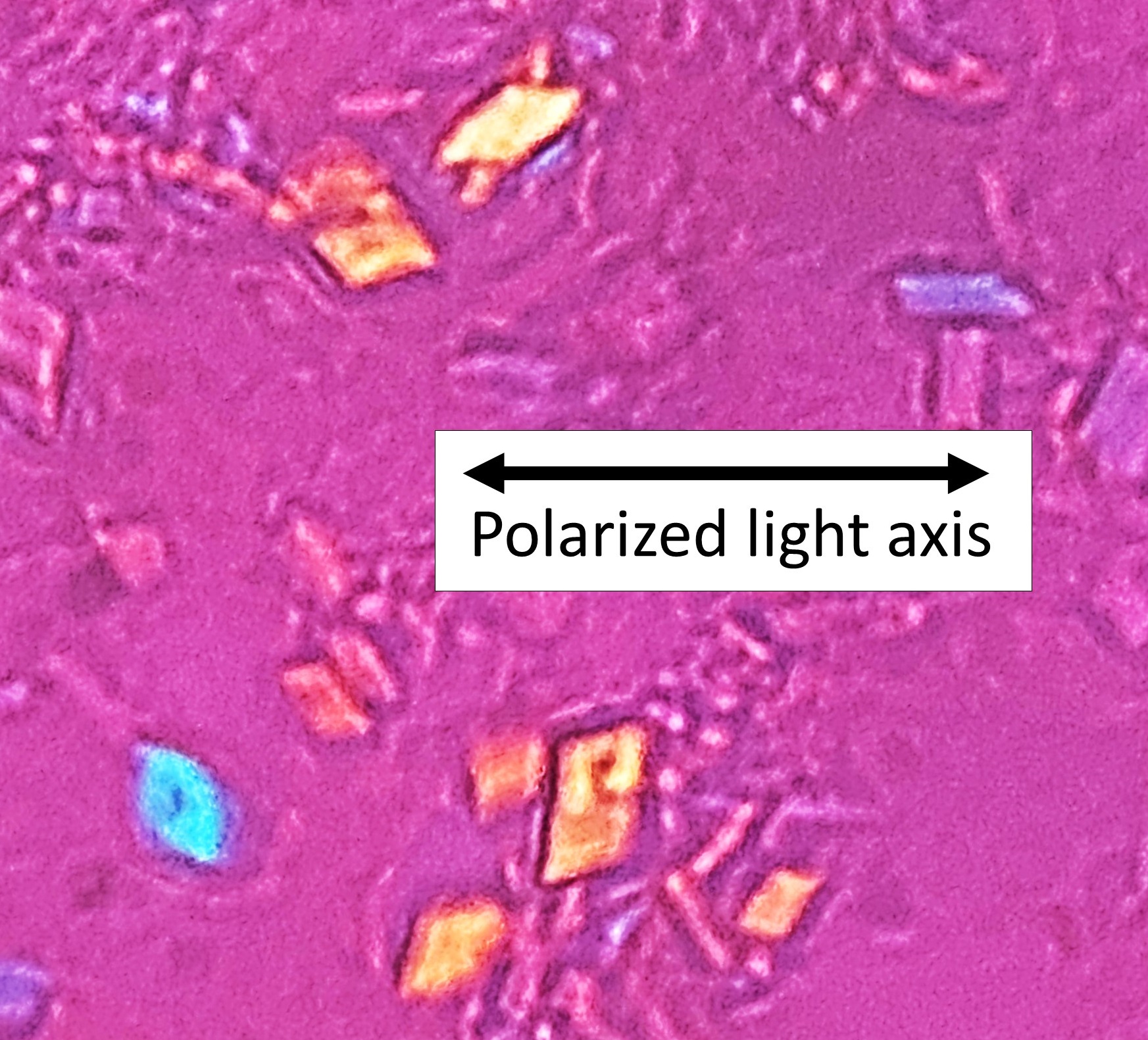
Calcium Pyrophosphate Dihydrate Crystal Deposition Disease
Wikipedia
There can also be findings of osteoarthritis . [4] [3] The white blood cell count is often raised. [3] In many instances, patients may also have signs of carpal tunnel syndrome . [3] This condition can also be associated with Milwaukee shoulder syndrome . [ citation needed ] Cause [ edit ] Calcium pyrophosphate The cause of CPPD disease is unknown. ... However, if an acute attack is already occurring, higher doses are administered. [3] If nothing else works, hydroxychloroquine or methotrexate may provide relief. [13] Research into surgical removal of calcifications is underway, however, this still remains an experimental procedure. [3] Epidemiology [ edit ] The condition is more common in older adults. [4] CPPD is estimated to affect 4% to 7% of the adult populations of Europe and the United States. [14] Previous studies have overestimated the prevalence by simply estimating the prevalence of chondrocalcinosis , which is found in many other conditions as well. [14] It may cause considerable pain, but it is never fatal. [3] Women are at a slightly higher risk than men, with an estimated ratio of occurrence of 1.4:1. [3] History [ edit ] CPPD crystal deposition disease was originally described over 50 years ago. [12] Terminology [ edit ] Calcium pyrophosphate dihydrate crystals are associated with a range of clinical syndromes , which have been given various names, based upon which clinical symptoms or radiographic findings are most prominent. [12] A task force of the European League Against Rheumatism (EULAR) made recommendations on preferred terminology . [5] Accordingly, calcium pyrophosphate deposition (CPPD) is an umbrella term for the various clinical subsets, whose naming reflects an emphasis on particular features. ... External links [ edit ] Classification D ICD - 10 : M11.1 - M11.2 ICD - 9-CM : [712.3 275.49 [712.3]] OMIM : 600668 118600 MeSH : D002805 DiseasesDB : 10832 External resources MedlinePlus : 000421 eMedicine : med/1938 radio/125 orthoped/382 emerg/221 v t e Diseases of joints General Arthritis Monoarthritis Oligoarthritis Polyarthritis Symptoms Joint pain Joint stiffness Inflammatory Infectious Septic arthritis Tuberculosis arthritis Crystal Chondrocalcinosis CPPD (Psudogout) Gout Seronegative Reactive arthritis Psoriatic arthritis Ankylosing spondylitis Other Juvenile idiopathic arthritis Rheumatoid arthritis Felty's syndrome Palindromic rheumatism Adult-onset Still's disease Noninflammatory Hemarthrosis Osteoarthritis Heberden's node Bouchard's nodes Osteophyte
-
Southeast Asian Ovalocytosis
Wikipedia
External links [ edit ] Classification D ICD - 10 : D58.1 ICD - 9-CM : 282.1 OMIM : 109270 MeSH : C566230 DiseasesDB : 9416 v t e Diseases of red blood cells ↑ Polycythemia Polycythemia vera ↓ Anemia Nutritional Micro- : Iron-deficiency anemia Plummer–Vinson syndrome Macro- : Megaloblastic anemia Pernicious anemia Hemolytic (mostly normo- ) Hereditary enzymopathy : Glucose-6-phosphate dehydrogenase deficiency glycolysis pyruvate kinase deficiency triosephosphate isomerase deficiency hexokinase deficiency hemoglobinopathy : Thalassemia alpha beta delta Sickle cell disease / trait Hereditary persistence of fetal hemoglobin membrane : Hereditary spherocytosis Minkowski–Chauffard syndrome Hereditary elliptocytosis Southeast Asian ovalocytosis Hereditary stomatocytosis Acquired AIHA Warm antibody autoimmune hemolytic anemia Cold agglutinin disease Donath–Landsteiner hemolytic anemia Paroxysmal cold hemoglobinuria Mixed autoimmune hemolytic anemia membrane paroxysmal nocturnal hemoglobinuria Microangiopathic hemolytic anemia Thrombotic microangiopathy Hemolytic–uremic syndrome Drug-induced autoimmune Drug-induced nonautoimmune Hemolytic disease of the newborn Aplastic (mostly normo- ) Hereditary : Fanconi anemia Diamond–Blackfan anemia Acquired: Pure red cell aplasia Sideroblastic anemia Myelophthisic Blood tests Mean corpuscular volume normocytic microcytic macrocytic Mean corpuscular hemoglobin concentration normochromic hypochromic Other Methemoglobinemia Sulfhemoglobinemia Reticulocytopenia
-
Aortic Aneurysm, Familial Thoracic 6
Omim
Despite the young age of death of some family members, the Kaplan-Meier survival curve of the cohort estimated a median survival of 67 years, suggesting that the disease is less deadly than Loeys-Dietz syndrome (see 609192) and similar to treated Marfan syndrome (MFS; 154700). ... In 40 German probands with thoracic aortic aneurysms, 21 of whom had clinical features suggestive of Marfan syndrome, but all of whom were negative for mutation in the FBN1 (134797) and TGFBR2 (190182) genes, Hoffjan et al. (2011) sequenced the ACTA2 gene and identified heterozygous mutations in 3 patients (see, e.g., 102620.0005 and 102620.0006).
-
Disordered Steroidogenesis Due To Cytochrome P450 Oxidoreductase Deficiency
Omim
Mutations in this gene also result in a form of Antley-Bixler syndrome (ABS1; 201750). Description This rare variant of congenital adrenal hyperplasia, caused by mutations in the POR gene, results in apparent combined deficiency of P450C17 (609300) and P450C21 (613815) and accumulation of steroid metabolites. ... Boys and girls can present with bone malformations, sometimes suggesting the pattern seen in patients with Antley-Bixler syndrome (see 207410) (summary by Arlt et al., 2004). ... In 1 family an affected sister showed bony changes similar to those seen in Antley-Bixler syndrome, as well as ambiguous genitalia; her brother with congenital adrenal hyperplasia had normal male genitalia at birth and no skeletal abnormalities.CYP17A1, HSD3B2, CYP21A2, POR, PDE8B, PRKAR1A, AVPR2, HTR4, AVPR1A, CYP11B1, STAR, CYP11B2, PDE11A, KCNJ5, PRKACA, USP8, CDH23, CYP21A1P, CYP2B6, POMC, NR3C1, GML, AIRE, CACNA1D, TBC1D24, TNXB, AR, HSD3B1, CHST3, CYP19A1, GLO1, HLA-A, CYP4F3, NR0B1, MC2R, HSD11B2, TNXA, SRY, REN, GIP, HLA-B, GH1, CYP2C19, CRH, RNU1-4, TWIST1, SDHD, TP53, PPIG, SCO2, VWF, TGM5, SULT2A1, ARMC5, COASY, CGB5, CGB8, RBM45, OTOA, EYS, ACADVL, SDHB, PROP1, ATP7B, AVP, AZF1, BCL2, C4B, CGA, CGB3, CRYGD, CYP3A7, CYP2B7P, CYP11A1, ENPEP, FH, GRB7, HADHA, HLA-DRB1, HTC2, TNC, LEP, MDH2, MEN1, SERPINA1, PRKACB, LOC110673972


